Artículo original
Micronutrients status along with hematological and biochemical
parameters in sickle dubtypes: preliminary report from India
Pandey S,* Ranjan R,* Toteja GS,** Rao S,** Mishra RM,*** Pandey Sw,* Saxena R*
RESUMEN
Antecedentes:
Objetivo: evaluar las concentraciones de varios micronutrientes en pacientes con anemia drepanocítica y estudiar los parámetros bio-químicos y hematológicos asociados con la gravedad de la enfermedad.
Material y método: estudioÖ..al que se incluyeron 25 pacientes con drepanocitosis y 30 sujetos controles. El diagnóstico de drepano-citosis se hizo por cromatografía líquida de alta resolución. En todos los pacientes y controles se analizaron las características clínicas y los parámetros hematológicos. Se midieron la cifras de glucosa, bilirrubina total, proteínas totales, transaminasas, fosfatasa alcalina,
SURWHtQD&UHDFWLYD\ORVPLFURQXWULHQWHVFLQFVHOHQLRFREUHYLWDPLQD$\YLWDPLQD(/RVSDFLHQWHVFRQGUHSDQRFLWRVLVVHVXEODVL¿FDURQ
en tres grupos: heterocigotos (n = 6), homocigotos (n = 8) y dobles heterocigotos para hemoglobinopatía S y talasemia beta (n =11). Las transaminasas hepáticas se encontraron elevadas, principalmente la AST. En los pacientes homocigotos la proteína C-reactiva, fosfatasa
DOFDOLQD\JOXFRVDVHHQFRQWUDURQHOHYDGDV\ODVFRQFHQWUDFLRQHVGHFLQFVHOHQLR\YLWDPLQD$HVWXYLHURQVLJQL¿FDWLYDPHQWHGLVPLQXLGDV
Las concentraciones de vitamina E disminuyeron en los pacientes dobles heterocigotos.
Conclusiones: las concentraciones de varios micronutrientes están disminuidas en los pacientes drepanocíticos de la India y asociadas
VLJQL¿FDWLYDPHQWHFRQSDUiPHWURVKHPDWROyJLFRV\ELRTXtPLFRVUHODFLRQDGRVFRQODJUDYHGDGGHOSDGHFLPLHQWR Palabras clave: micronutriente, SCD, talasemia beta, dobles heterocigotos
ABSTRACT
The objective of the study was toestimate the level of various micronutrients in sickle cell patients and to study the various biochemical and haematological parameters associated with the disease severity.7ZHQW\¿YHVLFNOHFHOOSDWLHQWVDQGWKLUW\FRQWUROVZHUHVHOHFWHGDV
the subjects of this study. The patients were diagnosed by HPLC. The clinical features and haematological parameters were recorded for all the patients and controls. Glucose (random), Total Bilirubin,Total protein, AST, ALT, Alkaline phosphate and CRP had been evaluated along with micronutrients; zinc, selenium ,Cu, vitamin A and Vitamin E in all the subjects.Sickle cell patients were sub typed into three groups sickle trait (N=6), sickle homozygous (N=8) and Sickle beta thal (N=11). Key enzymes of liver functioning (AST and ALT) were
HOHYDWHGZKLOH$67ZDVVWDWLVWLFDOO\VLJQL¿FDQWEXW$/7ZDVQRQVLJQL¿FDQW3YDOXH&53$ONDOLQHSKRVSKDWDVHDQGUDQGRP JOXFRVHOHYHOVZHUHLQFUHDVHGLQVLFNOHKRPR]\JRXVSDWLHQWV=Q6HODQG9LWD$ZHUHVLJQL¿FDQWO\ORZHULQVLFNOHKRPR]\JRXVSDWLHQWV 9LWDPLQ(ZDVVLJQL¿FDQWO\UHGXFHGLQVLFNOHEHWDWKDOSDWLHQW>@7KXVLWZDVFRQFOXGHGWKDWWKHOHYHOVRIYDULRXVPLFUR
-QXWULHQWV=Q&X6HO9LWD$ (ZHUHUHGXFHGLQ,QGLDQ6&'SDWLHQWVDQGDVVRFLDWHGVLJQL¿FDQWO\ZLWKWKHELRFKHPLFDO KHPDWRORJLFDO
parameters and enhance the disease severity.
Key words: Micronutrient, SCD, Sickle beta thalassemia, Sickle homozygous
* Department of Hematology, AIIMS, New Delhi, India. ** ICMR, New Delhi, India.
*** APS University Rewa MP
Correspondence: Dr. Renu Saxena. Department of Hematology, I.R.C.H. Building (1stÀRRU$OO,QGLD,QVWLWXWHRI0HGLFDO6FLHQFHV Ansari Nagar, New Delhi - 110029, India.
E-mail: renusax@hotmail.com
Recibido: Julio 2011. Aceptado: Julio 2011
M
icronutrients are chemical entities, which
in-clude minerals and vitamins, required in trace
amount in living organisms for maintaining
JRRGKHDOWK7KHGH¿FLHQF\RIWKHVHPLFURQXWULHQWVUH
-sults in varying the severity of various disorders. Sickle
cell disease (SCD) is such an inherited hemoglobin
di-sorder characterized by sickled red blood cells in which
the severity markedly increases due to micronutrient
GH¿FLHQF\%ORRGOHYHOVRIVHYHUDOYLWDPLQVDQGPLQHUDOV
are often low in individuals with SCD, including
vita-min A and carotenoids,
vitamin B6, vitamin C, vitamin
E, magnesium and zinc.
1-87KHVH GH¿FLHQFLHV FDXVH D
VLJQL¿FDQW GHSUHFLDWLRQ LQ EORRGDQWLR[LGDQW VWDWXV LQ
these patients
and the resulting oxidative stress may
precipitate vaso-occlusion related acute chest syndrome.
9-10
Studies indicate that vitamin-mineral supplements of
certain nutrients (vitamins C and E, zinc, magnesium)
or treatment with a combination of high-dose
antioxi-dants can reduce the percentage of irreversibly sickled
cells.
5,11-14Zinc is known as an important nutrient for
JURZWKDQGGHYHORSPHQW$GH¿FLHQF\RI]LQFLQSDWLHQWV
with sickle cell disease results in growth retardation
GZDU¿VP K\SRJRQDGLVP LQ PDOHV URXJK VNLQ SRRU
appetite, mental lethargy and recurrent infections.
7Prasad
HWDO¿UVWUHSRUWHG]LQFGH¿FLHQF\LQDGXOWSDWLHQWVZLWK
sickle cell disease.
15The biological importance of iron is well known in the
synthesis of hemoglobin. Copper is known to be essential
in the proper functioning of different metalloenzymes,
which include ceruloplasmin involved in iron metabolism
ZKHUHDVLWVGH¿FLHQF\
is known to cause anemia.
16Certain
minerals, copper, iron, magnesium selenium as well as
some antioxidants and vitamins (C, E, folate, B6, B12)
have been found to effectively relieve the oxidative stress
that prevails in SCD.
17-19The pathology of sickle cell hemoglobin is considerably
variable. The clinical expression of HbS is reported to be
variable even within the same population. The effect on
hematological and biochemical parameter values is also
YDULDEOHDQGLVLQÀXHQFHGE\WKHVHYHULW\RIWKHGLVHDVHDQG
the occurrence of the crisis. In the heterozygous state, the
presence of HbS in low concentration in red cells does not
result in the sickling phenomenon under normal conditions
and so the carriers of HbS are generally asymptomatic with
normal hematological parameters values. The homozygous
VWDWHKDVEHHQGH¿QHGDVDQLQFDSDFLWDWLQJGLVHDVHLQZKLFK
the hematological and several biochemical parameter
values are abnormal.
20-22Acute phase reactants such as C-reactive protein (CRP)
is one of the important biochemical parameter which is
mo-derately increased in stable symptom free sickle patients
DQGVLJQL¿FDQWO\LQFUHDVHGGXULQJSDLQIXOYDVRRFFOXVLYH
crisis and C-reactive protein (CRP) indicated
LQÀDPPD
-tion.
23-25Serum bilirubin levels is markedly increased due
to increased rate of breakdown of hemoglobin and serves
as an important biochemical indicator of SCD. In
addi-tion, hepatic dysfunctions may occur due to sequestration
of the liver by the sickled cells. Bone infarction due to
YDVRFFOXVLRQRIWKHFDSLOODULHVLVDFRPPRQ¿QGLQJDQG
is associated with bone pains in SCD which results in
in-creased alkaline phosphatase level due to constant damage
repair by osteoblasts.
26,27The rate of glucose consumption
and pyruvate and lactate production in red blood cells of
QRUPDOVLFNOHFHOOWUDLWDQG66GLVHDVHVXEMHFWVZHUHPHD
-sured and Glucose consumption was found to be increased
VLJQL¿FDQWO\ LQ UHG FHOOV RI VLFNOH FHOO SDWLHQWV WKDQ WKH
sickle cell trait and normal persons.
28Aspartate
amino-transferase (AST) and Alanine aminoamino-transferase (ALT)
levels are reported to be elevated in SCD patients with
liver enlargement. In Indian scenario there are very few
UHSRUWHGOLWHUDWXUHRQWKHVXEMHFWRIPLFURQXWULHQWVWDWXVLQ
sickle cell patients and its relationship with hematological
and biochemical parameters so in this study it was aimed
DW¿QGLQJWKHOHYHOVDQGVWDWXVRIWKHVHPLFURQXWULHQWVLQ
Indian SCD patients.
MATERIAL AND METHOD
Sickle cell Patients attending haematology OPD (Out
Pa-tient Department), All India Institute of Medical Sciences,
1HZ'HOKLIRUURXWLQHIROORZXSZHUHVHOHFWHGDVVXEMHFW
of the study after getting the signed consent form. Twenty
¿YH VLFNOH FHOO SDWLHQWV ZKLFK ZHUH GLDJQRVHG E\ +LJK
Performance Liquid Chromatography (HPLC) along with
30 healthy individuals were selected as the control for the
study. Control population were chosen from the healthy
relatives of patients had never diagnosed any disease. All
the patients and control were adults with similar age group.
Duration of the study was one year.
and red cell indices were measured by automated
Analy-zer (SYSMEX K-4500, Kobe Japan) using Transasia
diagnostic kit. Quantitative assessment of hemoglobin
variants; HbF, HbA, HbA2, and HbS was performed by
HPLC (Bio-Rad-Variant
TMfrom Bio Rad, CA, USA)
using Bio-Rad diagnostic kit. All biochemical
investi-gation were done by modular-P-800 auto analyzer from
Roche (USA) using Roche diagnostic kit. Serum samples
were diluted 50 folds using 1% nitric acid for the
analy-VLVRI]LQFFRSSHUDQGVHOHQLXPE\7KHUPR6FLHQWL¿F
ICPMS X-Series -2 (Bremen, Germany). Gallium was
used as the internal standard and seronorm trace element
serum (Level 1 and 2) was used as the reference material
for ensuring accuracy of analysis. Analysis of Vitamin
A and E was carried out on LC-6AD Binary Gradient
HPLC System (Shimadzu, Kyoto, Japan) using the Bieri
et. al method.
29CRP was analyzed using Immulite-1000
(Siemens, USA).
Statistical analysis were done by Pearson chi square and
Fisher’s exact test. P- values below 0.05 were considered
DVVWDWLVWLFDOO\VLJQL¿FDQW
All haematological and biochemical parameters were
evaluated in the department of haematology while micro
nutrient parameters evaluated in the ICMR reference
laboratory campus -2.
RESULTS
7ZHQW\ ¿YH VLFNOH FHOO SDWLHQWV ZHUH VXEW\SHG RXW RI
which 6 were sickle trait [HbS=37.81(10-45.2)], 8 sickle
homozygous [HbF =8.8(6-14.6) and HbA2= 2.65(1.
9-3)] and 11 were sickle beta thal [HbF= 22.5(20-39.7)
and HbA2 = 4.5(3.3-6.8]. Patients were different states
from India i.e Karnataka (6), Bihar (5), Orissa (4)
Mad-hya Pradesh (4) Jharkhand (3) and Delhi (3). In study
design we distributed them in 4 group, group -1 sickle
trait, group 2- sickle homozygous, group -3 sickle beta,
group -4 healthy control. We evaluated hematological,
biochemical, micronutrient and clinical parametrs in all
above mentioned groups.
The demographic variables are given in table 1, which
VKRZHGZHLJKWDQGKHLJKWWREHVWDWLVWLFDOO\VLJQL¿FDQW
(P = 0.0003 and 0.0079 respectively)
The biochemical variables (Table 2) showed that
random glucose level (104 g%) was high in sickle
homozygous. Total Bilirubin (2.2 mg %), Aspartate
aminotransferase (AST = 68 IU) and Alanine
amino-transferase (ALT 37 I.U.) was high in sickle beta thal
patients but ALT was non-significant (P = 0.4598)
whereas AST and total bilirubin were statistically
sig-nificant. (P = 0.0012 and 0.0001 respectively). Alkaline
phosphate (367 IU) was high in sickle homozygous
patients among all the groups and the P-value was in
the border range (0.05). C-reactive protein (CRP) (10.96
mg/L) was significantly increased in sickle homozygous
patients. (P=0.0292)
Hematological parameters were evaluated in all patents
and control group (Table-3). All the parameters were
sta-WLVWLFDOO\VLJQL¿FDQWH[FHSW0&+0&+&DQG:%&+&7
value was similar across all the patient groups. Low RBC
levels were seen in sickle homozygous (3.365 millions/
ȝ/DQGVLFNOHEHWDWKDOPLOOLRQVȝ/6LPLODUO\+H
-moglobin level were also low in sickle homozygous (9.1
g/dL) and sickle beta thal (9.1 g/dL). MCV was lowest in
VLFNOHEHWDWKDOÀZKHUHDV
MCHC value was
hig-hest in sickle beta thal (33.1 g/dL). Lowest platelets were
UHFRUGHGLQVLFNOHEHWDWKDO7KVȝ/
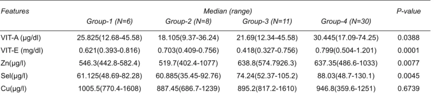
Micronutrient variables (VIT-A, VIT-E, Zn, Sel and
Cu) were evaluated in all the groups and the results are
tabulated in Table 4. All the variables were statistically
VLJQL¿FDQW H[FHSW &X LQ WKH SDWLHQW JURXS 9LWDPLQ$
(18.105 μg/dL), Zn (519.7 μg/L) and Selenium (60.885
μg/L) were low in sickle homozygous patients whereas
Vitamin E (0.418 mg/dL) was lowest in sickle beta that
patients.
Various clinical features associated with sickle cell
disease were recorded for all the patients. (Table 5)
Anemia and pain (33.33 %) were the main clinical
findings among the sickle trait patients whereas two
patients had normal profile. Half of the Sickle
homo-zygous patients were having painful episode and fever
DV WKH PDMRU FOLQLFDO ILQGLQJ ZKHUHDV WKUHH SDWLHQWV
were reported to have anemia. In the third group,
the most important findings were splenomegaly and
MDXQGLFHDIIHFWLQJRIWKHVHSDWLHQWV1RQHRI
the controls showed any clinical symptoms associated
with the sickle disease.
The demographic variables showed that the height and
ZHLJKWRIWKHVLFNOHFHOOSDWLHQWVZHUHVLJQL¿FDQWO\ORZHU
when compared to control, which is well known.
7Next we
looked at the biochemical parameters in which sickle beta
that patients had high levels of the key enzymes AST and
$/7ZKLFKKHOSVLQ¿QGLQJRXWWKHQRUPDOIXQFWLRQLQJRI
DISCUSSION
This study was a preliminary investigation of the valuable
micronutrient status in association with hematological and
biochemical parameter’s that affects the clinical
manifes-tation in sickle cell patients.
Table 1. Demographic variables
)HDWXUHV 0HGLDQUDQJH P-value
*URXS1 *URXS1 *URXS1 *URXS1
Age(Yrs) 25(15-38) 22(18-32) 21(17-35) 26(15-40) 0.0859
Sex M 3(50) 7(87.50) 7(63.64) 18(60)
F 3(50) 1(12.50) 4(36.36) 12(40)
Weight (Kg) 50(45-67) 49(40-61) 45(40-65) 60(40-70) 0.0003
Height (Cm) 162.5(130-170) 162.5(130.5-180) 155(140-170) 170.5(150-177.5) 0.0079
Table 2. Biochemical variables
)HDWXUHV 0HGLDQUDQJH P-value
*URXS1 *URXS1 *URXS1 *URXS1
Glucose (random) (gm %) 92(63-131) 104(80-130) 100(50-151) 68.5(28-106) 0.0003 Total Bilrubin (mg %) 1.2(0.6-1.9 ) 1.4(0.7-1.6) 2.2(1.1-6.1) 0.6(0.2-2.2) 0.0001 Total protein (gm%) 7.15(6.4-7.9) 7.25(5.9-7.7) 7.1(6.2-7.8) 7.4(6-7.9) 0.9067
AST (IU) 58.5(24-102) 45(33-86) 68(26-111) 34(12-72) 0.0012
ALT (IU) 32(20-55) 28.5(17-59) 37(16-66) 28.5(7-50) 0.4598
Alkaline Phosphate (IU) 301(104-628) 367(178-596) 336.5(169-792) 224(139-346) 0.0500 CRP (mg/l) 0.69(0.379-1.5) 10.96(0.3-17.4) 3.15(0.427-9.78) 0.683(0-1.2) 0.0292
Table 3. Hematological variables
)HDWXUHV 0HGLDQUDQJH P-value
*URXS1 *URXS1 *URXS1 *URXS1
:%&7KVȝO 7.4(5.3-11.5) 9.2(4.321.1) 7.4(3.9-20.13) 8.39(3.94-12.75) 0.4431
5%&PLOOLRQVȝO 4.18(4.26-6.02) 3.365(2.58-3.82) 3.3(2.11-5.59) 4.99(3.96-7.78) 0.0001 HGB (g/dl) 12.8(10.1-15.1) 9.1(4.7-13.1) 9.1(5.6-12.3) 14.45(13.3-17.2) 0.0001
HCT (%) 28.55(24.3-42.70) 28.6(20.6-38.6) 28.3(17.8-35.2) 47.15(29.7-56.1) 0.0001
0&9À 82.15(64.6-90.8) 73.4(13.6-97.6) 70.5(25.6-85.3) 95.45(80.5-100) 0.0001
MCH (pg) 26.75(25.3-30) 30.75(20.4-42) 26.1(20.6-86.2) 29.55(26.9-32.8) 0.1748
MCHC(g/dl) 31.55(25.3-33) 31.45(26.1-39.9) 33.1(30.3-39.9) 29.9(33-35.3) 0.0212
Table 4. Micronutrient variables
)HDWXUHV 0HGLDQUDQJH P-value
*URXS1 *URXS1 *URXS1 *URXS1
VIT-A (μg/dl) 25.825(12.68-45.58) 18.105(9.37-36.24) 21.69(12.34-45.58) 30.445(17.09-74.25) 0.0388
VIT-E (mg/dl) 0.621(0.393-0.816) 0.703(0.409-0.756) 0.418(0.327-0.756) 0.799(0.504-1.201) 0.0001
Zn(μg/l) 546.3(442.8-582.4) 519.7(402.4-1077) 638.8(574.7926.3) 637.35(486.6-1033) 0.0077
Sel(μg/l) 61.125(48.69-82.28) 60.885(35.45-92.76) 74.24(52.37-105.2) 88.03(48.7-130.1) 0.0045
Cu(μg/l) 1005.5(770.4-1608) 887.45(686.7-1239) 895.2(817.2-1610) 946.8(359.6-1251) 0.6739
Table 5.&OLQLFDO¿QGLQJV
&OLQLFDOIHDWXUHV )UHTXHQF\ 7RWDO
*URXS1 *URXS1 *URXS1 *URXS1
Anemia 2(33.33) 3(37.50) 2(18.18) 0 7(12.73)
Fever 1(16.67) 4(50) 2(18.18) 0 7(12.73)
Pain 2(33.33) 4(50) 4(36.36) 0 10(18.18)
Splenomegaly 0 1(12.50) 5(45.45) 0 6(10.91)
Jaundice 0 2(25) 5(45.45) 0 7(12.73)
Chest pain 0 1 (12.50) 2(18.18) 0 3(5.45)
Osteonecrosis 0 1 (12.50) 0 0 1(1.82)
G6PD 0 1 (12.50) 0 0 1(1.82)
Joint pain 0 1 (12.50) 0 0 5(9.09)
Weakness 0 1 (12.50) 1(9.09) 0 2(3.64)
Hyper – pigmentation 0 0 1(9.09) 0 1(1.82)
Hepato-megaly 0 0 2(18.18) 0 2(3.64)
Pallor 0 0 2(18.18) 0 2(3.64)
Stones 0 0 1(9.09) 0 1(1.82)
normal 2(33.33) 0 1(9.09) 30(100) 33(60)
liver. Since these patients have liver dysfunction, the levels
of these enzymes are elevated. Total bilirubin was elevated
in sickle beta thal as expected since the heavy burden of
pigment resulting from increased red cell destruction lead
to extreme hyper-bilirubinemia with SCD, when combined
with acute
hepatic damage or common bile duct
obstruc-tion. Under similar circumstances,
increased haemolysis
from any cause may lead to markedly elevated levels of
serum bilirubin.
30The Alkaline phosphatase was high in
the sickle homozygous as bone complications are more
common in this subtype of SCD. Alkaline phosphatase
levels is considered as a sensitive marker of bone turnover
and could be especially useful as valuable non-invasive
biochemical marker for identifying sickle cell patients
with bone complications.
31CRP was elevated in the sickle homozygous patients
because these patients suffer from vaso-occlusion, which
results in damage to the endothelial walls of the blocked
FDSLOODULHVZKLFKLQLWLDWHVDQLQÀDPPDWRU\UHVSRQVHOHD
-ding to the increased levels of this protein.
32This study
mainly focused on the micronutrient levels in the various
subtypes of SCD. Vitamin E levels were reduced in sickle
beta thal patients due to higher consumption of this
vita-min.
33, 346SOHQRPHJDO\ MDXQGLFH VWRQHV DQG FKHVW SDLQ
and may be associated with vitamin E. Supplementation
with this
vitamin has been shown to restore plasma vitamin
E levels, improve
clinical outcome, and reduce the number
of irreversible sickled
red cells
.
18, 35-379LWDPLQ$ ZDV VLJQL¿FDQWO\ UHGXFHG LQ DOO VXEW\SHV
whereas sickle homozygous patients had the lowest level
of this vitamin. Vitamin A Level has been reported to be
low and associated with increased hospitalizations, poor
growth and hematological status in patients with sickle
cell disease. It is important to note that zinc is required
for the synthesis of retinol binding protein (RBP), and the
decreased level of zinc found in patients with Sickle cell
anemia may affect the level of RBP and vitamin A in these
patients despite adequate intake.
38-40,QWKLVVWXG\]LQFZDVVLJQL¿FDQWO\SUHVHQWLQORZOHYHOV
in the sickle homozygous subtype. This may be attributed
WRWKHIDFWWKDW]LQFGH¿FLHQF\LVDVVRFLDWHGZLWKVLFNOH
cell anemia and appears to play a role in various aspects
of the illness. The preliminary research has correlated low
zinc levels with poor growth in children with sickle cell
anemia.
41Zinc supplementation is reported to decrease the
number of infections in adults with sickle cell anemia.
42Copper was reduced in the sickle homozygous subtype but
when compared to controls its levels were not
statistica-OO\VLJQL¿FDQW$OD\DVKHWDOKDYHUHSRUWHGWKDW]LQFDQG
copper levels in patients were found to be close to those
RIWKHFRQWUROVXEMHFWVZKLFKLVVLPLODUWRRXU¿QGLQJEXW
a contrasting situation exists in North American Black
VXEMHFWVZLWKVLFNOHFHOODQHPLD
43Selenium is another important micronutrient associated
with SCD and it has been seen from earlier studies that the
OHYHOVRIVHOHQLXPZDVVLJQL¿FDQWO\ORZHULQ6&'WKDQ
those of controls.
447KLV ¿QGLQJV DOVR VKRZ FRQVLVWHQW
decrease in selenium levels in all the subtypes of SCD.
Another important association of SCD is with the
various hematological parameters, which help in
asses-sing the severity of the disease. Red blood cell indices
(MCV, MCHC and MCH) help classify types of anemia,
a decrease in the oxygen carrying capacity of the blood.
,QWKLVVWXG\WKHUHVXOWVVKRZWKDW0&9LVVLJQL¿FDQWO\
decreased in the sickle beta thal patient, which is the
characteristic feature of microcytic anemia. Patients with
Sickle cell Anemia have reduced hemoglobin or
hemato-crit (HCT)
levels which were observed in the results. The
sickle homozygous subtype has increased WBC count.
It is known that WBC contribute to SCD by adhering to
blood vessel walls and obstructing the lumen, aggregating
with other blood cells with more effective blockage of the
lumen, stimulating the vascular endothelium to increase
its expression of ligands for adhesion molecules on blood
FHOOVDQGFDXVLQJWLVVXHGDPDJHDQGLQÀDPPDWRU\UHDFWLRQ
which predispose to vaso-occlusion.
45CONCLUSION
From this study it was concluded that the levels of various
micronutrients (Zn ,Cu,Sel, Vita-A and E) were reduced in
indian SCD patients. It was also found the biochemical and
KHPDWRORJLFDO SDUDPHWHUV ZHUH DVVRFLDWHG VLJQL¿FDQWO\
ZLWKWKHPLFURQXWULHQWVOHYHODQGSOD\VLJQL¿FDQWUROH
to modulate disease severity in SCD patients. A larger
SRSXODWLRQ VWXG\ LV UHTXLUHG WR FRQ¿UP WKH SUHOLPLQDU\
¿QGLQJLQWKLVVWXG\
REFERENCES
1. Gray NT, Bartlett JM, Kolasa KM, et al. Nutritional status and dietary intake of children with sickle cell anemia. Am J Pediatr Hematol Oncol 1992;14:57-61.
2. Tangney CC, Phillips G, Bell RA, et al. Selected indices of micronutrient status in adult patients with sickle cell anemia (SCA). Am J Hematol 1989;32:161-166.
3. Segal JB, Miller ER III, Brereton NH, et al. Concentrations of B vitamins and homocysteine in children with sickle cell anemia. South Med J 2004;97:149-155.
4. Westerman MP, Zhang Y, McConnell JP, et al. Ascorbate levels in red blood cells and urine in patients with sickle cell anemia. Am J Hematol 2000;65:174-175.
5. Marwah SS, Blann AD, Rea C, et al. Reduced vitamin E an-tioxidant capacity in sickle cell disease is related to transfusion status but not to sickle crisis. Am J Hematol 2002; 69:144-146. 6. Zehtabchi S, Sinert R, Rinnert S, et al. Serum ionized magne-sium levels and ionized calcium-to-magnemagne-sium ratios in adult patients with sickle cell anemia. Am J Hematol 2004;77:215-222.
7. Zemel BS, Kawchak DA, Fung EB, et al. Effect of zinc sup-plementation on growth and body composition in children with sickle cell disease. Am J Clin Nutr 2002;75:300-307. 8. Riddington C, De Franceschi L. Drugs for preventing red blood
cell dehydration in people with sickle cell disease. Cochrane Database Syst Rev 2002;CD003426.
9. Blann AD, Marwah S, Serjeant G, et al. Platelet activation and endothelial cell dysfunction in sickle cell disease is unrelated to reduced antioxidant capacity. Blood Coagul Fibrinolysis 2003;14:255-259.
11. Jaja SI, Ikotun AR, Gbenebitse S, et al. Blood pressure, hema-tologic and erythrocyte fragility changes in children suffering from sickle cell anemia following ascorbic acid supplementa-tion. J Trop Pediatr 2002;48:366-370.
12. Ohnishi ST, Ohnishi T, Ogunmola GB. Sickle cell anemia: a potential nutritional approach for a molecular disease. Nutrition 2000;16:330-338.
13. De Franceschi L, Bachir D, Galacteros F, et al. Oral magnesium supplements reduce erythrocyte dehydration in patients with sickle cell disease. J Clin Invest 1997;100:1847-1852. 14. Muskiet FA, Muskiet FD, Meiborg G, et al. Supplementation of
patients with homozygous sickle cell disease with zinc,
alpha-WRFRSKHUROYLWDPLQ&VR\EHDQRLODQG¿VKRLO$P-&OLQ1XWU
1991;54:736-744.
15. Prasad AS, Schoomaker EB, Ortega J, Brewer GJ, et al. Zinc
GH¿FLHQF\LQVLFNOHFHOOGLVHDVH&OLQLFDOFKHP
587.
16. Prasad AS. Zinc and Trace minerals. Workshop on Nutrient Metabolism in genetic Anemias, NHLBI 1999, Bethesda MD. 17. Natta CL, Machlin LJ, Brin M. A decrease in irreversibly
sickle cell anemia patients given vitamin E. Am J Clin Nutr 1980;33:968-971.
18. Whethers DL. Introduction and overview of the problem. WHO Expert Committee on Malaria (1998). Technical report Series1999;889.
19. Sies H, Stahl W, Sevanian M. Nutritional, dietary and postpran-dial oxidative stress. J Nutr 2005;135(5):969-972.
20. Serjeant GR. The clinical features of sickle cell disease. In: Clinical Studies, ed Bearn AG, Black DAK, Hiatt HH. North-Holland Publishing Company, 4:59, Amsterdam, 1974. 21. Weatherall DJ, Clegg JB, Blankson J, McNeil JR. A new
sickling disorder resulting from interaction of the genes of hemoglobin S and alpha-thalassaemia. Br J Hematol 1969; 17:517-526.
22. Afonja OA. Osteoblastic activity in sickle cell disease. Clin Chem Newsletter 1982; 3:161-163.
23. Singhal A, Doherty JF, Raynes JG, McAdam KPWJ, et al. Is there an acute-phase response in steady state sickle cell disease? Lancet 1993;341:651-653.
24. Stuart J, Stone PCW, Akinola NO, Gallimore JR, et al. Moni-toring the acute phase response to vaso-occlusive crisis in sickle cell disease. J Clin Pathol 1994;47:166-169.
25. Jacqueline M, et al. Proinflammatory Cytokines and the Hypermetabolism of Children with Sickle Cell Disease. Experimental Biology and Medicine 2005;230:68-74. 26. Barrett-Conner E. Bacterial infections and sickle cell ane-mia: An analysis of 250 infections in 166 patients and a review of the literature. Medicine 1971;50:97-112.
27. Kotila T, Adedapo K, Adedapo A, Oluwasola O, et al. Liver dysfunction in steady state sickle cell disease. Ann Hepatol 2005;4(4):261-263.
28. Dash BP, Mitra A, Kar BC. A study on the glucose uptake, pyruvate and lactate formation in red blood cells of normal,
sickle cell trait and sickle cell patients. Indian Journal of Clinlcal Biochemistry 1992;7(2):134-137.
29. Bieri JG, Tolliver IJ, Catiguiani GL. Simultaneous determina-tion of alpha tocopherol and retinol in plasma and red cells by high-pressure liquid chromatography. Am J Clin Nutr 1979;32: 2143-2149.
30. Hargrove MD. Marked increase in serum bilirubin in sickle cell anemia. A report of 6 Patients. Digestive Diseases 1970;15(5):437-442.
$]LQJH(&%RODULQ'02VWHRFDOFLQDQGERQHVSHFL¿FDONDOLQH
phosphatase in sickle cell haemoglobinopathies. Nigerian Journal of Physiological Sciences 2006;2(1-2):21-25. 32.
http://hdl.handle.net/1961/3586,http://dspace.wrlc.org/bits-tream/1961/3586/1/Raluca%20Tavaluc%20et%20al..ppt. 33. Reed JD, Redding-Lallinger R, Orringer EP. Nutrition and sickle
cell disease. Am J Hematol 1987;24:441-455.
5DFKPLOHZLW]($6KLIWHU$.DKDQH,9LWDPLQ(GH¿FLHQF\LQ
ß-thalassemia major: Changes in hematological and bioche-mical parameters after a therapeutic trial with -tocopherol. Am J Clin Nutr 1979;32:1850-1858.
35. Bowie LJ, Carreathers SA, Wright AG. Lipid peroxidation, vitamin E, and the generation of irreversibly sickled cells in sickle cell anemia. Clin Chem 1979;25:1076-1084.
36. Chiu D, Vichinsky E, Yee M, Kleman K, et al. Peroxidation, vitamin E, and sickle cell anemia. Ann N Y Acad Sci 1982;82:323-335. 37. Leonard PJ, Losowsky MS. Effect of -tocopherol
adminis-WUDWLRQRQUHGFHOOVXUYLYDOLQYLWDPLQ(GH¿FLHQWVXEMHFWV$P
J Clin Nutr 1971;24:388-393.
38. Finan AC, Elmer MA, Sasanow SR, McKinney S, et al. Nutri-tional factor sand growth in children with sickle cell disease. Am J Dis Child 1988;142(2):237-240.
39. Schall JI, Zemel BS, Kawchak DA, Ohene-Frempong K, Sta-lling VA. A status hospitalization , and other outcome in young children with sickle cell disease. J Pediatr 2004;145(1):99-106. 40. Tangney CC, Phillips G, Bell RA, Fernandes P, Hopkins R
, et al. Wu.Selected indices of micronutrient status in adult patients with sickle cell anemia (SCA). American Journal of Hematology 2006;32(3):161-166.
41. Pellegrini Braga JA, Kerbauy J, Fisberg M. Zinc, copper and iron and their interrelations in the growth of sickle cell patients. Arch Latinoam Nutr 1995;45:198-203.
42. Bao B, Prasad AS, Beck FWJ, et al. Zinc supplementation decreases oxidative stress, incidence of infection, and
gene-UDWLRQRILQÀDPPDWRU\F\WRNLQHVLQVLFNOHFHOOGLVHDVHSDWLHQWV
Transl Res 2008;152:67-80.
43. Alayash AI, Dafallah A, Al-Quorain A, Omer A H, Wilson MT. Zinc and copper status in patients with sickle cell anemia. Acta Haematologica 1987;77(2):87-89.
44. Natta CL, Chen LC, Chow CK. Selenium and glutathione peroxidase levels in sickle cell anemia. Acta Haematol 1990;83:130-132.