Computer Vision and Medical Image Processing:
A brief survey of application areas
Jorge Antonio P´arraga- ´Alava1
Departamento de Ingenier´ıa Inform´atica. Universidad de Santiago de Chile. Santiago, Chile
jorge.parraga@usach.cl
Abstract. Every day is greater the number of images obtained to char-acterize the anatomy and functions of the human body, because of this the automation of the medical image processing has become a practice to improve the diagnosis and treatment of certain diseases. In this study the main areas of application of computer vision to the digital processing of medical images are reviewed. It begins with the selection of the three edges with more publications available in Springer, ScienceDirect, Wiley, and IEEE which are: segmentation of organs and lesions, feature extrac-tion in optical images and labelling machine on x-ray images. Over them, latest algorithms, techniques and methods for medical imaging process-ing are analyzed exposprocess-ing its main characteristics and ways of use.
Keywords: Medical Image, Segmentation, Feature Extraction, Labelling, Application Areas.
1
Introduction
The digital image processing, includes a set of techniques that operate on the digital representation of an image to highlight some of the elements of the scene, to facilitate future analysis, either by a user or a machine visi´on system. In general, image processing techniques are applied when necessary to enhance an image to highlight some aspect of the information contained in it, or when required, measure, or classify an item contents in the same. [1]
Medical images, for its part, consider a set of techniques, processes and art of creating visual representations (images) inside a body for clinical analysis and medical intervention. [2] Using these images, it has become a basic tool in both medical diagnosis and biomedical research. Almost all specialties of a modern hospital handled some kind of images on your clinical routine. The increasing introduction of digital imaging modalities allows efficient storage thereof, and what is more important, the possibility of image processing and analysis to obtain quantitative data from them. [3]
applications, among which are: - Tomodensitometry (x-rays) or scanner, - Mag-netic resonance, - Tomography, - Echocardiography, - Angiography. Although these images provide information on the morphology and function of the organs, their objective and quantitative interpretation is still difficult to perform, as it requires extensive knowledge of the subject and ability to manipulate vast wealth of images and information about the same. [5] Hence, this study aims to review the main techniques, algorithms and methods of medical image processing, in different application areas in order to facilitate the task of clinical interpretation and provide insight to future researchers of the state of the art in this area of computer science.
2
Main Application Areas
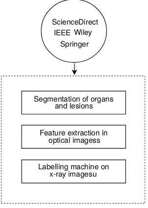
Today, the medical image processing (MIP) It is an area of very specific research in computing and is closely related to the digital signal processing. This rela-tionship stems from the fact that essentially the MIP is a very special form of digital signal processing in two or three dimensions. [6] The application of signal processing techniques to the field of medical imaging takes several decades to produce progress in assisting the diagnosis. Main areas of application include theSegmentation of organs and lesions,Feature extraction in optical images; and Labelling Machine on x-ray images. [7]. In Fig. 1 flowchart of this review is shown.
Segmentation of organs and lesions
Feature extraction in
Labelling machine on x-ray imagesu optical imagess
ScienceDirect IEEE
[image:2.612.233.384.410.622.2]Springer Wiley
2.1 Segmentation of organs and lesions
The medical image analysis by visual inspection by a specialist presents many difficulties, due to the subjectivity of the physician, data complexity and variety of procedures for image acquisition. One application that has received more interest in recent years is the segmentation of organs or injuries (lesions).
Research in [8] [9] [10] [11] use interesting approaches through different meth-ods and techniques to perform the segmentation of magnetic resonance imaging (MRI) of the head. In [8] propose a fuzzy c-means (FCM)-based algorithm with incorporated spatial neighborhood information. This is determined using a fac-tor that represents the spatial influence of the neighboring pixels on the current pixel achieving robust to noisy images even at increased levels of noise, thereby enabling effective segmentation of noisy medical images. In [11] propose a novel method considering the hidden Markov random field model (HMRF) to model the image class labels. To combine the spatial coherency modeling capabilities of the HMRF model and the enhanced flexibility obtained by fuzzy c-means (FCM) algorithm, they use a fuzzy clustering expectation maximization (FCEM) algo-rithm. Finally, both model parameters as well as class labels of medical images are estimated recursively using proposed algorithm until the model parameters converge to the optimal ones. Another approach in [9] use K-means clustering to produce a primary segmentation of the input image, after that apply the improved watershed segmentation algorithm to the primary segmentation to ob-tain the final segmentation map. TThis improve use a automated thresholding on the gradient magnitude map and post-segmentation merging on the initial partitions to reduce the number of false edges and over-segmentation. In [10], [12] also, propose a segmentation algorithm based on watershed. The first, uses this algorithm together with rough set theory but without consider some clus-tering algorithm. they partitioned the image into the edge-detail sub-image and smooth sub-image according to indiscernibility relation of rough set theory. Two enhancement methods are designed for the two sub-images, and watershed trans-formation is used for the further segmentation in the smooth sub-image. Finally, combine the two processed sub-images to obtain the segmentation result. While the second is based on graph theory that reconstructs gradient before watershed segmentation, based on the reconstruction, a floating-point active-image is in-troduced as the reference image of watershed transform. Finally, a graph theory based algorithm Grab Cut is used for fine segmentation. False contours of over-segmentation are effectively excluded and total over-segmentation quality significant improved as suitable for medical image segmentation.
At last, of trachea segmentation is achieved. In [15] [16] use segmentation based in histograms. First, proposes a new technique to increase the resolution of the medical images to identify the features and edges of the medical images by using multi-class histogram based segmentation method. While the second proposes a dualistic sub-image histogram equalization based enhancement and segmenta-tion techniques. They subdivide an image into its constituent objects. Technique is based on directional homogeneity using modified metric in [17].
[image:4.612.135.484.254.415.2]The table 1 shows a summary of the revised approaches.
Table 1: Summary of approaches of segmentations of organs and lesions.
Method Type of Image Improvement Type Study FCM MRI of the head Spatial neighborhood
information
Organ [8]
FCM, HMRF Cancerous kidney FCEM Lesion [11] K-means, Watershed MRI of the head Watershed
improve-ment
Organ [9]
Watershed MRI of the head Rough set theory Organ [10] Watershed MRI of the head Graph theory Organ [12] GMM MRI of the head Bayesian inference
crite-rion
Lesion [13]
Wavelet transform Image of trachea - Organ [14] Histogram Image of breast Multi-class histogram Lesion [15] Histogram MRI of the head
and legs
Improvement direc-tional homogeneity
Lesion [16]
2.2 Feature extraction in optical images
Optical images are photographs of the eye used by optometrists to measure characteristics such as tear film thickness or the amount of red in the eyeball, and diagnosis of diseases. In this type of images relevant structures such as the eyelashes or eyelids, they should be isolated so as not to influence the further processing. [18]
Other approaches for feature extraction can be find in [21], [22] and [23]. In [21] propose a edge detection method customized on characteristics of optical coherence tomography images for stable feature extraction. They use a local win-dow holding many pixels for tracking structural tendencies, edges are detected on reliably limited areas in reduced noise effect. Finally through clustering based on Gaussian mixture model (GMM) the features are obtained. In [22], propose a feature-based method for video mosaic with super-resolution for optical medical images through build of a minimal cost graph path for mosaic using topology inference. Then a mosaicing image with super-resolution is created by way of maximum a posterior (MAP) estimation and selective initialization. And last approach in [23] address the problem of feature extraction for automated classi-fication of optical projection tomography images of colorectal polyp. 3D patches are classified using the bag of visual words framework and support vector ma-chines (SVM) getting the feature extraction of colorectal polyp images.
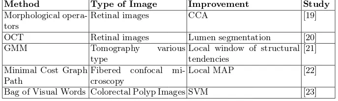
[image:5.612.136.480.340.443.2]The table 2 shows a summary of the revised approaches.
Table 2: Summary of approaches of feature extraction in optical images.
Method Type of Image Improvement Study
Morphological opera-tors
Retinal images CCA [19]
OCT Retinal images Lumen segmentation [20] GMM Tomography various
type
Local window of structural tendencies
[21]
Minimal Cost Graph Path
Fibered confocal mi-croscopy
Local MAP [22]
Bag of Visual Words Colorectal Polyp Images SVM [23]
2.3 Labelling machine on x-ray images
patches of different sizes to extract the arteries. While in [26] propose a region growing segmentation method which implements using morphological image pro-cessing operations and flood fill method. It can extract the boundary of main CA for after to be labelling. Furthermore in [27] a method to increase the average density of microfibrillated cellulose (MFC) in X-ray microtomographic images is proposed. Labeling is based on attaching metals to the surface of MFC fibres. This is characterized using scanning electron microscope (SEM), X-ray fluores-cent (XRF) measurements, electron energy dispersive scattering (EDS) analysis and inductively coupled plasma optical emission spectrophotometry (ICP-OES) measurements.
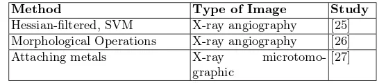
[image:6.612.169.447.290.350.2]The table 3 shows a summary of the revised approaches.
Table 3: Summary of approaches of labelling machine on x-ray images.
Method Type of Image Study
Hessian-filtered, SVM X-ray angiography [25] Morphological Operations X-ray angiography [26] Attaching metals X-ray
microtomo-graphic
[27]
3
Conclusions
At the end of this study, conclusions are:
- Application areas of medical image processing are varied, highlighting the radiology, nuclear medicine, endoscopy, thermography, angiography, magnetic resonance, ultrasound and microscopy; in all these fields the positive impact on the daily lives of human beings it is invaluable due to can be used as non-invasive methods of looking inside the human body and help to doctors in diagnosis diseases.
- Computational methods of segmentation facilitate the delimitation of tis-sues from noisy data, and allow quick and automatic extraction of parameters such as diameter, surface, or volume; aspects relating to the diagnosis and mon-itoring of diseases.
- The task of segmentation arteries is of great importance in the context of cardiovascular imaging, as the accuracy with which this work is done has a direct influence on diagnosis and therapy, or other clinical decisions associated with dangerous situations for life of patients.
- Automatic methods of detection and segmentation of retinal blood vessels are important for the automatic detection of diabetic retinopathy because allow diagnosis of abnormal lesions in this part of the human eye.
References
1. Ayala, M., Toro, J., Medina, R.: Iris images based personal identification. In Muller-Karger, C., Wong, S., La Cruz, A., eds.: IV Latin American Congress on Biomedical Engineering 2007, Bioengineering Solutions for Latin America Health. Volume 18 of IFMBE Proceedings. Springer Berlin Heidelberg (2008) 346–350 2. Miller, A., Blott, B., hames, T.: Review of neural network applications in medical
imaging and signal processing. Medical and Biological Engineering and Computing 30(1992) 449–464
3. Malpica, N., Tamames, J.H., Mora, I., Quiros, A., Santiago, R.: An´alisis avanzado de im´agenes m´edicas. Universidad Rey Juan Carlos (2013)
4. Reducindo, I., Arce-Santana, E., Campos-Delgado, D., Alba, A.: Evaluation of multimodal medical image registration based on particle filter. In: Electrical Engi-neering Computing Science and Automatic Control (CCE), 2010 7th International Conference on. (2010) 406–411
5. Cifor, A., Pridmore, T., Pitiot, A.: Smooth 3-d reconstruction for 2-d histological images. In Prince, J., Pham, D., Myers, K., eds.: Information Processing in Medical Imaging. Volume 5636 of Lecture Notes in Computer Science. Springer Berlin Heidelberg (2009) 350–361
6. Dominguez, A.: Procesamiento digital de im´agenes. Instituto de Investigaciones sobre la Universidad y Educaci´on (1996)
7. Chittajallu, D., Shah, S., Kakadiaris, I.: A shape-driven mrf model for the segmen-tation of organs in medical images. In: Computer Vision and Pattern Recognition (CVPR), 2010 IEEE Conference on. (2010) 3233–3240
8. Beevi, S., Sathik, M., Senthamaraikannan, K., Jaseema Yasmin, J.: A robust fuzzy clustering technique with spatial neighborhood information for effective med-ical image segmentation: An efficient variants of fuzzy clustering technique with spatial information for effective noisy medical image segmentation. In: Comput-ing Communication and NetworkComput-ing Technologies (ICCCNT), 2010 International Conference on. (2010) 1–8
9. Ng, H., Ong, S., Foong, K., Goh, P., Nowinski, W.: Medical image segmentation using k-means clustering and improved watershed algorithm. In: Image Analysis and Interpretation, 2006 IEEE Southwest Symposium on. (2006) 61–65
10. Li, R.: Medical image segmentation based on watershed transformation and rough sets. In: Bioinformatics and Biomedical Engineering (iCBBE), 2010 4th Interna-tional Conference on. (2010) 1–5
11. Patra, D., Pradhan, S.: Development of fuzzy clustering based unsupervised scheme for medical image segmentation using hmrf model. In: Industrial Electronics, Con-trol Robotics (IECR), 2010 International Conference on. (2010) 225–229
12. Zhang, Y., Cheng, X.: Medical image segmentation based on watershed and graph theory. In: Image and Signal Processing (CISP), 2010 3rd International Congress on. Volume 3. (2010) 1419–1422
13. Sudre, C., Cardoso, M., Bouvy, W., Biessels, G., Barnes, J., Ourselin, S.: Bayesian model selection for pathological neuroimaging data applied to white matter lesion segmentation. Medical Imaging, IEEE Transactions onPP(2015) 1–1
14. Gongwen, X., Zhijun, Z., Weihua, Y., Li’Na, X.: On medical image segmenta-tion based on wavelet transform. In: Intelligent Systems Design and Engineering Applications (ISDEA), 2014 Fifth International Conference on. (2014) 671–674 15. Ganga, T., Karthikeyani, V.: Medical image segmentation using multi resolution
16. Mohan, K., Thirugnanam, G.: A dualistic sub-image histogram equalization based enhancement and segmentation techniques for medical images. In: Image Informa-tion Processing (ICIIP), 2013 IEEE Second InternaInforma-tional Conference on. (2013) 566–569
17. Chaudhuri, D., Murthy, C., Chaudhuri, B.: A modified metric to compute distance. Pattern Recognition25(1992) 667 – 677
18. Navarro, R.: The optical design of the human eye: a critical review. Journal of Optometry 2(2009) 3 – 18
19. Miri, M., Mahloojifar, A.: Retinal image analysis using curvelet transform and multistructure elements morphology by reconstruction. Biomedical Engineering, IEEE Transactions on58(2011) 1183–1192
20. Celi, S., Berti, S.: In-vivo segmentation and quantification of coronary lesions by optical coherence tomography images for a lesion type definition and stenosis grading. Medical Image Analysis18(2014) 1157 – 1168
21. Cha, Y.M., Han, J.H.: Efficient edge detection method for anatomic feature ex-traction of neuro-sensory tissue image based on optical coherence tomography. In: Brain-Computer Interface (BCI), 2013 International Winter Workshop on. (2013) 65–66
22. Hu, M., Penney, G., Rueckert, D., Edwards, P., Bello, F., Figl, M., Casula, R., Cen, Y., Liu, J., Miao, Z., Hawkes, D.: A robust mosaicing method with super-resolution for optical medical images. In Liao, H., Edwards, P., Pan, X., Fan, Y., Yang, G.Z., eds.: Medical Imaging and Augmented Reality. Volume 6326 of Lecture Notes in Computer Science. Springer Berlin Heidelberg (2010) 373–382
23. Li, W., Coats, M., Zhang, J., Mckenna, S.J.: Comparative analysis of feature extraction methods for colorectal polyp images in optical projection tomography (2013)
24. Kawathekar, P., Karande, K.: Severity analysis of osteoarthritis of knee joint from x-ray images: A literature review. In: Signal Propagation and Computer Technology (ICSPCT), 2014 International Conference on. (2014) 648–652 25. Plourde, M., Duong, L.: Multi scale classification approach for coronary artery
detection from x-ray angiography. In: Information Science, Signal Processing and their Applications (ISSPA), 2012 11th International Conference on. (2012) 181–186 26. Kulathilake, K., Ranathunga, L., Constantine, G., Abdullah, N.: Region grow-ing segmentation method for extractgrow-ing vessel structures from coronary cine-angiograms. In: Moratuwa Engineering Research Conference (MERCon), 2015. (2015) 142–147