TítuloAcute effects of a prooxidant herbicide on the microalga Chlamydomonas reinhardtii: Screening cytotoxicity and genotoxicity endpoints
39
0
0
Texto completo
(2) Acute effects of a prooxidant herbicide on the microalga Chlamydomonas reinhardtii: screening cytotoxicity and genotoxicity endpoints.. Marta Esperanza, Ángeles Cid, Concepción Herrero and Carmen Rioboo*carmen.rioboo@udc.es. Laboratorio de Microbiología, Facultad de Ciencias, Universidad de A Coruña, Campus de A Zapateira s/n, 15071 A Coruña, SPAIN *. Corresponding author. Tel.: +34 981 167 000.. 1.
(3) Highlights. Mitochondrial membrane potential constituted the most sensitive parameter assayed Several genotoxicity methods were applied for first time in ecotoxicological studies Oxidative DNA base damage (8-OHdG) was induced by paraquat exposure Cells with DNA strand breakage and subG1-nuclei increased in treated cultures Typical apoptosis hallmarks were observed in microalgal cells exposed to paraquat. 2.
(4) Abstract Since recent evidence has demonstrated that many types of chemicals exhibit oxidative and/or genotoxic potential on living organisms, reactive oxygen species (ROS) formation and DNA damage are currently the best accepted paradigms to assess the potential hazardous biological effects of a wide range of contaminants. The goal of this study was to compare the sensitivity of different cytotoxicity and genotoxicity responses on the model microalga Chlamydomonas reinhardtii exposed to the prooxidant herbicide paraquat. In addition to the growth endpoint, cell viability, mitochondrial membrane potential and presence of reactive oxygen species (ROS) were assayed as potential markers of cytotoxicity using flow cytometry (FCM). To study the effects of paraquat on C. reinhardtii DNA, several genotoxicity approaches were implemented for the first time in an ecotoxicological study on microalgae. Oxidative DNA base damage was analysed by measuring the oxidative DNA lesion 8‐OHdG by FCM. DNA fragmentation was analysed by different methods: comet assay, and cell cycle analysis by FCM, with a particular focus on the presence of subG1‐nuclei. Finally, effects on morphology of nuclei were monitored through DAPI staining.. The. evaluation of these endpoints showed that several physiological and biochemical parameters reacted to oxidative stress disturbances with greater sensitivity than integrative parameters such as growth rates or cell viability. The experiments revealed concentration‐dependent cytotoxicity (ROS formation, depolarization of mitochondrial membrane), genotoxicity (oxidative DNA damage, DNA strand breakage, alterations in nuclear morphology), and cell cycle disturbances (subG1‐nuclei, decrease of 4N population) in paraquat‐treated cells. Overall, the genotoxicity results indicate that the production of ROS caused by exposure to paraquat induces oxidative DNA damage followed by DNA single‐ and double‐strand breaks and cell cycle alterations, possibly leading to apoptosis in C. reinhardtii cells. This is supported by the observation of typical hallmarks of apoptosis, such as mitochondrial membrane depolarization, alterations in nuclear morphology and subG1 nuclei in cells exposed to the highest assayed concentrations. To our knowledge, this is the first study that provides a comprehensive analysis of oxidative DNA base damage in unicellular algal cells exposed to a prooxidant pollutant, as well as of its possible relation with other physiological effects. These results reinforce the need for additional studies on the genotoxicity of environmental pollutants on ecologically relevant organisms such as microalgae that can provide a promising basis for the characterization of potential pollutant hazards in the aquatic environment.. Keywords herbicide, flow cytometry, genotoxicity, cytotoxicity, microalgae, oxidative stress. 3.
(5) Abbreviations DAPI: 4',6‐diamidino‐2‐phenylindole; FCM: flow cytometry; FICT: fluorescein isothiocyanate ; FS: forward scatter light; HE: hydroethidine; JC‐1:. 5,5′,6,6′‐tetrachloro‐1,1′,3,3′‐tetraethylbenzimidazolcarbocyanine. iodide; MMP: mitochondrial membrane potential; PI: propidium iodide; PS: photosystem; ROS: reactive oxygen species ; SS: side scatter light; 8‐OHdG: 7,8‐dihydro‐8‐oxo‐2'‐deoxyguanosine. 4.
(6) 1. Introduction Chemicals are used extensively and intensively in our technological society, and their use is continuously increasing. This large amount of different chemicals can reach surface and groundwater systems, and have unpredictable environmental and health effects in the long term (Guillén et al., 2012). This serious threat to aquatic life has prompted the creation of new regulations related to chemical safety, with the aim of assessing the environmental risk of individual substances, commonly after ‐ but increasingly and ideally before ‐ the substance is available on the market. The pesticide directive EC No 1107/2009, the Regulation on Registration, Evaluation and Assessment of Chemicals (REACH), the Water Framework Directive (WFD) and the Marine Strategy Framework Directive (MSFD) are the main pieces of European legislation that regulate chemicals in the environment (Gregorio and Chévre, 2014; Segner, 2011). In order to protect biological/ecological systems, the study of potential toxic impact imposed by particular chemicals has become a priority field in current environmental research. Nowadays, the existing knowledge gap in the biological effects of contaminants, particularly for ecologically relevant aquatic species, constitutes a real bottleneck in risk assessment of exposure to potentially harmful substances, given the wide variety of chemicals, and their mixtures, that need to be assessed. This realization has resulted in a shift from (chemical‐based) contaminant monitoring towards reliance on (effect‐based) ecotoxicity testing using biological assays (Lam and Gray, 2003). Among the intracellular toxicity alterations that can be induced by chemicals, reactive oxygen species (ROS) production and DNA damage are currently the best accepted paradigms to assess and compare the toxicity of different environmental contaminants, since recent evidence has shown that many types of chemicals exhibit oxidative and/or genotoxic potential on living organisms such as unicellular algae (Cirulis et al., 2013, Hess, 2000; Prado et al., 2009, 2015). Among aquatic organisms, the use of unicellular algae as test species in cell‐based bioassays is clearly justified in terms of environmental protection. Microalgae are a key component of food chains in aquatic environments due to their fundamental participation in energy conversion and ecosystem food web maintenance. Therefore, it is essential to have early assessment tools for cytotoxic and genotoxic effects at the cellular level, which could lead to disturbance in structure and productivity of the algae community which, in turn, could induce direct structural changes in the rest of the ecosystem (Martinez et al., 2014). In this regard, the green freshwater microalga Chlamydomonas reinhardtii, extensively used in molecular biology and photosynthesis research (Harris, 2001), has recently gained importance in ecotoxicological research (Fischer et al., 2010; Jamers and De Coen, 2010). This unicellular alga is sensitive to different contaminants, is commonly found in freshwater and soils, has a short generation time, is easy to culture and its genome has been sequenced. In current toxicity bioassays with microalgae, integrative endpoints such as growth and cell viability are the most widely monitored parameters. These population‐based parameters are highly relevant ecologically. 5.
(7) because they provide information about the likely outcome of a particular chemical at the cellular level, potentially compromising structure and function of higher trophic levels in aquatic ecosystems (Campanella et al., 2001, Prado et al., 2009; Rioboo et al., 2011). However, these conventional endpoints do not provide information about the toxic mechanisms by which environmental chemicals disrupt biological processes in algal cells. In the last years, considerable research efforts have been directed towards incorporating a suite of more specific molecular, biochemical and physiological endpoints which can provide early warning signals for possible hazardous effects induced by chemical exposure in unicellular algae. In this regard, previous studies have reported the implementation of flow cytometry (FCM) in toxicity bioassays with microalgae as an alternative to traditional techniques, assessing the biological effects of several environmental pollutants (Adler et al., 2007, Franklin et al., 2004, Franqueira et al., 2000, Hankamer et al., 2014, Jamers and De Coen, 2010; Prado et al., 2009, 2012a, 2015). Despite all points mentioned above, current genotoxicity data using microalgae as a model organism for aquatic chemicals are still scarce. Among the genotoxicity tests available, the comet assay has recently attracted much attention, being increasingly used to study DNA strand breaks in ecotoxicological studies (Park and Choi, 2007). Since this method primarily detects high molecular‐weight DNA resulting from single and double‐strand breaks and alkali‐labile sites in cells, unicellular algae could be a reliable model for the assessment of genotoxic effect of aquatic pollutants. To date, only a few genotoxicity studies based on the comet assay have been conducted with microalgae (Akcha et al., 2008, Aoyama et al., 2003, Desai et al., 2006, Erbes et al., 1997, Li et al., 2009, Martinez et al., 2014, Prado et al., 2009; Sastre et al., 2001). The bipyridyl herbicide paraquat is included in the EU priority database of potentially dangerous compounds that must be monitored in environmental samples (Commission Regulation (EC) No 1272/2008; access date: December 2014). Paraquat (1,1′‐dimethyl 4,4′‐bipyridinium ion) is a quaternary nitrogen herbicide used to control grasses and broadleaf weeds for a variety of agricultural crops. In the present work, paraquat was selected from the available diversity of aquatic contaminants as a model chemical for a specific toxic mechanism: paraquat catalyses the generation of superoxide radicals, thereby causing oxidative stress (Jamers and De Coen, 2010, Knauert and Knauer, 2008, Suntres, 2002; Szivák et al., 2009). Although its application is partially restricted and even banned in several countries (Baltazar et al., 2014), paraquat is one of the most widely used herbicides in the world (Nanseu‐Njiki et al., 2010; Santos et al., 2013). Furthermore, the risk of contamination by paraquat is enhanced by its high water solubility (620 g L‐ 1. ) (Tomlin, 2009), having often been detected in surface and drinking waters in maximum concentrations of. 97.8 μg L‐1 (Amondham et al., 2006; Zhou et al., 2009). Despite the inconclusive evidence regarding the carcinogenic effects of paraquat, the US Cal EPA (1993) has classified this herbicide as a possible human carcinogen. For these reasons, paraquat represents a serious risk not only for aquatic environments but also for human health. The goal of this study was to evaluate the sensitivity of different cytotoxicity and genotoxicity responses on. 6.
(8) the model microalga Chlamydomonas reinhardtii to exposure to the prooxidant herbicide paraquat, assaying similar concentrations to those previously reported in the environment (Amondham et al., 2006; Zhou et al., 2009). In addition to the traditional growth endpoint, several physiological and biochemical parameters such as cell viability, mitochondrial membrane potential and ROS production were monitored by flow cytometry (FCM) as potential markers of cytotoxicity. To study the potential effects of paraquat on C. reinhardtii DNA, we performed a comprehensive analysis of herbicide‐induced DNA damage using several genotoxicity approaches. Since oxidative DNA base damage can be a reliable marker of oxidative stress at the DNA level, the formation of 8‐OHdG was analysed by FCM. In addition to the aforementioned comet assay, DNA fragmentation was also studied by cell cycle analysis using FCM, with a particular focus on the presence of subG1‐nuclei, indicative of apoptosis phenomena. Finally, the potential effects on nuclei morphology were also monitored using DAPI staining. These genotoxicity methods have been successfully applied in various clinical and toxicological studies using mouse and human cell lines (Cambi et al., 2013; Liu et al., 2013) but have never, to our knowledge, been used in ecotoxicological studies with microalgae.. 7.
(9) 2. Materials and methods Reagents All chemicals were purchased from Sigma‐Aldrich (Spain) unless stated differently. 2.1. Chlamydomonas strain and culture conditions The unicellular green alga Chlamydomonas reinhardtii Dangeard (strain CCAP 11/32A mt+) was obtained from the Culture Collection of Algae and Protozoa of Dunstaffnage Marine Laboratory (Scotland, UK). C. reinhardtii cells were cultured photoautotrophically on a rotary shaker set at 150 rpm in Tris‐minimal phosphate medium (Harris, 1989) at 22 ± 1°C under a 12:12 light:dark cycle at an intensity of photosynthetically active radiation (PAR) of 100 μmol photon m‐2 s‐1. For experimental cultures, cells in mid‐ logarithmic growth phase were used as inoculum and suspended at an initial density of 2 x 10⁵ cells mL‐1. 2.2. Experimental design Before each experiment, fresh stock solutions of paraquat (Sigma‐Aldrich; MW: 257.2) were prepared by dissolving the herbicide in distilled water and filtering through 0.2‐μm membrane filters. At time zero, paraquat was added to C. reinhardtii cultures at three final nominal concentrations determined by previously carried out concentration–growth rates response assays: 0.1 μM (growth inhibition equal to 0%), 0.25 μM (close to the 96‐h EC50 value) and 0.5 μM (growth inhibition equal to 100%). Control cultures were also included, to which no paraquat was added. All cultures were set up in triplicate for 24 h and at least three independent experiments were carried out for each parameter analysed. 2.3. Flow cytometry acquisition and analysis Flow cytometric analyses of C. reinhardtii cells were performed on a Beckman‐Coulter Gallios flow cytometer fitted with 488‐nm and 633‐nm excitation lasers and interfaced to Gallios acquisition software version 1.2 (Beckman‐Coulter, Fullerton, CA). In order to exclude non‐microalgal particles, Chlamydomonas cells which present a cell size of 10 μm x 6 μm (Harris, 1989), were gated by their morphological features as displayed in dot‐plots of forward scatter (FS, an estimation of cell size) versus red autofluorescence (FL4 channel, 675/20 nm, an estimation of cell chlorophyll a content). At least 10.000 gated cells per sample were collected and analysed using Kaluza software version 1.1 (Beckman Coulter). All flow cytometric experiments were performed at least three times and duplicate samples were run on the flow cytometer. The 488‐nm argon‐ion laser was used as excitation source for all the probes assayed. Prior to the investigation, extensive experiments were conducted to optimize the probe titters, the incubation conditions and the concentration of cells to be used for flow cytometry (FCM) in order to obtain significant and stable staining of cells without toxicity being developed. All probe stock solutions were made up in dimethyl sulfoxide (DMSO) and stored in 20‐μl aliquots at −20°C. Further dilu ons were made in phosphate buffered saline (PBS) to a final concentration of DMSO of less than 0.01%. Fluorescent probe JC‐1. 8.
(10) was from Molecular Probes (Scotland, UK). 2.3.1. Absolute cell counting by FCM For absolute cell counting after 24 h of paraquat exposure, fluorescent polystyrene microspheres (Flow‐ Count Fluorospheres; Beckman‐Coulter) were added as an internal reference to all cell samples. Absolute C. reinhardtii cell counts were derived from forward scatter (FS, an estimation of cell size) versus side scatter (SS, an estimation of internal granularity) dot‐plot analysis as well as the ratio of the gated algal cells and corresponding reference beads with known concentration (provided by the manufacturer). Growth rates (µ) expressed as day−1 were calculated via the formula μ=[ln(Nt)−ln(N0)]/ln2(t−t0) where Nt is the cell density at time t and N0 is the cell density at time 0. 2.3.2. Determination of plasma membrane integrity as viability marker with propidium iodide (PI) Membrane integrity is a good indicator for the loss of cell viability, because cells with damaged membranes cannot maintain stable conditions of their molecular structures and usually die. Therefore, most fluorescence dyes for cell viability measurements work with the dye exclusion method. In the present work, the fluorescent dye propidium iodide (PI) was used to evaluate cell membrane damage as a viability marker in C. reinhardtii cells exposed to paraquat. As other typical viability dyes, PI is excluded by viable cells but can penetrate the damaged cell membrane of dead cells and binds to nucleic acid structures, increasing there by the orange fluorescence of cells (Ormerod, 1990). For this purpose, C. reinhardtii cells (2 × 10⁵ cells mL−1) were incubated with 4 μM of PI for 15 min at room temperature (RT) and in the dark prior to analysing the samples by FCM. Cells excluding propidium iodide, identified as PI negative (PI‐) for orange fluorescence in the FL3 channel (620/20 nm), were regarded to be alive. Thermal death by microwave heating for 5 s was included as a control of the probe. 2.3.3. Fluorescence measurement of intracellular reactive oxygen species (ROS) Oxidative stress in C. reinhardtii was evaluated by FCM using determinations of intracellular levels of superoxide anion radical (O2•‐) with an oxidation‐sensitive fluorescent dye. Moreover, we also investigated the ROS‐mediated DNA damage using a direct fluorescent protein binding method targeting 8‐oxoguanine moieties (as part of the oxidized nucleotide 8‐oxoguanosine) in cellular DNA. Hydroethidine (HE) or dihydroethidium, a sodium borohydride‐reduced derivative of ethidium bromide (EB), was used to evaluate the generation of ROS on exposure to paraquat in C. reinhardtii cells. HE, a specific and sensitive indicator of O2•‐ (Benov et al., 1998), is cell permeant and can be directly oxidized to EB by O2•‐ produced by the cell. DNA‐binding EB is fluorescent (610 nm) on excitation with the 488‐nm argon‐ion laser. HE was added at a final concentration of 16 μM to the cell suspensions (2 x 10⁵ cells ml‐1) for dye loading. Thirty minutes after incubation at RT and in darkness, cells with high intracellular levels of superoxide anion (HE+) were identified as positive for orange‐EB fluorescence in the FL3 channel (600/20 nm).. 9.
(11) The presence of oxidative DNA damage was detected by FCM using the fluorescent OxyDNA assay. The OxyDNA Assay (OxyDNA Assay Kit, Fluorometric, Calbiochem) contains a fluorescent binding protein with high affinity for 7,8‐dihydro‐8‐oxo‐2'‐deoxyguanosine (8‐OHdG), an important indicator of free radical‐ induced DNA damage and oxidative stress (Liu et al., 2013; Zribi et al., 2010). The method was used according to the manufacturer's instructions with some modifications. Briefly, 106 cells were fixed with 1% (p/v) paraformaldehyde on liquid nitrogen for 15 min and washed with PBS and the wash solution provided by the manufacturer. C. reinhardtii cells were then incubated with a FITC‐conjugated probe (binding protein labelled to Fluorescein Isothiocyanate) that specifically binds to 8‐OHdG‐DNA adducts in ROS‐damaged cells, for 1 h, at RT and in the dark. Finally, the FITC‐labelled probe was removed and oxidative DNA damage was analysed in the FL1 channel (525/10 nm). ROS‐damage levels were expressed as mean values of green‐ FITC fluorescence intensity (normalized to cell size values estimated using FS detector). To validate all these oxidative stress markers, pre‐stained cells were exposed to H2O2, a commonly used oxidant, at a final concentration of 5 mM for 10 min prior to analysis by FCM as control (Prado et al., 2012a) 2.3.4. Mitochondrial membrane potential (MMP) assay with JC‐1 Changes in mitochondrial membrane potential (MMP) of C. reinhardtii cells after treatment with paraquat were evaluated by staining cells with the lipophilic cationic probe JC‐1 (5,5′,6,6′‐tetrachloro‐1,1′,3,3′‐ tetraethylbenzimidazolcarbocyanine iodide). JC‐1 is a ratiometric, dual emission potential‐sensitive probe that is internalized and concentrated by respiring mitochondria, where it exits in a monomeric form emitting at 525 nm (green fluorescence) after excitation at 490 nm. In response to MMP (implicating a high dye concentration), JC‐1 forms the so called J‐aggregates that are associated with a large shift in fluorescence emission from green to orange (590 nm) (Reers et al., 1991). C. reinhardtii cells (2 x 10⁵ cells mL−1) were incubated with 3 μM of JC‐1 for 20 min at RT in the dark. Afterwards, cells were washed twice and finally resuspended in PBS before FCM analysis. JC‐1 was excited at 488 nm, and both green (JC‐1 monomers) and orange (JC‐1 oligomers) emission were determined at FL1 (525/10 nm) and FL3 (600/20 nm) channels, respectively. MMP results were expressed as the mean orange (JC‐1 oligomers)/green (JC‐1 monomers) fluorescence intensity ratio which is dependent only on the mitochondrial membrane potential (Cassart et al., 2007). Carbonylcyanide m‐chlorophenylhydrazone (CCCP), a known mitochondrial uncoupler, was the control for the MMP assay, C. reinhardtii cells being co‐ incubated with CCCP at a final concentration of 49 μM and JC‐1 prior to analysis in the flow cytometer. 2.3.5. Cell‐cycle distribution analysis using PI A flow cytometric assay was performed to assess effects of paraquat on C. reinhardtii cell cycle using propidium iodide (PI), a popular fluorescent dye that binds to double‐stranded nucleic acids as described above. For the measurement of cellular DNA content using PI labelling, Chlamydomonas cells (106 cells) were harvested and the pellet was fixed with 70% (v/v) cold ethanol for 2 h at 4°C. After fixation, cells were. 10.
(12) washed, pelleted and resuspended in PI solution (0.01 mg mL‐1) containing RNase A (0.10 mg mL‐1) and Triton X‐100 0.1% (p/v) and incubated at 37°C in the dark for 30 min. Cell DNA content analysis was then performed recording PI‐fluorescence in a linear mode by FL3 detector (620/20 nm) to generate DNA cytograms and to determine percentage of cells in each phase of the cell cycle. 2.4. Comet assay by single‐cell gel electrophoresis A number of techniques are available for detecting the effects of genotoxic agents in organisms. Of these, alkaline single‐cell gel electrophoresis or comet assay is a well‐established method for detecting DNA strand breaks (Prado et al., 2009; Singh, 1994). In the present work, the comet assay was conducted to detect the DNA damage after 24 h of exposure to paraquat on C. reinhardtii according to Erbes et al. (1997), except for that DNA staining was performed with SYBR Green I nucleic acid stain (Molecular Probes, Scotland, UK). Microscope slides were pre‐coated with a base layer of 100 μL of 1% (p/v) normal melting point agarose (NMA). The cells were resuspended in 0.7% low melting point agarose (LMA). A volume of 80 μL of the cell suspension (approximately 2 × 10⁴ cells) was dropped on a pre‐coated slide, and a glass coverslip placed on top to spread the gel, which was left to set at 4°C. After solidification of the agarose, slides were covered with another 80 μL of 0.7% (p/v) LMA and then placed in lysis solution (300 mM NaOH, 30 mM Na2EDTA and 0.1% (p/v) sodium dodecyl sulphate (SDS)) at RT for 10 min in darkness to remove cellular membranes, cytoplasm, and histones, leaving DNA as nucleoids. After lysis, the slides were transferred to an electrophoresis tank containing alkaline solution (300 mM NaOH, 1 mM Na2EDTA, pH > 13) for 5 min at 4°C for DNA unwinding. For electrophoresis of the DNA, an electric current of 25 V/300 mA was applied for 10 min at 4°C. The slides were removed and washed three times with a neutralizing buffer (0.4 M Tris, pH 7.5) for 5 min at 4°C before staining with SYBR Green I stain. Two slides were prepared from each replicate culture (n=2). Besides, Chlamydomonas cells treated with a reference genotoxic, H2O2, (10 mM, 2 h in darkness) were used as control of the assay. The diameter of C. reinhardtii nucleus is 3 μm (Harris, 1989). The prepared slides were subsequently examined at X400 magnification using a Nikon Eclipse E400 epifluorescence microscope (Nikon Instruments Europe BV) equipped with FITC filters (excitation 450–490, dichroic minor 505, band pass 520) to detect SYBR Green fluorescence emission. One hundred nucleoids per culture were randomly analysed (50 images per slide), counting the number of comet‐positive nuclei for determination of the percentage of DNA‐damaged cells, as previously described in Prado et al. (2009). 2.5. Fluorescent staining of nuclei by DAPI One of the most common DNA stains is DAPI (4',6‐diamidino‐2‐phenylindole) which binds to A‐T rich regions of the DNA double helix. In this study, this fluorescent dye was applied for depicting changes in nuclear morphology and chromatin distribution (Darehshouri et al., 2008) of C. reinhardtii. Control and treated cells suspensions were stained with 7.2 μM of DAPI for 10 min at RT and then were mixed in 0.7% (p/v) low. 11.
(13) melting point agarose (LMA). The prepared slides were subsequently examined at X400 magnification using a Nikon Eclipse E400 epifluorescence microscope (Nikon Instruments Europe BV) equipped with UV‐2A filters (excitation 330–380 nm, dichroic mirror 400 nm, band pass 420 nm) to detect DAPI fluorescence emission. One hundred cells per culture were randomly analysed (50 cells per slide), counting the number of cells affected. Photographs were taken with a high‐definition cooled colour camera Nikon DS‐5Mc. 2.6. Statistical analysis Data were calculated as mean values ± standard deviation (SD) of at least three independent experiments and statistically analysed by an overall one‐way analysis of variance (ANOVA) using SPSS Statistical software (version 21.0, SPSS, IBM). A P‐value < 0.05 was required for the results to be considered statistically significant. When significant differences were observed, means were compared using the multiple‐range Duncan test. EC50 values for viability, subG1 population, intracellular reactive oxygen species levels (O2−.), cells with intact DNA and cells without nuclear alterations were calculated using the Probit analysis, since these parameters are not continuous variables. This statistical method was carried out using the SPSS software. EC50 values for growth, oxidative DNA base damage (8‐OHdG levels) and mitochondrial membrane potential were calculated using the computer program Compusyn (Chou and Martin, 2005; Compusyn Inc, USA).. 12.
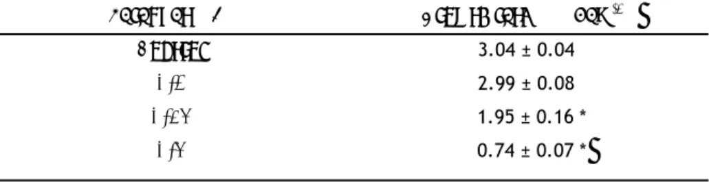
(14) 3. Results 3.1. C. reinhardtii growth is inhibited in response to paraquat exposure To determine the potential effects of paraquat on C. reinhardtii cultures, growth rates of C. reinhardtii cells exposed to different paraquat concentrations were calculated after 24 h of herbicide exposure as shown in Table 1. Growth data analysis indicated that paraquat treatment induced a noticeable inhibitory effect on the proliferation of C. reinhardtii in a concentration‐dependent manner, which resulted in significantly lower growth rates in cultures exposed to the two highest concentrations assayed, 0.25 and 0.5 μM of paraquat: about 65% and 25% of the growth rate in control cells, respectively (P < 0.05; Table 1). EC50 value after 24 h was 0.33 μM (Table 2).. 3.2. Viability of C. reinhardtii cells after paraquat treatment To determine whether the significant decrease in growth of C. reinhardtii at the highest concentrations of paraquat was associated with the loss of cell viability, a FCM assay based on dye exclusion of the probe PI was used to identify cells with intact plasma membrane. As shown in Figure 1, the percentage of C. reinhardtii cells with intact plasma membrane (PI‐ cells) after 24 h of paraquat exposure remained close to 99% for control and for all herbicide concentrations assayed (P > 0.05), except in cells exposed to the highest concentration assayed. Viability of C. reinhardtii cells exposed to 0.5 μM of paraquat significantly dropped when compared to non‐treated cells (P < 0.05), presenting lower percentages of viable (PI‐) cells (96.05 ± 1.11%) than control (99.13 ± 0.22%) cells. EC50 value for this parameter after 24 h of paraquat exposure was 1.20 μM (Table 2).. 3.3. Cell cycle perturbations in C. reinhardtii cells after treatment with paraquat To gain insight into the above‐mentioned growth‐inhibitory effects and their potential relation with DNA damage, we were interested in determining whether paraquat also induced cell cycle perturbations and apoptosis in C. reinhardtii cells by measuring cellular DNA content using FCM. C. reinhardtii is a haploid organism (Harris, 1989). Figure 2 shows the result of a flow cytometric measurement of DNA content in C. reinhardtii cells. After 24 h of culture, the profile in non‐treated C. reinhardtii cells showed three discrete peaks of propidium iodide (PI) fluorescence, consisting of cells with 1N cellular DNA content (G1 phase, 80% of cells), 2N cellular DNA content and even 4N cellular DNA content (G2 phase, about 20% of cells) nuclei. Only a small fraction of cells with DNA less than 1N (subG1) nuclei were observed, representing a percentage lower than 2% of the control cells. After 24 h of treatment, no significant changes in the fractions with 1N DNA and with 2N DNA nuclei were found in the cells exposed to paraquat when compared. 13.
(15) to control cells (P > 0.05). However, paraquat treatment resulted in a considerable decrease in the number of C. reinhardtii cells with 4N DNA nuclei compared to non‐treated cells (P < 0.05). The fraction of cells with 4N DNA nuclei in control cells was 10%, whereas the percentage of these 4 N cells significantly decreased (5%, 2%, and 1% cells) after exposure to 0.1, 0.25, and 0.5 μM concentrations of paraquat, respectively (Figure 2). Exposure to herbicide also induced accumulation of C. reinhardtii cells in subG1 (Figure 2). When incubated with paraquat for 24 h, the fraction of the subG1 cell population of C. reinhardtii cultures significantly increased at all the herbicide concentrations assayed (P < 0.05), which is consistent with the presence of DNA damage. EC50 for population with subG1‐nuclei after 24 h of paraquat exposure was 1.14 μM (Table 2).. 3.4. Paraquat exposure induces intracellular ROS overproduction in C. reinhardtii In order to test the relationship between paraquat treatment and oxidative stress induction in C. reinhardtii, FCM analysis of ROS generation using the probe HE was performed in algal cells. As shown in Figure 3, exposure to paraquat caused a remarkable increase in ROS levels in C. reinhardtii cells, resulting in a substantial increase in the population of cells with high intracellular ROS levels (HE‐positive) in the cultures treated with herbicide. After 24 h of treatment, the percentage of cells with high intracellular ROS levels (HE+) in the presence of 0.1, 0.25, and 0.5 μM of paraquat exhibited significant increases of around 2.5‐, 3.5‐ and 9.0‐fold, respectively, of those in the control (P < 0.05). In fact, when C. reinhardtii cells were exposed to the highest concentration assayed, the percentage of cells with high intracellular ROS levels rose to 99.10% (Figure 3). EC50 value after 24 h of treatment was 0.27 μM (Table 2).. 3.5. Paraquat exposure provokes formation of 8‐OHdG‐DNA adducts in C. reinhardti cells In view of the increase in ROS levels detected in C. reinhardtii cells as consequence of paraquat treatment, we used FCM to further monitor the presence of 8‐OHdG‐DNA adducts, one of the most abundant oxidative products of cellular DNA, in C. reinhardtii cells exposed to this herbicide. FCM analysis revealed that paraquat exposure for 24 h provoked the formation of oxidative 8‐OHdG adducts in the DNA of C. reinhardtii (Figure 4). As shown in Figure 4B, the levels of 8‐OHdG adducts in cells treated with the lowest concentration assayed, 0.1 μM of herbicide, were similar (P > 0.05) to those recorded in non‐treated cells, whereas significant 1.6‐ and 3.5‐fold increases (P < 0.05) in the formation of 8‐OHdG residues were recorded when C. reinhardtii cells were incubated with 0.25 and 0.5 μM of paraquat, respectively. EC50 value after 24 h of paraquat exposure was 1.02 μM (Table 2).. 3.6. Mitochondrial membrane depolarization in C. reinhardtii cells exposed to paraquat. 14.
(16) To assess whether paraquat‐mediated ROS production was accompanied by mitochondrial dysfunction, C. reinhardtii cells were stained with the mitochondria‐specific dye, JC‐1, which is widely used to monitor mitochondrial membrane potential. Paraquat exposure caused a remarkable loss of MMP after 24 h of treatment, as evidenced by the lower orange (JC‐1 oligomers)/green (JC‐1 monomers) fluorescence ratio values recorded in paraquat‐treated cells relative to those in non‐treated controls (Figure 5). The fluorescence ratio signal of JC‐1 exhibited a significant concentration‐dependent decrease in the presence of all paraquat concentrations assayed in C. reinhardtii cells (P < 0.05). That is, the higher the paraquat concentration, the lower the fluorescence ratio values observed after 24 h of exposure, which suggests a concentration‐dependent depolarization of paraquat‐exposed mitochondria. C. reinhardtii cells exposed to the two highest concentrations assayed, 0.25 and 0.5 μM, showed a marked collapse of the mitochondrial membrane potential, as indicated by the fluorescence ratio signal of JC‐1 decreasing up to 50% of values in control cells (Figure 5). EC50 value for mitochondrial membrane potential after 24 h of herbicide exposure was 0.13 μM (Table 2).. 3.7. Paraquat exposure induces DNA fragmentation in C. reinhardtii cells Because paraquat treatment induced oxidative damage of DNA in C. reinhardtii cells, the effect of this herbicide on DNA integrity was also examined. DNA strand breaks in C. reinhardtii nuclei were assessed using comet assay (Figure 6). Exposure to paraquat for 24 h caused detectable DNA strand breaks in C. reinhardtii as comet‐positive nuclei were developed in cells exposed to this herbicide. Compared to control cells, the percentage of comet nuclei in C. reinhardtii cultures showed significant 2.5‐, 3.2‐ and 5.4‐fold increases after a 24‐hour treatment with 0.1, 0.25 and 0.5 μM of paraquat, respectively. The percentage of cells with comet‐positive nuclei reached 86.47 ± 2.44% in cultures exposed to the highest concentration assayed (Figure 6). EC50 value after 24 h was 0.28 μM (Table 2).. 3.8. Nuclear morphological alterations in C. reinhardtii cells after treatment with paraquat To confirm the damage of paraquat on the DNA of C. reinhardtii, nuclei in treated and non‐treated cells were analysed by DAPI staining. A significantly increased number of DAPI‐positive cells with intense fluorescence staining nuclei (Figure 7B) were observed in all cultures treated with paraquat for 24 h compared to control (Figure 7A). The percentage of DAPI‐positive cells reached 16 ± 4.24, 50.75 ± 5.62, 67.15 ± 1.63 in cultures exposed to 0.1, 0.25 and 0.5 μM of paraquat for 24 h, respectively (Figure 7C). EC50 value after 24 h of paraquat exposure was 0.36 μM (Table 2).. 15.
(17) 4. Discussion The use of biological assays has become an increasingly popular tool for modern environmental assessment as they can theoretically help predict the biological effects of exposure to potentially harmful chemicals in the environment. In this context, several standardized toxicity assays with microalgae have been developed (e.g. according to ISO (2012) and OECD (2011) guidelines) and are currently required by authorities for notifications of new chemicals. To date, the response of microalgae to environmental contaminants is basically measured using population‐ based endpoints, such as specific growth rate. In the present work, growth of C. reinhardtii was affected by paraquat in a concentration‐dependent manner after 24 h of exposure (Table 1). However, growth rates were only significantly lower than those of the control when algae were exposed to paraquat concentrations higher than 0.1 µM. Similar threshold values (> 0.1 µM) for paraquat toxicity on growth of C. reinhardtii were reported in previous studies (Jamers and De Coen, 2010; Nestler et al., 2012). As shown in Table 2, the EC50 value obtained for this endpoint after 24 h of treatment also suggests that growth rate inhibition in C. reinhardtii is not the most sensitive parameter to paraquat exposure at concentrations usually detected in freshwater ecosystems (Amondham et al., 2006; Zhou et al., 2009). Furthermore, since inhibition of microalgal growth can be caused by chemical interference with a wide range of subcellular processes, this integrative endpoint alone is not sufficient for evaluating the toxicological effects of chemicals on unicellular algae. As an alternative to these standard algal endpoints, flow cytometry technique allows simultaneous measurements of multiple cellular and subcellular parameters which can be made separately on each cell within the suspension, under near in vivo conditions. However, the potential of this technique as a tool in unicellular algal toxicity bioassays has not yet been fully exploited. Among the toxicity endpoints that can be monitored in biological assays, the change in the redox status of the cell is of major interest since most classes of chemicals act as prooxidant stressors, increasing the intracellular generation of ROS and causing oxidative conditions in the cell (Hess, 2000). In this study, the percentage of gated cells from HE staining (Figure 3) was analysed by FCM to assess the extent of ROS production as a control of the well‐known toxicity mechanism of paraquat. Upon entering photosynthetic cells, the bipyridinium compound paraquat diverts electrons primarily from PS I and transfers them to molecular oxygen. The superoxide radicals produced by this process lead to the subsequent generation of other ROS, notably hydrogen peroxide and hydroxyl radicals (Hess, 2000). Results showed a significant increase in the population of cells with high intracellular ROS levels (HE‐positive) in the cultures with increasing paraquat concentration (Figure 3). In previous ecotoxicological studies in unicellular algae, intracellular ROS measurements in response to paraquat indicated mechanisms of toxic action involving reactive oxygen species (ROS) formation and oxidative stress (Jamers and De Coen, 2010, Nestler et al., 2012; Prado et al., 2012a, 2012b). Copper and other metals also induced an increase in levels of reactive species in microalgal cells (Melegari et al., 2013).. 16.
(18) Our results confirm that this parameter, related to oxidative activity measurement, could serve as a suitable indicator of exposure to prooxidant stressors, showing one of the lowest EC50 values obtained in response to paraquat (Table 2). It is well‐known that when a certain threshold of ROS production has been surpassed, the cellular antioxidant defence mechanisms are no longer able to respond sufficiently. At this point, oxidative stress increases rapidly, leading, among other effects, to membrane disruption and ultimately cell death (Hess, 2000). Based on this direct involvement of oxidative stress in the toxicity mechanism of paraquat, a strong relation between intracellular ROS levels and the loss of cell viability associated with membrane damage in C. reinhardtii cultures could be expected. Furthermore, many recent studies have evidenced that cell viability is a sensitive parameter for cytotoxicity monitoring of several contaminants in algae (Ebenezer et al., 2012, Lelong et al., 2012; Melegari et al., 2013). However, FCM analysis of C. reinhardtii cells with intact plasma membrane (PI‐ cells) showed no indication of massive cell death. We only found a significant cell viability decrease in C. reinhardtii cultures exposed to the highest concentration assayed of paraquat compared to control cultures (Figure 1). A potential explanation for this observation could be the stimulation of antioxidant defence mechanisms, such as antioxidant enzyme activities, as reported before for green algae exposed to herbicides (Chankova et al., 2014; Dewez et al., 2005), or the dependence on exposure time of the cells to paraquat, since only short‐term effects (24 h) were assessed in the present study. In fact, these results align with previous studies (Jamers and De Coen, 2010; Prado et al., 2009) that reported a decrease in C. reinhardtii viability after 48 h of exposure to paraquat. In conclusion, the cell viability endpoint could be limited by dependence on exposure time, indicating that membrane integrity is altered only after other cellular parameters, such as ROS levels or mitochondrial dysfunction, have already been strongly affected by the herbicide. In this regard, the EC50 value for cell viability was the highest of the various parameters assayed in the present study (Table 2, EC50 value of 1.20 µM). Because paraquat can act as an alternative acceptor of electrons from the respiratory chain, the study of perturbations on mitochondrial respiratory chain could be a suitable indicator of paraquat toxicity on C. reinhardtii cells (Drechsel and Patel, 2009; Mohammadi and Ghazi, 2008). The analysis of mitochondrial membrane potential (MMP), using FCM assay with fluorochrome JC‐1, has been widely used as an approach to measure the activity of the mitochondrial respiratory chain in animal cells (Yang et al., 2013 and Binet et al., 2014). Previous studies in animal and vegetal cells have observed a proton‐phobic effect exerted by paraquat on mitochondria, i. e., this herbicide causes a depolarization of the inner mitochondrial membrane and an uncoupling of the oxidative phosphorylation (Costantini et al., 1995, Palmeira et al., 1995; Vicente et al., 2001). This finding is in accordance with our results, where a significant depolarization of the MMP was observed in C. reinhardtii cells after 24 h of exposure to all the assayed concentrations of paraquat. It is noteworthy that the adverse effects of paraquat were also concentration‐dependent (Figure 5). Several authors reported that this depolarization effect provoked by paraquat could be due to a permeabilization of. 17.
(19) the inner membrane of the mitochondria, as a consequence of the peroxidation of lipids due to ROS generation (Palmeira et al., 1995) as well as a result of the opening of calcium‐dependent pores specific of this membrane (Costantini et al., 1995). Recently, an inhibitory effect of paraquat on ATP synthase activity of the inner mitochondrial membrane has been described, which can also result in membrane depolarization (Gómez et al., 2007). Since the collapse of the MMP can be attributed to indirect or direct oxidative damage generated by paraquat in the mitochondria, our results indicate that paraquat induces oxidative disturbance not only in the chloroplast but also in the mitochondria of the alga C. reinhardtii. In fact, the MMP constituted one of the most promising markers of toxicity for paraquat exposure among those assayed, showing the lowest EC50 value reported in the present study (0.13 μM). In spite of this, the MMP parameter is rarely implemented as endpoint in ecotoxicity bioassays. With respect to the potential genotoxic mode of action of paraquat, ROS are considered to be the major source of spontaneous damage to DNA. Oxidative attack on DNA results in mutagenic structures such as 8‐ hydroxyadenine and 8‐hydroxyguanine, which induce instability of repetitive sequences. In this context, oxidative DNA base damage in C. reinhardtii cells was analysed via the formation of 8‐OHdG by FCM, a method that has extensively been used to assess the quality of human sperm (Cambi et al., 2013; Liu et al., 2013). Results showed a concentration‐dependent increase in oxidative DNA damage in algal cells exposed to paraquat (Figure 4) with respect to control. This is the first study, to the authors' knowledge, that provides quantitative measurements of oxidative DNA damage in microalgal cells exposed to environmental contaminants, indicating that oxidative stress is a likely cause of paraquat genotoxicity in C. reinhardtii. ROS‐mediated DNA oxidation in paraquat‐treated C. reinhardtii cells indicated the possibility of DNA damage where an early effect could be evidenced on the cell cycle, particularly on the potential activation of the pathways underlying DNA repair, cell cycle progression, and apoptosis. Using FCM, changes in cell cycle perturbations can be analysed by categorizing the percentage of cell populations residing in each phase of the cell cycle and apoptotic cascade phenomena (Anbumani and Mohankumar, 2015). However, no studies have addressed the potential genotoxic effects of contaminant exposure on microalgal toxicity by analysing the cell cycle distribution through DNA content. In the classical cell division cycle, a growing cell duplicates all of its components and divides them into two nearly identical daughter cells. A cell cycle can be separated into two phases: a pre‐ and a post‐commitment phase. The pre‐commitment phase (G1) consists of growth processes leading to an increase in cell size, the building of cellular structures and the accumulation of energy reserves. This phase is dependent on energy and thus, in the case of microalgae, dependent on photosynthesis. At a certain point during growth in the pre‐commitment phase, algae reach a threshold size that triggers a sequence of events leading to cell division. This point is called the commitment point (CP). As it was found in previous works (Donnan et al., 1985, John, 1987; Setlik and Zachleder, 1984), some Chlorococcal and Volvocean algae are able to divide into more than two daughter cells by a non‐ canonical mechanism called multiple fission. When growth is allowed to continue during the post‐. 18.
(20) commitment phase, cells can grow in G1 up to many times their original size and therefore attain additional commitment points, which makes them divide into multiple daughter cells at the end of the cell cycle. In the multiple fission cell cycle, the size of the mother cell and, subsequently, the number of commitment points determines n, generating 2n daughter cells of uniform size after a rapid series of subsequent S and M cycles. In the present work, the DNA content of C. reinhardtii cells was analyzed by measuring fluorescence due to PI staining on the flow cytometer. In control cultures, three populations of cells containing 1, 2 or 4 copies of DNA were observed (Figure 2A), indicating that C. reinhardtii cells were dividing by multiple fission and releasing 2 to 4 daughter cells per mother cell. This finding suggests that, under the culture conditions assayed in this study, C. reinhardtii cells were be able to grow until attaining the first and second commitment points. However, the exposure of algal cells to paraquat concentrations resulted in a concentration‐dependent decrease in 4N cell population compared to control cultures (Figure 2), suggesting that cells in cultures exposed to paraquat would not be able to divide into 4 daughter cells. Following this reasoning, the exposure to this photosynthesis‐inhibiting herbicide could result in a lower amount of daughter cells per mother cell, since less cells would be able to reach the second commitment point, which would cause the lower growth rates recorded in paraquat‐treated cultures (Table 1). These results are in line with previous findings reported in another Chlorococcal microalga exposed to a triazine herbicide, where terbutryn altered the normal number of daughter cells (4 autospores) obtained from each mother cell, being only two in the cultures treated with 250 nM of herbicide (Rioboo et al., 2009). Furthermore, cells with DNA fragmentation can be identified in DNA content frequency cytograms by the appearance of a subG1 cell population (Ebrahimi Nigjeh et al., 2013, Kajstura et al., 2007, Kim et al., 2013, Kim et al., 2014; Stanojković et al., 2013). In this study, a significant subpopulation of C. reinhardtii cells treated with paraquat seemed to suffer severe DNA damage, which led to a concentration‐dependent increase in the percentage of cells observed in the subG1 phase with regard to control cultures. The lower DNA content suggests that algal cells were breaking down their DNA, which is considered an indicator of apoptosis (Moharikar et al., 2006; Segovia et al., 2003). This concentration‐dependent increase in cell population in subG1 suggests that DNA damage in C. reinhardtii cells increases in the presence of all the paraquat concentrations assayed, probably due to the observed oxidative DNA damage (Figure 4). With regard to these data, it is well‐known that genotoxic damage during DNA replication can transiently delay progression through the S phase. The existence of DNA damage in C. reinhardtii cells suggests that this prooxidant herbicide could cause a delay in the timing of the G1 phase of the cell cycle, resulting in an increase in the doubling time of the algal cells. This increase in the duration of G1 could be interpreted as a situation where cells gain extra time to repair DNA damage and could also explain the negative effect of paraquat on the 4N population of C. reinhardtii cultures (Figure 2). Overall, the cell cycle analysis by FCM confirms that this method can be extremely helpful to elucidate the different modes of action of contaminants in cells exposed to acute or chronic toxicity. However, our findings indicate the need to. 19.
(21) determine the timing of each phase of the cell cycle during all the experimental period in order to gain an in‐depth knowledge of the impact of paraquat on cell cycle progression in C. reinhartdtii. Furthermore, genome stability in C. reinhardtii cells was investigated using comet assay. This genotoxicity technique has been successfully applied to detect ROS‐induced strand breaks in the primary structure of the DNA of microalgal cells exposed to several contaminants (González et al., 2007, Liu et al., 2006; Prado et al., 2009). In agreement with these previous works, the comet assay results showed an extensive and concentration‐dependent damage to DNA after treatment of C. reinhardtii cells with the prooxidant herbicide paraquat. Paraquat‐treated algal cells showed a concentration‐dependent increase in the percentage of comet nuclei in cells exposed to all the assayed concentrations of the herbicide, as compared to control cells (Figure 6). In cultures exposed to the highest paraquat concentration, the percentage of damaged nuclei reached values of 86.47 ± 2.44% of C. reinhardtii cells, which indicated the extent of DNA damage. In the current work, it is also noteworthy that the comet assay results, expressed as percentage of comet nuclei in the algal cells exposed to paraquat, could be related to oxidative stress data, expressed as percentage of cells with high intracellular ROS levels (HE‐positive) in the C. reinhardtii cultures treated with herbicide (Figure 3). Taking these observations into account, this result indicates that this parameter is valuable to further study damage at the DNA level caused by the oxidative activity of paraquat in C. reinhardtii cells, and could constitute a reliable and sensitive indicator for genotoxicity caused by contaminants capable of redox cycling, with an EC50 value of 0.28 μM after 24 h of paraquat exposure (Table 2). In addition to the DNA fragmentation damage found by the comet assay in C. reinhardtii, the DAPI‐ morphology nuclei results further corroborated the alterations in paraquat‐treated cells (Figure 7B) as compared to untreated cells (Figure 7A). Several authors have related these changes in nuclei morphology to chromatin condensation (Figure 7B), which is frequently described as an apoptotic hallmark (Darehshouri et al., 2008, Elmore, 2007, Ferradás et al., 2014, Giri et al., 2013, Jiménez et al., 2009; Zuppini et al., 2009, 2010). In accordance to this, C. reinhardtii cells exposed to paraquat presented irregular DAPI staining as well as a slight degree of chromatin clumping in their nuclei (Figure 7B). Since DAPI can stoichiometrically bind to DNA and C. reinhardtii cultures were not synchronized (Figure 2), an increase of dye fluorescence could exclusively be related to the different DNA content in the cells. However, in DAPI‐positive cells, chromatin aggregation was not only restricted to the nuclear area, with blue‐stained granules also observed in the centre of the cell. These observations suggest some degree of karyolysis (Jiménez et al., 2009), which would be in coherence with the severe DNA damage observed on the cell cycle and, particularly, in the comet assay results (Figures 2 and 6). In this regard, it is worth noting the similarity in EC50 values obtained for DAPI‐positive and comet‐positive cells, 0.36 and 0.28 μM, respectively (Table 2). All these data reinforce the need for additional studies on the genotoxicity of environmental contaminants on ecologically relevant organisms such as microalgae, which, by linking genotoxic activity to other biological responses, could provide further understanding of adverse effects on organisms living in aquatic. 20.
(22) environments.. 5. Conclusions. The evaluation of the endpoints of the present study showed that several physiological and biochemical parameters reacted to oxidative stress disturbances with greater sensitivity than integrative parameters such as growth rates or cell viability. Among the different subcellular endpoints assessed in this work, the mitochondrial membrane potential parameter constituted the most sensitive indicator for herbicide exposure, showing the lowest EC50 value reported after 24 h of treatment. FCM analyses indicated that ROS were generated in the presence of paraquat, which could explain the cellular disturbances as well as other specific toxicological outcomes. Our experiments revealed a concentration‐dependent cytotoxicity (ROS production, depolarization of mitochondrial membrane), genotoxicity (oxidative DNA base damage, DNA strand breakage, alterations in nuclear morphology), and cell cycle disturbances (presence of subG1‐nuclei, decrease of 4N population) in paraquat treated cells. These results confirmed that parameters associated with oxidative stress and DNA damage are highly sensitive endpoints in unicellular algae exposed to different concentrations of prooxidant pollutants, which provides a promising basis for the characterization of potential pollutant hazards in the aquatic environment.. Acknowledgements M.E. acknowledges a pre‐doctoral fellowship from the Spanish Xunta de Galicia.. 21.
(23) References Adler NE, Schmitt‐Jansen M, Altenburger R (2007) Flow cytometry as a tool to study phytotoxic modes ofaction. Environmental and Toxicology Chemistry 26: 297‐306 Akcha F, Arzul G, Rousseau S, Bardouil M (2008) Comet assay in phytoplankton as biomarker of genotoxic effects of environmental pollution. Marine Environmental Research 66: 59‐61 Amondham W, Parkpian P, Polprasert C, DeLaune RD,. Jugsujinda A (2006) Paraquat adsorption,. degradation, and remobilization in tropical soils of Thailand. Journal of Environmental Science and Health B 41: 485‐507 Anbumani S, Mohankumar MN (2015) Gamma radiation induced cell cycle perturbations and DNA damage in Catla Catla as measured by flow cytometry. Ecotoxicology and Environmental Safety 113: 18‐22 Aoyama K, Iwahori K, Miyata N (2003) Application of Euglena gracilis cells to comet assay: evaluation of DNA damage and repair. Mutation Research 538: 155‐162 Baltazar MT, Dinis‐Oliveira RJ, Martins A, Bastos MDL, Duarte JA, Guilhermino L, Carvalho F (2014) Lysine acetylsalicylate increases the safety of a paraquat formulation to freshwater primary producers: a case study with the microalga Chlorella vulgaris. Aquatic Toxicology 146: 137‐43 Benov L, Sztejnberg L, Fridovich I (1998) Critical evaluation of the use of hydroethidine as a measure of superoxide anion radical. Free Radical Biology and Medicine 25: 826‐831 Binet MT, Doyle CJ, Williamson JE, Schlegel P (2014) Use of JC‐1 to assess mitochondrial membrane potential in sea urchin sperm. Journal of Experimental Marine Biology and Ecology 452: 91‐100 Bisova K, Krylov DM, Umen JG (2005) Genome‐wide annotation and expression profiling of cell cycle regulatory genes in Chlamydomonas reinhardtii. Plant Physiology 137: 475‐491 Cal EPA. 1993. Summary of toxicology data: paraquat dichloride. Medical Toxicology Branch, Department of Pesticide Regulation, California Environmental Protection Agency, file name: T931006. www.panap.net Cambi M, Tamburrino L, Marchiani S, Olivito B, Azzari C, Forti G, Muratori M (2013) Development of a specific method to evaluate 8‐hydroxy, 2‐deoxyguanosine in sperm nuclei: relationship with semen quality in a cohort of 94 subjects. Reproduction 145: 227‐235 Campanella L, Cubadda F, Sammartino MP, Saoncella A (2001) An algal biosensor for the monitoring of water toxicity in estuarine enviroments. Water Research 35: 69‐76 Cassart D, Fett T, Sarlet M, Baise E, Coignoul F, Desmecht D (2007) Flow cytometric probing of mitochondrial function in equine peripheral blood mononuclear cells. BMC Veterinary Research 3: 25‐31. 22.
(24) Chankova SG, Dimova EG, Mitrovska Z, Miteva D, Mokerova DV, Yonova PA, Yurina NP (2014) Antioxidant and HSP70B responses in Chlamydomonas reinhardtii genotypes with different resistance to oxidative stress. Ecotoxicology and Environmental Safety 101: 131‐137 Chioccioli M, Hankamer B, Ross IL (2014) Flow cytometry pulse width data enables rapid and sensitive estimation of biomass dry weight in the microalgae Chlamydomonas reinhardtii and Chlorella vulgaris. PloS One 9: e97269 Chou TC, Martin N (2005) CompuSyn for Drug Combinations: PC Software and User’s Guide: A Computer Program for Quantification of Synergism and Antagonism in Drug Combinations and the Determination of IC50 and ED50 and LD50 Values. ComboSyn, Inc., Paramus, NJ. Cirulis JT, Scott JA, Ross GM (2013) Management of oxidative stress by microalgae. Canadian Journal of Physiology and Pharmacology 91: 15‐21 Costantini P, Petronilli V, Colonna R, Bernardi P (1995) On the effects of paraquat on isolated mitochondria. Evidence that paraquat causes opening of the cyclosporine A‐sensitive permeability transition pore synergistically with nitric oxide. Toxicology 99: 77‐88 Darehshouri A, Affensellez M, Lütz‐Meindl U (2008) Cell death upon H2O2 induction in the unicellular green alga Micrasterias. Plant Biology 10: 732‐745 Desai SR, Verlecar XN, Nagarajappa, Goswami U (2006) Genotoxicity of cadmium in marine diatom Chaetoceros tenuissimus ussing the alkaline Comet assay. Ecotoxicology 15: 359‐363 Dewez D, Geoffroy L, Vernet G, Popovic R (2005) Determination of photosynthetic and enzymatic biomarkers sensitivity used to evaluate toxic effects of copper and fludioxonil in alga Scenedesmus obliquus. Aquatic Toxicology 74: 150‐159 de Winter L, Klok AJ, Cuaresma Franco M, Barbosa MJ, Wijffels RH (2013) The synchronized cell cycle of Neochloris oleoabundans and its influence on biomass composition under constant light conditions. Algal Research 2: 313‐320 Donnan L, Carvill EP, Gilliland TJ, John PCL (1985) The cell cycles of Chlamydomonas and Chlorella. The New Phytologist 99: 1‐40 Drechsel DA, Patel M (2009) Differential contribution of the mitochondrial respiratory chain complexes to reactive oxygen species production by redox cycling agents implicated in Parkinsonism. Toxicology Science 112: 427‐434 Ebenezer V, Nancharaiah YV, Venugopalan VP (2012) Chlorination‐induced cellular damage and recovery in marine microalga, Chlorella salina. Chemosphere 89: 1042‐1047 Ebrahimi Nigjeh S, Yusoff FM, Mohamed Alitheen NB, Rasoli M, Keong YS, Omar ARB (2013) Cytotoxic effect. 23.
(25) of ethanol extract of microalga, Chaetoceros calcitrans, and its mechanisms in inducing apoptosis in human breast cancer cell line. BioMed Research International 2013: 783690 EC, Regulation 1907/2006. Registration, Evaluation, Authorisation and Restriction of Chemicals (REACH). European Commission, 2006 EC, Regulation No. 1272/2008 of the European parliament and of the council of 16 December, 2008. On classification, labelling and packaging of substances and mixtures, amending and repealing Directives 67/548/EEC and 1999/45/EC, and amending Regulation (EC) No 1907/2006. In: Official Journal of the European Union, European Commission, Brussels, Belgium, 2008 EC, Regulation No. 1107/2009 of the European Parliament and of the Council of 21 October, 2009. Concerning the placing of plant protection products on the market and repealing Council Directives 79/117/EEC and 91/414/EEC. In: Official Journal of the European Union, European Commission, Brussels, Belgium, 2009 EC, Water Framework Directive 2000/60/EC. European Commission, 2000 EC, Marine environmental policy (Marine Strategy Framework Directive 2008/56/EC). European Commission, 2008b. Elmore S (2007) Apoptosis: A Review of Programmed Cell Death. Toxicology Pathology 35: 495‐516 Erbes M, Webler A, Obst U, Wild A (1997) Detection of primary DNA damage in Chlamydomonas reinhardtii by means of modified microgel electrophoresis. Environmental and Molecular Mutagenesis 30: 448‐458 Ferradás Y, López M, Rey M, González MV (2014) Programmed cell death in kiwifruit stigmatic arms and its relationship to the effective pollination period and the progamic phase. Annals of Botany 114: 35‐45 Fischer BB, Rüfenacht K, Dannenhauer K, Wiesendanger M, Eggen RIL (2010) Multiple stressor effects of high light irradiance and photosynthetic herbicides on growth and survival of the green alga Chlamydomonas reinhardtii. Environmental Toxicology and Chemistry 29: 2211‐2219 Franklin NM, Stauber JL, Lim RP (2004) Development of multispecies algal bioassays using flow cytometry. Environmental Toxicology and Chemistry 23: 1452‐1462 Franqueira D, Orosa M, Torres E, Herrero C, Cid A (2000) Potential use of flow cytometry in toxicity studies with microalgae. Science of the Total Environment 247: 119‐126 Giri BR, Roy B, Sinha‐Babu SP (2013) Evidence of apoptosis in Raillietina echinobothrida induced by methanolic extracts of three traditional medicinal plants of Northeast India. Experimental Parasitology 134: 466‐73 Gómez C, Bandez MJ, Navarro A (2007) Pesticides and impairmnt of mitochondrial function in relation with the parkinsonian syndrome. Frontiers in Bioscience 12: 1079‐1093. 24.
(26) González NV, Soloneski S, Larramendy ML (2007) The chlorophenoxy herbicide dicamba and its commercial formulation banvel induce genotoxicity and cytotoxicity in Chinese hamster ovary (CHO) cells. Mutation Research 634: 60‐8 Gregorio V, Chèvre, N (2014) Assessing the risks posed by mixtures of chemicals in freshwater environments: case study of Lake Geneva, Switzerland. Wiley Interdisciplinary Reviews: Water 1: 229‐247 Guillén D, Ginebreda A, Farré M, Darbra RM, Petrovic M, Gros M, Barceló D (2012) Prioritization of chemicals in the aquatic environment based on risk assessment: Analytical, modeling and regulatory perspective. Science of the Total Environment 44: 236‐252 Harris EH (1989) The Chlamydomonas sourcebook: A comprehensive Guide to biology and laboratory use. Academic Press, San Diego Harris EH (2001) Chlamydomonas as a model organism. Annual Review of Plant Physioly and Plant Molecular Biology 52: 363‐406 Hess FD (2000) Light‐dependent herbicides: an overview. Weed Science 48: 160‐170 ISO 8692:2012. Water quality‐Fresh water algal growth inhibition test with unicellular green algae. International Organization for Standardization, Suiza Jamers A, De Coen W (2010) Effect assessment of the herbicide paraquat on a green alga using differential gene expression and biochemical biomarkers. Environmental Toxicology and Chemistry 29: 893‐901 Jiménez C, Capasso JM, Edelstein CL, Rivard CJ, Lucia S, Breusegem S, Berl T, Segovia M (2009) Different ways to die: Cell death modes of the unicellular chlorophyte Dunaliella viridis exposed to various environmental stresses are mediated by the caspase‐like activity DEVDase. Journal of Experimental Botany 60: 815‐828 John PCL (1987) Control points in the Chlamydomonas cell cycle. In Wiesnar W, Robinson DG, Starr RC (eds), Algal Development. Molecular and Cellular Aspects. Springer‐Verlag, Berlin, 9‐16 pp Kajstura M, Halicka HD, Pryjma J, Darzynkiewicz Z (2007) Discontinuous Fragmentation of Nuclear DNA During Apoptosis Revealed by Discrete “Sub‐G 1” Peaks on DNA Content Histograms. International Society for Analytical Cytology, Cytometry Part A 71A: 125‐131 Kim AD, Lee Y, Kang SH, Kim GY, Kim HS, Hyun JW (2013) Cytotoxic effect of clerosterol isolated from Codium fragile on A2058 human melanoma cells. Marine Drugs 11: 418‐430 Knauert S, Knauer K (2008) The role of reactive oxygen species in copper toxicity to two freshwater green algae. Journal of Phycology 44: 311‐319 Lam PKS, Gray JS (2003) The use of biomarkers in environmental monitoring programmes. Marine Pollution Bulletin 46: 182‐186. 25.
(27) Lelong A, Jolley DF, Soudant P, Hégaret H (2012) Impact of copper exposure on Pseudo‐nitzschia spp. physiology and domoic acid production. Aquatic Toxicology 118‐119: 37‐47 Li M, Hu C, Gao X, Xu Y, Qian X, Brown MT, Cui Y (2009) Genotoxicity of organic pollutants in source of drinking water on microalga Euglena gracilis. Ecotoxicology 18: 669‐676 Liu Y, Zhang Y, Liu J, Huang D (2006) The role of reactive oxygen species in the herbicide acetochlor‐induced DNA damage on Bufo raddei tadpole live. Aquatic Toxicology 78: 21‐26 Liu C, Duan W, Xu S, Chen C, He M, Zhang L, Yu Z, Zhou Z (2013) Exposure to 1800MHz radiofrequency electromagnetic radiation induces oxidative DNA base damage in a mouse spermatocyte‐derived cell line. Toxicology Letters 218: 2‐9 Martinez RS, Di Marzio WD, Sáenz ME (2014) Genotoxic effects of commercial formulations of Chlorpyrifos and Tebuconazole on green algae. Ecotoxicology 24: 45‐54 Melegari SP, Perreault F, Costa RHR, Popovic R, Matias WG (2013) Evaluation of toxicity and oxidative stress induced by copper oxide nanoparticles in the green alga Chlamydomonas reinhardtii. Aquatic Toxicology 142‐143: 431‐440 Mohammadi A, Ghazi M (2008) Alternative electron acceptors: propose mechanism of paraquat mitochondrial toxicity. Environmental Toxicology Pharmacology 26: 1‐5 Moharikar S, Souza JSD, Kulkarni AB, Rao BJ (2006) Apoptotic‐like cell death pathway is induced in unicellular Chlorophyte Chlamydomonas reinhardtii (Chlorophyceae) cells following uv irradiation: detection and functional analyses. Phycological Society of America 42: 423‐433 Murray MG, Thompson WF (1980) Rapid isolation of high molecular weight plant DNA. Nucleic Acids Research 8: 4321‐4325 Nanseu‐Njiki CP, Dedzo GK, Ngameni E (2010) Study of the removal of paraquat from aqueous solution by biosorption onto Ayous (Triplochiton schleroxylon) sawdust. Journal of Hazardous Materials 179: 63‐71 Nestler H, Groh KJ, Schönenberger R, Behra R, Schirmer K, Eggen RIL, Suter MJF (2012) Multiple‐endpoint assay provides a detailed mechanistic view of responses to herbicide exposure in Chlamydomonas reinhardtii. Aquatic Toxicology 110‐111: 214‐224 OECD (2011) Alga, Growth Inhibition Test (201). OECD Guideline for Testing of Chemicals. Organization for Economic Cooperation and Development, Paris, France. Ormerod MG (1990) Analysis of DNA. General methods. In: Ormerod, M.G. (Ed.), Flow Cytometry. A practical Approach. Oxford Univesity Prees, Oxford, pp. 69‐87 Palmeira CM, Moreno AJ, Madeira VMC (1995) Mitochondrial bioenergetics is affected by the herbicide paraquat. Biochimica et Biophysica Acta 1229: 187‐192. 26.
(28) Park SY, Choi J (2007) Cytotoxicity, genotoxicity and ecotoxicity assay using human cell and environmental species for the screening of the risk from pollutant exposure. Environment International 33: 817‐822 Prado R, García R, Rioboo C, Herrero C, Abalde J Cid A (2009) Comparision of the sensitivity of different toxicity test endpoints in microalga exposed to the herbicide paraquat. Environment International 35: 240‐ 247 Prado R, Rioboo C, Herrero C, Suarez‐Bregua P, Cid A (2012a) Flow cytometric analysis to evaluate physiological alterations in herbicide‐exposed Chlamydomonas moewusii cells. Ecotoxicology 21: 409‐420 Prado R, Rioboo C, Herrero C, Cid A (2012b) Screening acute cytotoxicity biomarkers using a microalga as test organism. Ecotoxicology and Enviromental Safety 86: 219‐226 Prado R, García R, Rioboo C, Herrero C, Cid A (2015) Suitability of cytotoxicity endpoints and test microalgal species to disclose the toxic effect of common aquatic pollutants. Ecotoxicology and Environmental Safety, 114: 117‐125 Reers M, Smith TM, Chen LB (1991) J‐agregate formation of a carbonyanine as a quantitative fluorescent indicator of membrane potentisal. Biochemistry 30: 4480‐4486 Rioboo C, O’Connor JE, Prado R, Herrero C, Cid A (2009) Cell proliferation alterations in Chlorella cells under stress conditions. Aquatic Toxicology 94: 229‐237 Rioboo C, Gonzalez‐Barreiro O, Abalde J, Cid A (2011) Flow cytometric analysis of the encystment process induced by paraquat exposure in Haematococcus pluvialis (Chlorophyceae). European Journal of Phycology 46: 89‐97 Santos MSF, Schaule G, Alves A, Madeira LM (2013) Adsorption of paraquat herbicide on deposits from drinking water networks. Chemical Engineering Journal 229: 324‐333 Sastre MP, Vernet M, Steinert S (2001) Single‐cell gel/comet assay applied to the analysis of UV radiation‐ induced DNA damage in Rhodomonas sp. (Cryptophyta). Photochemistry and Photobiology 74: 55‐60 Segner H (2011) Moving beyond a descriptive aquatic toxicology: The value of biological process and trait information. Aquatic Toxicology 105: 50‐55 Segovia M, Haramaty L, Berges JA, Falkowski PG (2003) Cell death in the unicellular chlorophyte Dunaliella tertiolecta. A hypothesis on the evolution of apoptosis in higher plants and metazoans. Plant Physiology 132: 99‐105 Setlik I, Zachleder V (1984) The fission cell reproductive patterns in algae. In Nurse P, Streiblova E (eds), The Microbial Cell Cycle. CRC Press, Boca Raton, FL, 253–279 pp Singh NP, Stephens RE, Schneider EL (1994) Modifications of alkaline microgel electrophoresis for sensitive detection of DNA damage. International Journal of Radiation Biology 66: 23‐28. 27.
(29) Stanojković TP, Šavikin K, Zdunić G, Kljajić Z, Anti J (2013) In vitro antitumoral activities of Padina pavonia on human cervix and breast cancer cell lines. Journal of Medicinal Plants Research 7: 419‐424 Suntres ZE (2002) Role of antioxidants in paraquat toxicity. Toxicology 180: 65‐77 Szivák I, Behra R, Sigg L (2009) Metal‐induced reactive oxygen species production in Chlamydomonas reinhardtii (Chlorophyceae). Journal of Phycology 45: 427‐435 Tomlin CDS (2009) The Pesticide Manual: a world compendium. Fifteenth edition. British Crop Protection Council Publications, UK. 1457 pp Vicente JAF, Peixoto F, Lopes ML, Madeira VMC (2001) Differential sensitivities of plant and animal mitochondria to the herbicide paraquat. Journal of Biochemical and Molecular Toxicology 15: 322‐330 Yang C, Zhou J, Liu S, Fan P, Wang W, Xia C (2013) Allelochemical induces growth and photosynthesis inhibition, oxidative damage in marine diatom Phaeodactylum tricornutum. Journal of Experimental Marine Biology and Ecology 444: 16‐23 Zachleder V, Schläfli O, Boschetti A (1997) Growth‐controlled oscillation in activity of histone h1 kinase during the cell cycle of Chlamydomonas reinhardtii (chlorophyta). Journal of Phycology 33: 673‐681 Zhou L, Chen H, Jiang X, Fang L, Zhou Y, Yin W, Ji X (2009) Modification ofmontmorillonite surfaces using a novel class of cationic Gemini surfactants. Journal of Colloid and Interface Science 332: 16‐21 Zribri N, Chakroun NF, El Euch H, Gargouri J, Bahloul A, Keskes LA (2010) Effects of cryopreservation on human sperm deoxyribonucleic acid integrity. Fertility and Stirility 93: 159‐166 Zuppini A, Gerotto C, Moscatiello R, Bergantino E, Baldan B (2009) Chlorella saccharophila cytochrome f and its involvement in the heat shock response. Journal of Experimental Botany 60: 4189‐4200 Zuppini A, Gerotto C, Baldan B (2010) Programmed cell death and adaptation: two different types of abiotic stress response in a unicellular chlorophyte. Plant & Cell Physiology 51: 884‐95. 28.
Figure
Documento similar
In this paper, a series of annealing experiments of ultrafine-grained WC–Co materials were conducted, and the effects of the annealing temperature and holding time on the
The Dwellers in the Garden of Allah 109... The Dwellers in the Garden of Allah
to inspect what were, at that time, the most advanced and sensitive research and industrial activities” (Fischer, 1997, pp. 3) As to the contents of inspections, the
The expansionary monetary policy measures have had a negative impact on net interest margins both via the reduction in interest rates and –less powerfully- the flattening of the
Jointly estimate this entry game with several outcome equations (fees/rates, credit limits) for bank accounts, credit cards and lines of credit. Use simulation methods to
In our sample, 2890 deals were issued by less reputable underwriters (i.e. a weighted syndication underwriting reputation share below the share of the 7 th largest underwriter
Our results, using freshwater fish cell lines and a set of relevant cytotoxicity endpoints including cell viability, oxidative stress, and DNA damage, provide additional mechanistic
Cervical cancer is one of the first 5 most common neoplasms in women in the world, derived from this over time that screening tests have emerged for its timely diagnosis
