Corneal biomechanical properties : Measurement, modification and simulation
Texto completo
Figure
![Figure 1.1: The eye, taken from Fuensanta et al [212].](https://thumb-us.123doks.com/thumbv2/123dok_es/6080371.175272/24.629.97.498.75.351/figure-eye-taken-fuensanta-et-al.webp)
Documento similar
We consider different methods aim to evaluate cellular uptake and localization in cells and tissues, and in vitro methods for the study of the toxicity induced by MNPs
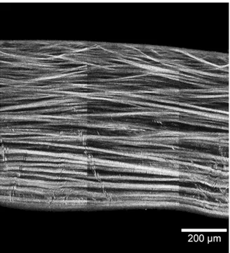
In our approach, we have defined and analyzed the following variables: the areas of the anterior and posterior corneal surfaces of the solid model generated (A ant , A post ), the
points used hereafter [3] (Figure 2): A ant is the area of the anterior corneal surface [1,2,4]; A post is the area of the posterior corneal surface [1,2,4]; CV is the total
Moreover, we evaluated the antioxidant capacity of CEO in vitro by applying the 1,1-diphenyl-2-picrylhydrazyl (DPPH) free radical scavenging method, and by determining the
In this study, a porcine in vitro fertilization (IVF) system and seminal quality parameters of frozen–thawed boar semen were used to assess the effectiveness of two different
For instance, (i) in finite unified theories the universality predicts that the lightest supersymmetric particle is a charged particle, namely the superpartner of the τ -lepton,
Heat map of the Pearson correlation coefficient established among observed in vitro effects and the concentration of different phytochemicals in coffee and cocoa by-products (A)
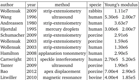
Our main aim was to perform a quantitative trait study analyzing the associ- ation of HGF variant rs12536657 with biometric ocular measurements and several corneal parameters
