RNPS 2235-145 © 2009-2013 Cardiocentro “Ernesto Che Guevara”, Villa Clara, Cuba. Todos los derechos reservados.
200
________________
Caso Clínico
Trauma valvular mitral en un lactante durante la valvuloplastia
aórtica y su tratamiento quirúrgico
Dr. Luis E. Marcano Sanz
a, Dr. Juan C. Ramiro Novoa
b, Dr. Gilberto Bermúdez Gutiérrez
a,
Dr. Francisco Díaz Ramírez
by Dra. Katia Rivera Ladino
aa Departamento de Cirugía Cardiovascular. Cardiocentro Pediátrico “William Soler”. La Habana, Cuba. b Departamento de Hemodinámica. Cardiocentro Pediátrico “William Soler”. La Habana, Cuba.
Full English text of this article is also available
INFORMACIÓN DEL ARTÍCULO
Recibido: 26 de noviembre de 2012 Aceptado: 14 de enero de 2013
Conflictos de intereses
Los autores declaran que no existen conflictos de intereses
Versiones On-Line:
Español
-
InglésK Rivera Ladino
Cardiocentro Pediátrico William Soler Ave. 100 y Perla, Alta Habana Boyeros, CP 10800
La Habana, Cuba Correo electrónico:
RESUMEN
La valvuloplastia con globo es ampliamente aceptada como tratamiento de elección de la estenosis aórtica congénita en recién nacidos y lactantes. Las complicaciones por el procedimiento son bien conocidas pero el daño valvular mitral es infrecuente. Se presenta un paciente masculino, de siete meses de edad, con el diagnóstico de coartación de la aorta y estenosis valvular aórtica a quién se le realizó dilatación de ambas lesiones por cateterismo intervencionista. A las 20 horas del procedimiento se diagnosticó insuficiencia cardíaca grave secundaria a lesión de la valva anterior mitral. Se intervino quirúrgicamente de urgencia y se observó un desgarro en forma de hendidura desde el borde libre hasta el anillo, el cual se reparó y se realizó anuloplastia en la comisura lateral. En el postoperatorio inmediato evolucionó sin complicaciones y al año de seguimiento presenta insuficiencia valvular mitral leve sin signos de recoartación ni de estenosis valvular aórticas.
Palabras clave: Valvuloplastia aórtica, Válvula mitral, Cirugía cardíaca
Mitral valve trauma in an infant during aortic valvuloplasty and its
surgical treatment
ABSTRACT
Balloon valvuloplasty is widely accepted as the treatment of choice for congenital aortic stenosis in newborns and infants. Complications from the procedure are well known but mitral valve damage is rare. This is the case of a 7-month-old male patient with the diagnosis of coarctation of the aorta and aortic valve stenosis who under-went dilation of both lesions via catheterization. At 20 hours of the procedure a severe heart failure secondary to an injury of the anterior mitral valve was diagnosed. The patient underwent emergency surgery and a slit-like tear from the free edge to the annulus was observed, which was repaired and annuloplasty in the lateral commissure was performed. In the immediate postoperative period the patient progressed without complications and at one year follow up he shows mild valve regurgitation without evidence of recoarctation or aortic valve stenosis.
Marcano Sanz LE, et al.
CorSalud 2013 Abr-Jun;5(2):200-203 201
Figura1.Cineangiografíaenvistalateralizquierda.A: Dilata-ción exitosa de la coartaDilata-ción aórtica. B: Ausencia de insufi-ciencia valvular aórtica.
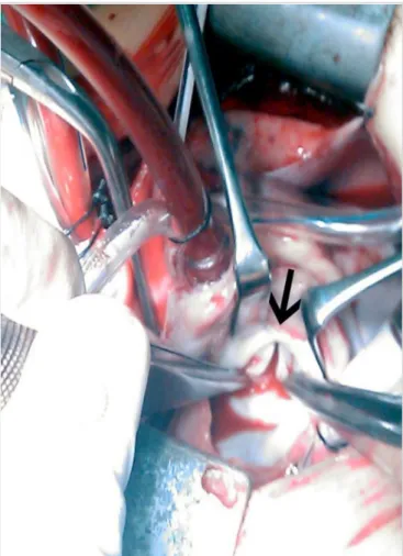
Figura 2. Imagen quirúrgica. Se expone a través de la
aurícu-la izquierda aurícu-la valva anterior mitral fisurada en sentido trans-versal (flecha).
INTRODUCCIÓN
La estenosis aórtica tiene una incidencia de 3 a 6 % entre las cardiopatías congénitas1. La valvuloplastia con globo (comúnmente conocida como valvuloplastia con balón) es ampliamente aceptada como trata-miento de elección para esta cardiopatía en recién nacidos y lactantes, por la corta estadía hospitalaria y los buenos resultados obtenidos con este tipo de intervención2. No obstante, este procedimiento no está libre de riesgos, se han descrito diferentes com-plicaciones que incluyen parada cardiorrespiratoria, perforación ventricular, taponamiento cardíaco e insuficiencia valvular aórtica aguda3. El daño valvular mitral es muy poco frecuente y se plantea que ocurre por la posición muy posterior de la guía metálica, por la insuflación del globo mal posicionado o por la presencia de ventrículos pequeños e hipoplásicos4,5. El objetivo de este informe de caso es presentar un paciente con insuficiencia cardíaca grave, al cual se le
realizó reparación quirúrgica de la válvula mitral secundaria al trauma provocado por el globo de la valvuloplastia aórtica.
CASO CLÍNICO
Paciente masculino de 7 meses de edad y 8 kg de peso corporal, con el diagnóstico clínico y ecocardiográfico de coartación de la aorta grave circunscrita y estenosis valvular aórtica crítica. La válvula aórtica era bicúspide y no mostraba insuficiencia.
Con anestesia general e intubación endotraqueal electiva se le realizó cateterismo cardíaco izquierdo a través de la arteria femoral derecha. Se constató un gradiente de 48 mmHg a nivel de la coartación y de 77 mmHg entre el ventrículo izquierdo y la aorta. El anillo aórtico medido por cineangiografía fue de 8,5 mm. Se realizó dilatación manual exitosa de la coartación aórtica, sin gradiente residual, y valvuloplastia de la lesión valvular aórtica con manómetro, a 8 atmósferas
CorSalud 2013 Abr-Jun;5(2):200-203
202
Figura3.Cineangiografía en vista lateral izquierda. A: Guía metálica correctamente colo-cada en la cavidad del ventrículo izquierdo (flecha). B: Se observa la guía metálica, con trayecto muy posterior, cuyo extremo está en la cavidad de la aurícula izquierda (flecha). El catéter-globo en el ventrículo izquierdo se encuentra por debajo del plano valvular mitral. de presión. El gradiente residual a ese nivel fue de 18
mm Hg y no se constató insuficiencia aórtica (Figura 1).
A las 20 horas del procedimiento, el paciente pre-sentó signos clínicos de shock cardiogénico y requirió el traslado a la Unidad de Terapia Intensiva, ventila-ción mecánica y apoyo inotrópico. La ecocardiografía transtorácica evidenció lesión valvular mitral traumá-tica con desgarro de la valva anterior e insuficiencia mitral aguda grave.
Se intervino quirúrgicamente de urgencia, me-diante circulación extracorpórea en normotermia, pin-zamiento aórtico y cardioplejia cristaloide fría. Se accedió a la válvula mitral a través de la aurícula izquierda, se reparó la hendidura desde el borde libre de la valva anterior hasta el anillo (Figura 2), con tres puntos de polipropileno 6/0 reforzados con pericardio autólogo; se realizó anuloplastia en la comisura lateral con un punto de polipropileno 5/0. El ecocardiograma transesofágico mostró insuficiencia mitral mínima y buena función sistodiastólica biventricular.
La evolución postoperatoria fue sin complicaciones, la estadía en la Unidad de Terapia Intensiva fue de dos días y egresó del hospital a los siete días. En el seguimiento, la ecocardiografía al año de operado
mostró insuficiencia valvular mitral leve, sin signos de recoartación de la aorta ni de estenosis valvular aórtica.
COMENTARIO
De las complicaciones que se pueden presentar duran-te la valvuloplastia aórtica, la lesión en la válvula mitral se ha informado en pocos pacientes, relacionada por una posición muy posterior de la guía metálica en el ventrículo izquierdo o en la aurícula izquierda (Figura 3), aunque se sugiere que esta mal posición es más probable que ocurra si el paciente tiene una cavidad ventricular pequeña o un aparato valvular mitral anor-mal2,5.
En este paciente, la lesión valvular presumible-mente se produjo al insuflar el globo de valvuloplastia posicionado a nivel del plano valvular mitral. En ese momento se obvió el detalle de que la guía metálica había alcanzado la cavidad de la aurícula izquierda, lo que podría haber alertado de la incorrecta posición del catéter-globo. La hipertrofia del ventrículo izquierdo, debido a la coartación de la aorta y con ello, el menor tamaño de dicha cavidad, pudo haber favorecido el trayecto anormal de la guía metálica.
La magnitud del daño, que involucró el anillo valvu- lar mitral, explica la precoci-dad de los síntomas. En la literatura revisada, los infor-mes sobre el momento en que se diagnosticaron estas lesiones, varían de las 10 se-manas a los 3 meses des-puésdelprocedimiento5;sin embargo, mientras más se demora la cirugía reparado-ra, el flujo de regurgitación produce daños en otras es-tructuras del aparato valvu-lar como lo es la elongación de las cuerdas tendinosas y la dilatación del anillo. De ahí que la reparación valvu-lar precoz, en el paciente que se presenta, haya mos-trado excelente resultado en el seguimiento clínico y ecocardiográfico un año después de operado.
Marcano Sanz LE, et al.
CorSalud 2013 Abr-Jun;5(2):200-203 203
como complicación de la valvuloplastia aórtica es muy poco frecuente pero puede tener consecuencias gra-ves para los pacientes. Evaluar la correcta posición de la guía metálica antes de utilizar el catéter-globo es imprescindible para evitar esta complicación. La repa-ración quirúrgica precoz previene un mayor daño valvular mitral y proporciona excelentes resultados funcionales.
REFERENCIAS BIBLIOGRÁFICAS
1. Kitchiner DJ, Jackson M, Walsh K. Incidence and prognosis of congenital aortic valve stenosis in Liverpool (1960-1990). Br Heart J. 1993;69(1):71-9. 2. Sarihoglu E, Erek E, Yalçinbaş YK, Öztürk N, Sa-rioğlu A, SaSa-rioğlu T. Mitral valve injury during aortic ballon valvuloplasty and its surgical
treat-ment. Turkish J Thorac Cardiovasc Surg. 2005; 13: 169-70.
3. Yamamoto Y, Shiraishi I, Yamagishi M, Hamaoka K. Mitral valve injury during ballon valvuloplasty for an infant with severe aortic stenosis: Spatial evaluation using three dimensional echocardio-graphy. Pediatr Cardiol. 2003;24(3):300-3.
4. McCrindle BW, Blackstone EH, Williams WG, Sitti-wangkul R, Spray TL, Azakie A, et al. Are outcomes of surgical versus transcatheter ballon valvotomy equivalent in neonatal critical aortic stenosis? Circulation. 2001;104(12 Suppl 1):152-8.
RNPS 2235-145 © 2009-2013 Cardiocentro “Ernesto Che Guevara”, Villa Clara, Cuba. All rights reserved.
200
________________
Case Report
Mitral valve trauma in an infant during aortic valvuloplasty
and its surgical treatment
Luis E. Marcano Sanz
a, MD; Juan C. Ramiro Novoa
b, MD; Gilberto Bermúdez Gutiérrez
a,
MD; Francisco Díaz Ramírez
b, MD; and Katia Rivera Ladino
a, MD
a Department of Cardiovascular Surgery. Cardiocentro Pediátrico “William Soler”. Havana, Cuba. b Department of Hemodynamics. Cardiocentro Pediátrico “William Soler”. Havana, Cuba.
Este artículo también está disponible en español
ARTICLE INFORMATION
Received: November 26, 2012 Accepted: January 14, 2013
Competing interests
Authors have no competing interests
On-Line versions:
Spanish
-
English K Rivera Ladino
Cardiocentro Pediátrico William Soler Ave. 100 y Perla, Alta Habana Boyeros, CP 10800
La Habana, Cuba E-mail address:
ABSTRACT
Balloon valvuloplasty is widely accepted as the treatment of choice for congenital aortic stenosis in newborns and infants. Complications from the procedure are well known but mitral valve damage is rare. This is the case of a 7-month-old male patient with the diagnosis of coarctation of the aorta and aortic valve stenosis who under-went dilation of both lesions via catheterization. At 20 hours of the procedure a severe heart failure secondary to an injury of the anterior mitral valve was diagnosed. The patient underwent emergency surgery and a slit-like tear from the free edge to the annulus was observed, which was repaired and annuloplasty in the lateral commissure was performed. In the immediate postoperative period the patient progressed without complications and at one year follow up he shows mild valve regurgitation without evidence of recoarctation or aortic valve stenosis.
Key words: Aortic valvuloplasty, Mitral valve, Heart surgery
Trauma valvular mitral en un lactante durante la valvuloplastia
aórtica y su tratamiento quirúrgico
RESUMEN
La valvuloplastia con globo es ampliamente aceptada como tratamiento de elección de la estenosis aórtica congénita en recién nacidos y lactantes. Las complicaciones por el procedimiento son bien conocidas pero el daño valvular mitral es infrecuente. Se presenta un paciente masculino, de siete meses de edad, con el diagnóstico de coartación de la aorta y estenosis valvular aórtica a quién se le realizó dilatación de ambas lesiones por cateterismo intervencionista. A las 20 horas del procedimiento se diagnosticó insuficiencia cardíaca grave secundaria a lesión de la valva anterior mitral. Se intervino quirúrgicamente de urgencia y se observó un desgarro en forma de hendidura desde el borde libre hasta el anillo, el cual se reparó y se realizó anuloplastia en la comisura lateral. En el postoperatorio inmediato evolucionó sin complicaciones y al año de seguimiento presenta insuficiencia valvular mitral leve sin signos de recoartación ni de estenosis valvular aórticas.
Marcano Sanz LE, et al.
CorSalud 2013 Apr-Jun;5(2):200-203 201
Figure1.Cineangiography in left lateral view. A: successful dilation of aortic coarctation. B: Absence of aortic valve regurgitation.
Figure 2. Surgical image. The anterior mitral leaflet, fissured
in transverse direction (arrow), is accessed through the left atrium.
INTRODUCTION
Aortic stenosis has an incidence of 3-6% among con-genital heart diseases1. Balloon valvuloplasty is widely accepted as the treatment of choice for this heart disease in newborns and infants due to the short hospital stay and the good results obtained with this type of intervention2. However, this procedure is not without risks. Different complications including cardio-respiratory arrest, ventricular perforation, cardiac tamponade, acute aortic valve regurgitation have been described3. Mitral valve damage is very rare and occurs because of the very posterior position of the guidewire, inflation of a malpositioned balloon or by the presence of small and hypoplastic ventricles4,5. The aim of this case report is to present a patient with severe heart failure, who underwent surgical repair of the mitral valve secondary to a trauma caused by the balloon of the aortic valvuloplasty.
CASE REPORT
This is the case of a 7-month-old male patient with 8 kg of body weight, with clinical and echocardiographic diagnosis of severe circumscribed coarctation of the aorta and critical aortic valve stenosis. The aortic valve was bicuspid and showed no regurgitation.
Under general anesthesia and elective endotra-cheal intubation a left heart catheterization via the right femoral artery was performed. A gradient of 48 mmHg at the level of the coarctation and of 77 mmHg between the left ventricle and the aorta was found. The aortic annulus measured by cineangiography was 8.5 mm. Successful manual dilation of aortic coarc-tation, without residual gradient, was performed as well as valvuloplasty of the aortic valve lesion with manometer at a pressure of 8 atmospheres. The re-sidual gradient at that level was 18 mm Hg and aortic regurgitation was not found (Figure 1).
CorSalud 2013 Apr-Jun;5(2):200-203
202
Figure 3. Cineangiography in left lateral view. A guidewire correctly placed in left ventricular cavity (arrow). B: The guidewire is observed with a very posterior pathway, whose end is in the left atrial cavity (arrow). The balloon catheter in the left ventricle is below the mitral valve plane.
At 20 hours of the procedure, the patient had clinical signs of cardiogenic shock and required trans-fer to the Intensive Care Unit (ICU), mechanical ven-tilation and inotropic support. Transthoracic echocar-diography showed a traumatic mitral valve lesion with tear of the anterior leaflet as well as severe and acute mitral regurgitation.
Emergency surgery was performed using cardio-pulmonary bypass at normothermia, aortic clamping and cold crystalloid cardioplegia. The mitral valve was accessed through the left atrium, the slit was repaired from the free edge of the anterior leaflet to the annulus (Figure 2), with three 6/0 polypropylene stit-ches reinforced with autologous pericardium; annulo-plasty was performed in the lateral commissure with one 5/0 polypropylene stitch. Transesophageal echo-cardiogram showed minimal mitral regurgitation and good biventricular systolic/diastolic function.
The postoperative course was uneventful, and the patient stayed in the ICU for two days and was dis-charged from hospital on the seventh day. At follow-up, echocardiography at one year from surgery showed mild mitral valve regurgitation with no signs of coarctation of the aorta or aortic valve stenosis.
COMMENT
Among the complications that may occur during aortic valvuloplasty, mitral valve injury has been reported in a few patients, and it has been related to a very posterior positioning of the guidewire into the left ventricle or the left atrium (Figure 3), although it has been suggested that this malpositioning is most likely to occur if the patient has a small ventricular cavity or abnormal mitral valve apparatus2,5.
In this patient, the valve lesion occurred pre-sumably by inflating the valvuloplasty balloon, po-sitioned at the level of mitral valve plane. At that time, the detail that the guidewire had reached the cavity of the left atrium was overlooked, which could have alerted to the malpositioning of the catheter-balloon. Left ventricular hypertrophy, due to coarctation of the aorta and thus the smaller size of such cavity, may have favored the abnormal course of the guidewire.
The extent of the damage, which involved the mi-tral valve annulus, explains the earliness of symptoms. In the literature reviewed, reports about the time these injuries were diagnosed range from 10 weeks to 3 months after the procedure5, but the more repair surgery is delayed, the more damages are produced by regurgitant flow in other
structures of the valve ap-paratussuchaschordaeten- dinae elongation and annu-lar dilation. Hence, early valve repair in the patient presented has shown ex-cellent results in clinical and echocardiographic follow-up one year after surgery.
The mitral valve lesion as a complication of aortic valve replacement is rare but can have serious conse-quences for patients. Asses-sing the correct position of the guidewire before using the balloon catheter is essential to avoid this com-plication. Early surgical re-pair prevents further mitral valve damage and provides excellent functional results.
Marcano Sanz LE, et al.
CorSalud 2013 Apr-Jun;5(2):200-203 203
REFERENCES
1. Kitchiner DJ, Jackson M, Walsh K. Incidence and prognosis of congenital aortic valve stenosis in Liverpool (1960-1990). Br Heart J. 1993;69(1):71-9. 2. Sarihoglu E, Erek E, Yalçinbaş YK, Öztürk N, Sa
-rioğlu A, Sa-rioğlu T. Mitral valve injury during
aortic ballon valvuloplasty and its surgical treat-ment. Turkish J Thorac Cardiovasc Surg. 2005; 13: 169-70.
3. Yamamoto Y, Shiraishi I, Yamagishi M, Hamaoka K. Mitral valve injury during ballon valvuloplasty for an infant with severe aortic stenosis: Spatial
evaluation using three dimensional echocardio-graphy. Pediatr Cardiol. 2003;24(3):300-3.
4. McCrindle BW, Blackstone EH, Williams WG, Sitti-wangkul R, Spray TL, Azakie A, et al. Are outcomes of surgical versus transcatheter ballon valvotomy equivalent in neonatal critical aortic stenosis? Circulation. 2001;104(12 Suppl 1):152-8.
5. Brierley JJ, Reddy TD, Rigby ML, Thanopoulous V,
Redington AN. Traumatic damage to the mitral