www.medigraphic.org.mx
Non-hyperacute synchronous cardio-cerebral
infarction treated by double intervensionist therapy
Infarto sincrónico cardio-cerebral no hiperagudo
tratado con doble terapia intervencionista
Juan Carlos Plata-Corona,* José Aurelio Cerón-Morales,** Beatriz Lara-Solís***
Keywords: Acute myocardial infarction, acute ischemic stroke, thrombolysis, thrombectomy, cardio-cerebral infarction.
Palabras clave:
Infarto agudo al miocardio, evento vascular cerebral isquémico, trombólisis, trombectomía, infarto cardiocerebral.
ABSTRACT
Introduction: The simultaneous appearance of the acute myocardial infarction (AMI) and acute ischemic stroke (AIS) is known as cardio-cerebral infarction (CCI); this condition is extremely rare, and thus complex to treat. There is normally a subjacent cause that triggers these two events to occur simultaneously. Nonetheless, the timely recognition of these conditions is challenging and most of the times is only confirmed by an autopsy. Objectives: This article investigates the epidemiology, the physiopathology and the treatment of CCI within the existing literature. Clinical case: A 46-years-old male, who is diagnosed of CCI after 5 hours his clinical condition starts. We decided to carry out a double percutaneous approach; intra-arterial thrombolysis plus mechanical thrombectomy of the middle-right cerebral artery (MRCA) was performed, in addition to this, a coronariography with angioplasty and stenting in the proximal segment of the left anterior descending artery (LAD). Results: The patient’s clinical recuperation was fast and favorable, discharged on the third day without any registry of cardiac failure and a modified Rankin score (mRS) of 2, and 0 at one month. Conclusion: CCI is a clinical case very uncommon and generally devastating. Further information is needed to establish an ideal treatment strategy that may lead to better results. Nonetheless, the rarity of the condition makes it difficult to perform clinical trials. Based on the results obtained in this particular case, a combined endovascular approach is suggested in patients with non-hyperacute synchronous CCI.
RESUMEN
Introducción: La aparición simultánea de infarto agudo
de miocardio y evento vascular cerebral isquémico es conocida como infarto cardiocerebral (ICC), esta asociación es extremadamente infrecuente y representa un escenario complejo de cara al tratamiento. En estos casos generalmente hay una causa subyacente que vincula ambos eventos trombóticos. El reconocimiento simultáneo de estas dos condiciones es difícil en términos clínicos, y en muchos de los casos se llega a demostrar únicamente mediante autopsia. Objetivos: En la presente revisión se
aborda la epidemiología, fisiopatología y el tratamiento
del ICC dentro de la literatura actual. Caso clínico: Masculino de 46 años, diagnosticado de ICC después de cinco horas de iniciado su cuadro clínico, decidimos realizar un doble abordaje percutáneo con trombólisis intraarterial más trombectomía mecánica de la arteria cerebral media derecha asociada a coronariografía con angioplastia y colocación de stent en el segmento proximal de la arteria descendente anterior. Resultados: La recuperación del paciente fue favorable, egresándose al tercer día sin datos de falla cardiaca y con Rankin
score modificado de 2, y de 0 al mes. Conclusión: El
ICC es poco común y con frecuencia devastador. Se necesitan más estudios para establecer estrategias adecuadas de tratamiento, sin embargo, la rareza de esta condición hace difícil establecer ensayos clínicos para su estudio. Basados en los resultados obtenidos en este caso en particular, sugerimos un abordaje endovascular combinado en pacientes con ICC sincrónico no hiperagudo.
* Internal medicine resident. ** Neurologist. *** Doctor of Medicine. Internal Medicine Department at «Eduardo Vázquez Navarro» Hospital, Puebla, Pue.
Received: 26/12/2018 Accepted: 07/05/2019
C
liniCal CaseVol. 30 No. 2 April-June 2019
Cardiovascular
andMetabolic Science
introduced by Omar et al. in 2010, was used to describe the simultaneous occurrence of AIS and AMI. This event is very rare and presents a diagnostic and therapeutic challenge.1,2
CCI can be diagnosed by the presence of acute onset of a focal neurological deficit INTRODUCTION
www.medigraphic.org.mx
which suggest AIS, associated to chest painor evidence of myocardial infarction such as electrocardiogram changes and the elevation of cardiac enzymes.3,4
AIS and AMI are both life-threatening medical conditions with narrow therapeutic time-window that carry grave prognosis if not addressed promptly, in this way exist a similarity between the heart and the brain; the time of restoration of blood flow represents a critical moment. Several studies have shown that the effectiveness of any therapy is time dependent. Rapid reperfusion stops the progress of necrosis and preserves viable tissue.
A delayed intervention of one infarcted territory for the other may result in permanent irreversible morbidity or disability of the infarcted area that received delayed intervention.2
CCI can be classified as «synchronous» which is a simultaneous infarction in the cerebral and coronary vascular territories, and «metachronous» which is one event preceding the other.4 In this article, only the synchronous
presentation will be described.
Recently, the term «hyperacute» CCI has been proposed to describe patients with CCI who arrived at the hospital within 4.5 hours of the thrombolytic therapeutic window. Patients who present symptoms for more than 4.5 hours are in a non-hyperacute state and cannot receive thrombolytic therapy for the treatment of AIS.5
AIS and AMI association was recognized some decades ago and over the years, the awareness of this rare combination has increased. The acute management of both conditions separately is well documented in the literature, however, in case of synchronous presentation, there are no clear recommendations for ideal treatment.
Epidemiology
Chin et al. reported the incidence of metachronous CCI as 12.7% in geriatric patients who were screened for AMI within 72 hours of admission for AIS. Findings from the Global Registry of Acute Coronary Event (GRACE) trial reported an incidence of in-hospital stroke as 0.9% in a cohort of patients presenting with acute coronary syndrome, and the incidence was much higher in patients with S-T elevation
Myocardial Infarction (STEMI) than the non-ST elevation.2,6
Although there is an increased risk of AMI following AIS and vice versa, CCI has rarely been reported, with a global incidence of 0.009%.4
According to the Austrian stroke unit registry, during treatment in the stroke unit (median duration three days), 1% of patients with transient ischemic attack or ischemic stroke and 0.3% of patients with hemorrhagic stroke suffered AMI.7
A prospective observational study by Mochmann et al. revealed that approximately 13.7% of patients with acute ischemic stroke had elevated level of cardiac troponin.8
Etiology
Pathophysiology of CCI can be classified into three categories: (1) conditions leading to concurrent cerebral–coronary infarction, (2) cardiac conditions leading to cerebral infarction, and (3) brain–heart axis dysregulation or cerebral infarction leading to myocardial infarction. Some conditions that lead to simultaneous AIS and AMI are reported below (Table 1).5
Current treatment of cardiocerebral infarction
There are no clinical trials that have addressed this dilemma likely due to its rarity, and there are also no evidenced-based guidelines on the sequence of approach to management. Intravenous thrombolysis with Alteplase (rt-PA), approved for the acute management of both conditions has been suggested as the best approach to the treatment of CCI if there is no contraindication, and both presentations are within the time frame for the administration of a thrombolytic.2
We need to remember that in patients with AIS the use of intravenous thrombolysis (a therapeutic option for both vascular territories) is contraindicated if there is a history of AMI in the previous three months.24 However,
this recommendation (class IIb; Level of evidence C) is not evidenced-based and the American Heart Association/American Stroke Association recommend further study of these circumstances.24,25 Despite the fact that several
www.medigraphic.org.mx
1% incidence.26,27 We need to keep in mindthe benefits and risks at the time of choose the best treatment option.
On the other hand, it is known that AMI treated with thrombolysis increases the risk for hemorrhagic conversion of AIS, which represents the worst complication of this approach. Antiplatelet therapy, GP IIb/IIIa inhibitors and anticoagulants used in coronary intervention for AMI also increase the risk of intracerebral bleeding.28
Although there are still not enough clinical trials to approve thrombolytic therapy to
treat CCI, we consider that treatment with thrombolytic therapy for both vascular territories is a reasonable option when patient present STEMI and AIS within the time frame (<4.5 hours).
Thrombolytic dose, duration and method of administration and time frame for initiating therapy in case of AIS, STEMI, and Pulmonary Embolism (PE) are well described (Table 2), however, there’s not exist the adequate dose and duration to treat CCI.29
The lack of a clear guideline on the unifying dose for CCI is a source of great controversy
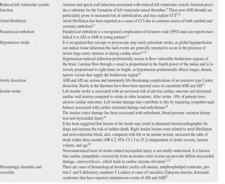
Table 1: Conditions that lead to cardio-cerebral infarction.
Reduced left ventricular systolic
function Anterior and apical wall infarction associated with reduced left ventricular systolic function provi-des a substrate for the formation of left ventricular mural thrombus.9 These post AMI thrombi are
particularly prone to increased risk of embolization, and may explain CCI9-11
Atrial fibrillation Atrial fibrillation has been reported as a cause of CCI due to common source of both cerebral and
coronary embolism12
Paradoxical embolism Paradoxical embolism is a recognized complication of foramen ovale (PFO) and case reports have linked it to AIS or AMI in young patients13
Hypotensive stroke It is recognized that syncope or presyncope may rarely potentiate stroke, as global hypoperfusion can induce tissue infarction but such events are generally reported to occur in the presence of severe large artery stenosis or during cardiac arrest14,15
Hypotension-induced infarction preferentially occurs in flow-vulnerable borderzone regions of the brain. Laminar flow through a vessel is proportional to the fourth power of the radius and is in -versely proportional to eight times its length, so hypotension preferentially affects longer, distant, narrow vessels that supply the borderzone region16
Aortic dissection AMI and AIS are serious and imminently life-threatening complications of an extensive type I aortic dissection. Rarely in the literature have there been reported cases of concurrent AMI and AIS17
Insular stroke Left insular stroke is associated with an increased risk of adverse cardiac outcome and decreased cardiac wall motion compared to stroke in other locations. After stroke, 10% of patients have adverse cardiac outcomes. Left insular damage may contribute to this by impairing sympathovagal balance associated with cardiac structural damage and arrhythmias18
The insular cortex damage has been associated with arrhythmia, blood pressure variation disrup-tion and myocardial injury19
It has been suggested that lesions in the insula may result in abnormal electrocardiographic fin
-dings and increase the risk of sudden death. Right insular lesions were related to atrial fibrillation
and atrioventricular block, also, compared with left or no insular lesions, increased the odds of death within three months (OR 6.2, 95% CI 1.5 to 25.2) independent of stroke severity, lesions volume, and age20
Neuroanatomical basis of stroke-related myocardial injury is not totally understood, It is known that cardiac sympathetic overactivity from an insular cortex lesion can provoke diffuse myocardial damage, «myocytolysis», which leads to cardiac enzyme elevation21,22
Hematologic disorders and
vasculitis There are cases of hematological disorders (sickle cell anemia, antiphospholipid syndrome, pro-tein C and S deficiency, mutation V Leiden) or cases of vasculitis (Takayasu arteritis, Kawasaki
www.medigraphic.org.mx
due to the fact that studies have shown that I.V.rt-PA as the definitive treatment of CCI is not possible because of the different dose of both vascular territories. Alteplase is administered at higher dose for AMI than AIS. (For example, for a 70 kg patient, the dose of AMI is 100 mg and that of acute ischemic stroke is 63 mg). An increased risk of hemorrhagic conversion of AIS when thrombolytic are administered at higher doses and administration of lower than recommended dose of a thrombolytic for AMI may be considered under-dosing, in any case, the percutaneous coronary intervention with stent is the first line therapy for AMI.4,30-33
According to the scientific statement from the American Heart Association/American Stroke Association (AHA/ASA), another reasonable approach to the acute management of CCI is a combined treatment of both vascular territories with administration of IV rt-PA at 0.9 mg/kg (maximum of 90 mg) infused for 60 minutes, with 10% of the total dose administered as an initial intravenous bolus for 1 min, followed by percutaneous coronary angioplasty (PCA) and stenting is reasonable (class IIa, level of evidence C), based on the fact that pretreatment with IV rt-PA does not decrease the coronary benefit of PCA and stenting.2,34
The ideal management of CCI is a treatment strategy that benefits both vascular territories, in that way when non hyperacute CCI is present (> 4.5 hours) and it is not allowed intravenous thrombolysis for AIS, mechanical thrombectomy, a procedure that is not available in most hospitals, could theoretically be combined with PCA.35
Advantages of this approach include the visualization of both coronary and cerebral artery occlusions, which confirms a definite CCI diagnosis, and the effectiveness in treating coronary and cerebral artery occlusion which carries significantly lower mortality than intravenous thrombolysis alone.4
To date, not a single CCI case has been reported in Mexico that has been treated in such way. Hence the benefits and complications of a double interventionist therapy are still unknown. In this article we made an approach with intra-arterial thrombolysis plus cerebral thrombectomy followed by percutaneous coronary angioplasty and stenting. This treatment shows promising results for the non-hyperacute synchronous CCI.
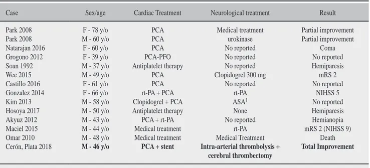
Few cases of the CCI have been reported worldwide, and each was treated in a particular way, thus finding a different clinical evolution in each case (Table 3).
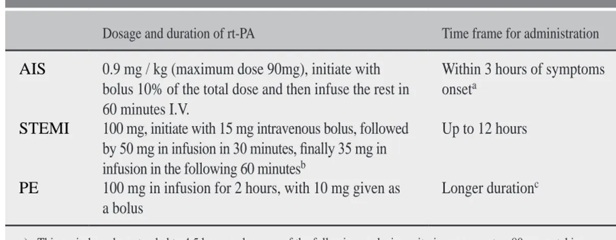
Table 2: The varying rt-PA dose, duration, and method of administration and time frame for initiating therapy in case of AIS, STEMI, and PE.
Dosage and duration of rt-PA Time frame for administration
AIS 0.9 mg / kg (maximum dose 90mg), initiate with bolus 10% of the total dose and then infuse the rest in 60 minutes I.V.
Within 3 hours of symptoms onseta
STEMI 100 mg, initiate with 15 mg intravenous bolus, followed
by 50 mg in infusion in 30 minutes, finally 35 mg in
infusion in the following 60 minutesb
Up to 12 hours
PE 100 mg in infusion for 2 hours, with 10 mg given as
a bolus Longer duration
c
a) This period can be extended to 4.5 hours unless any of the following exclusion criteria are present: > 80 years, taking oral anticoagulants regardless of the international normalized ratio, National Institute of Health Stroke Scale (NIHSS) > 25, and patients with a prior history of stroke and diabetes.
b) For patients weighing 67 kg or less, 15 mg are administered in bolus, followed by 0.75 mg / kg infusion in 30 minutes without exceeding 50 mg, and subsequent 0.50 mg/kg for the next 60 minutes without exceeding 35 mg.
www.medigraphic.org.mx
OBJECTIVESThe present review aims at examining the pathophysiology, etiology, and the current treatment options of CCI, in addition we suggest an innovative therapy by double percutaneous approach. We also exposing a successful case with this therapy which is the first case in Mexico described with this favourable results.
CLINICAL CASE
A 46-years-old male, active smoker, obese with a BMI of 32 kg/m2 and diabetic with 10 years
of evolution, currently under treatment with metformin and insulin.
The clinical case starts with left hemiparesis, deviation of the lips to the right, dysarthria, dyslalia, impossibility to walk, disorientation and nausea. He is taken into the hospital under the presumed diagnosis of AIS. He arrives to emergency department five hours later. The following vital signs are measured: blood pressure (140/90 mmHg), pulse (110 beats per minute), respiratory rate (19 breaths per minute), oxygen saturation (93%), body temperature (37.3 oC), clinically disoriented,
left-sided facial hemiparesis, positive left Babinski sign, no data regarding meningeal irritation. He referred precordial oppressive pain in a scale of EVA 5/10, diaphoresis, not spread elsewhere. General laboratories are carried out which show not significant results.
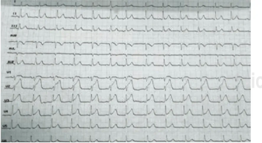
Weighing the possibility of AIS a cranial CT scan is immediately done. The results showed a hypodense area in the right temporoparietal region, which suggested AIS of MRCA. After this, an axial magnetic resonance (MRI) was done. We saw a hyperintensity in the territory of MRCA (Figure 1), what guided us to the diagnosis of AIS of the MRCA in acute state. There was an evident mismatch flair-diffusion (DWI) (Figure 2). The patent was ranked with NIHSS of 15 points, which catalogues him as an optimum candidate for a mechanical thrombectomy therapy. On a later stage, following the diagnosis protocol and because he referred precordial pain, an electrocardiogram is carried out, showing V1-V4 S-T elevation
(Figure 3). Further blood tests are carried out to get the cardiac enzymes, which were six times above the normal upper limit. These results made possible the diagnosis of anteroseptal STEMI. A basal transthoracic echocardiogram
Table 3: CCI Results and cases reported worldwide.
Case Sex/age Cardiac Treatment Neurological treatment Result
Park 2008 F - 78 y/o PCA Medical treatment Partial improvement Park 2008 M - 60 y/o PCA urokinase Partial improvement Natarajan 2016 F - 60 y/o PCA No reported Coma Grogono 2012 F - 39 y/o PCA-PFO No reported No reported Soan 1992 M - 37 y/o Antiplatelet therapy No reported Hemiparesis Wee 2015 M - 49 y/o PCA Clopidogrel 300 mg mRS 2 Castillo 2016 F - 61 y/o PCA No reported No reported Gonzalez 2014 F - 66 y/o rt-PA + PCA rt-PA NIHSS 5
Kim 2013 M - 58 y/o Clopidogrel + PCA ASA1 No reported
Hosoya 2017 M - 50 y/o Antiplatelet therapy None Hemiparesis Akyuz 2012 M - 43 y/o PCA + rt-PA No reported Hemianopia Maciel 2015 M - 44 y/o Medical treatment rt-PA mRS 2 (NIHSS 9) Omar 2010 M - 48 y/o Medical treatment Medical Treatment Death Cerón, Plata 2018 M - 46 y/o PCA + stent Intra-arterial thrombolysis +
cerebral thrombectomy
Total Improvement
www.medigraphic.org.mx
showed ejection fraction of 60%, left ventricular hypokinesia and the presence of left apical thrombus (Figure 4). Later on, the patient was subjected to a coronariography, which showed a subocclusion of the proximal LAD.
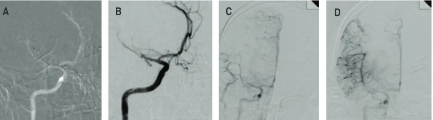
Non-hyperacute CCI was diagnosed and a double interventionist approach was carried out. A cerebral angiography was performed, showing occlusion of the MRCA in the M2 segment. We decided to start with intra-arterial thrombolysis with rt-PA 5 mg in bolus, followed by mechanic thrombectomy with which proves to be successful
(Figure 5). On a cardiac level, coronariography with angioplasty was performed by placing a drug-eluting stent in the proximal segment of the LAD reaching flow TIMI-3 (Figure 6).
Figure 1:
MRI DWI sequence, where the right insular lesion is evident.
Figure 2: MRI DWI sequence where the right insular lesion is evident, and MRI
T2 FLAIR sequence shows no evident lesion (mismatch FLAIR-DIFUSION). This suggests that the ischemic lesion had less than 6 hours, evolution.
Figure 3: V1-V4 S-T elevation showed in ECG.
Figure 4: Transthoracic echocardiogram apical
www.medigraphic.org.mx
Even though the diagnosis of the CCI is normally incidental or diagnosed by autopsy, this time it was hinted by the clinic profile of the patient. He showed chest pain and left hemiparesis, and the occlusion of both vascular territories was confirmed immediately by angiography.
Patients who cannot refer the exact time of the start of their symptoms, a good indicative of the elapsed time is the MRI. If the MRI shows a mismatch DWI-FLAIR, a time lapse of less than 4.5-6 hours can be inferred with high specificity. If the FLAIR sequence shows evidence of lesion, then a time-lapse of more than 6 hours can be inferred. In fact, to some neurointerventionists the occurrence of a mismatch DWI-FLAIR is enough to start thrombolytic therapy when the patient cannot clearly state when started presenting symptoms.25-28
The regular treatment offered to these patients is focused on angioplasty and the use of stents, at a cardiac level, leaving AIS in observation, leading to severe neurological deficit.
The treatment offered to our patient is unique because very few patients come in the therapeutic window necessary for an interventionist approach. Furthermore, not all hospitals have the right facilities to take such approach. PCA and colocation of drug-eluting stent in LAD, plus intra-arterial thrombolysis with rt-PA associated with mechanic thrombectomy of MRCA M2 segment was performed.
The majority of the clinical essays about endovascular therapy in AIS talk about the administration of IV rt-PA alongside the use Figure 5: Cerebral angiography which shows the occlusion of the MRCA M2 segment (A y B), reperfusion sequence (C y D).
B
A
C
D
A
B
Figure 6: Coronariography shows occlusion of the proximal segment of LAD (A),
with reperfusion sequence (B).
Both procedures were carried out without any eventualities. Medication with antiplatelet, statins, ACEIs and warfarin was started after both procedures. The patient was discharged on the third day showing an electrocardiogram without ischemic changes and a mRS of 2.
DISCUSSION
This article talks about a 46-years-old male, which is below the age mean of registered cases. Nonetheless, the patient has prothrombotic risk factors such as: smoking, obesity and diabetes, making him vulnerable to vascular damage at an early age.
www.medigraphic.org.mx
of endovascular therapy. Nonetheless, theanalysis of the subgroups of some clinical essays showed that patients who received thrombectomy therapy without IV rt-PA (due to contraindication), had functional independence and a more rapid rate of recovery than patients who were administered IV rt-PA with endovascular therapy.36,37
Because of this and because our patient was out of the therapeutic window, we decided not to apply intravenous thrombolytic therapy, and to administer intra-arterial thrombolytic therapy, as multiple studies recommend.
It is known that intra-arterial thrombolysis may provide multiple benefits in acute stroke treatment, including extending the treatment therapeutic window, tailored thrombolytic dosage and delivery, salvage therapy for IV rt-PA non responders, and combined use with other endovascular techniques, even in several series, mechanical approach in conjunction with intra-arterial thrombolysis has been shown to achieve higher rates of stroke recanalization and excellent functional outcome can be achieved.38,39
About the dosage, intra-arterial rt-PA is commonly used during mechanical thrombectomy for acute ischemic stroke in patients with large-vessel occlusion. In MR RESCUE TRIAL, a maximum of 14 mg of intra-arterial rt-PA was allowed, the mean dose given was 5.1 mg (2 mg-12 mg). MR CLEAN TRIAL allowed for intra-arterial thrombolytic use, with maximum rt-PA doses of 30 mg. Some studies administer 5 mg at 3 time points during mechanic thrombectomy: (1) upon catheterization of the cervical internal carotid artery, (2) at stentriever clot engagement, and (3) postrecanalization. The theory behind this dosing strategy is that the initial dose helps to further soften the proximal end of the thrombus for crossing with the microwire and microcatheter, the second dose is to assist the stent in absorbing into the clot before retrieval, and the third is to help dissolve microemboli that may occur despite aspiration.40
After therapy the patient was discharged showing an electrocardiogram without ischemic changes and a mRS of 2. The patient is asked to come for an appointment fifteen and thirty days after his discharge. A new transthoracic
echocardiogram is done showing ejection fraction of 65% without valvular disease or intracavitary thrombus, no pulmonary hypertension, no hypokinetic areas either. The patient shows a favorable neurological evolution, reclassifying at day 15 with mRS of 1, and 0 at day 30 after his discharge. The evolution of our patient with the therapy used was optimal, without neurological deficit or cardiohemodynamic compromise.
CONCLUSION
We recommend that every single patient who arrives under presumptive diagnostics of AIS must be protocolized for probable concomitant cardiac compromise with an electrocardiogram and cardiac enzymes. If positive, the next step will be to know how long ago the symptoms started: if < 4.5 hours, the approach will be the administration of IV rt-PA followed by percutaneous coronary angioplasty and stenting. If > de 4.5 hours (non-hyperacute state) we suggest an approach with cerebral thrombectomy alongside coronary angioplasty and stenting. The advantages of addressing non-hyperacute CCI in this way include the visualization of coronary and cerebral arterial occlusions that confirm the diagnosis; and the possibility to start treatment by endovascular approach at the same time. We suggest intra-arterial therapy with rt-PA when patients are no candidates for IV thrombolytic therapy. Further studies are needed to determine the benefits and complications of this approach.
ACKNOWLEDGEMENT
Alicia Gloria Grayeb Grayeb, for helping to translate this article.
REFERENCES
1. Jong Kyu P, Sang-Hak L, Seonghoon C, Jae-Hun J, Namho L. Two patients with acute myocardial infarction presenting with simultaneous acute ischemic stroke. Korean J Med. 2008; 74 (6): 672-625. 2. Akinseye OA, Shahreyar M, Heckle MR, Khouzam RN.
Simultaneous acute cardio-cerebral infarction: is there a consensus for management? Ann Transl Med. 2018; 6 (1): 7.
www.medigraphic.org.mx
cerebrovascular stroke; possible explanations. Int Arch Med. 2010; 3: 25.
4. Yeo LLL, Andersson T, Yee KW, Tan BYQ, Paliwal P, Gopinathan A et al. Synchronous cardiocerebral infarction in the era of endovascular therapy: which to treat first? J Thromb Thrombolysis. 2017; 44 (1): 104-111.
5. Kijpaisalratana N, Chutinet A, Suwanwela NC. Hyperacute simultaneous cardiocerebral infarction: rescuing the brain or the heart first? Front Neurol. 2017; 8: 664.
6. Budaj A, Flasinska K, Gore JM, Anderson FA, Dabbous OH, Spencer FA et al. Magnitude of and risk factors for in-hospital and postdischarge stroke in patients with acute coronary syndromes. Circulation. 2005; 111 (24): 3242-3247.
7. Gattringer T, Niederkorn K, Seyfang L, Seifert-Held T, Simmet N, Ferrari J et al. Myocardial infarction as a complication in acute stroke: results from the austrian stroke unit registry. Cerebrovasc Dis. 2014; 37 (2): 147-152.
8. Mochmann H-C, Scheitz JF, Petzold GC, Haeusler KG, Audebert HJ, Laufs U et al. Coronary angiographic findings in acute ischemic stroke patients with elevated cardiac troponin. Circulation. 2016; 133 (13): 1264-1271.
9. Gianstefani S, Douiri A, Delithanasis I, Rogers T, Sen A, Kalra S et al. Incidence and predictors of early left ventricular thrombus after ST-elevation myocardial infarction in the contemporary era of primary percutaneous coronary intervention. Am J Cardiol. 2014; 113 (7): 1111-1116.
10. Delewi R, Zijlstra F, Piek JJ. Left ventricular thrombus formation after acute myocardial infarction. Heart. 2012; 98 (23): 1743-1749.
11. Vaitkus PT, Barnathan E. Embolic potential, prevention and management of mural thrombus complicating anterior myocardial infarction: a meta-analysis. 1993; 1004-1009.
12. Tokuda K, Shindo S, Yamada K, Shirakawa M, Uchida K, Horimatsu T et al. Acute embolic cerebral infarction and coronary artery embolism in a patient with atrial fibrillation caused by similar thrombi. J Stroke Cerebrovasc Dis. 2016; 25 (7): 1797-1799.
13. Grogono J, Fitzsimmons S, Shah B, Rakhit D, Gray H. Simultaneous myocardial infarction and ischaemic stroke secondary to paradoxical emboli through a patent foramen ovale. 2012; 391-392.
14. Bladin CF, Chambers BR. Frequency and pathogenesis of hemodynamic stroke. 1994. pp. 2179-2182. 15. Dobkin B. Orthostatic hypotension as a risk factor for
symptomatic occlusive cerebrovascular disease. 1989. pp. 30-34.
16. Ryan D, Kenny R, Christensen S, Meaney J, Fagan A, Harbison J. Ischaemic stroke or TIA in older subjects associated with impaired dynamic blood pressure control in the absence of severe large artery stenosis. 2015.
17. Nguyen TL, Rajaratnam R. Dissecting out the cause: a case of concurrent acute myocardial infarction and stroke. 2011;
18. Laowattana S, Zeger SL, Lima JAC, Goodman SN, Wittstein IS, Oppenheimer SM. Left insular stroke is
associated with adverse cardiac outcome. Neurology. 2006; 66 (4): 477-4783.
19. Nagai M, Hoshide S, Kario K. The insular cortex and cardiovascular system: a new insight into the brain-heart axis. J Am Soc Hypertens. 2010; 4 (4): 174-182. 20. Christensen H, Boysen G, Christensen AF, Johannesen HH. Insular lesions, ECG abnormalities, and outcome in acute stroke. J Neurol Neurosurg Psychiatry. 2005; 76 (2): 269-271.
21. Ay H, Koroshetz WJ, Benner T, Vangel MG, Melinosky C, Arsava EM et al. Neuroanatomic correlates of stroke-related myocardial injury. Neurology. 2006; 66 (9): 1325-1329.
22. Cheshire WP, Saper CB. The insular cortex and cardiac response to stroke. Neurology. 2006; 66 (9): 1296-1297.
23. Hosoya H, Levine JJ, Henry DH, Goldberg S. Double the trouble: acute coronary syndrome and ischemic stroke in polycythemia vera. The Am J Med. 2017; 130 (6): e237-e240.
24. Jauch EC, Saver JL, Adams HP, Bruno A, Connors JJ, Demaerschalk BM et al. Guidelines for the early management of patients with acute ischemic stroke. Stroke. 2013; 44 (3): 870-947.
25. De Silva DA, Manzano JJF, Chang HM, Wong MC. Reconsidering recent myocardial infarction as a contraindication for IV stroke thrombolysis. Neurology. 2011; 76 (21): 1838-1840.
26. Bueno H, Martínez-Sellés M, Pérez-David E, López-Palop R. Effect of thrombolytic therapy on the risk of cardiac rupture and mortality in older patients with first acute myocardial infarction. 2005; 1705-1711. 27. Chang RY, Tsai HL, Hsiao PG, Tan CW, Lee CP, Chu IT
et al. Comparison of the risk of left ventricular free wall rupture in Taiwanese patients with ST-elevation acute myocardial infarction undergoing different reperfusion strategies: a medical record review study. 2016; e5308. 28. Patel M, J Meine T, Lindblad L, Griffin J, Granger C,
Becker RC et al. Cardiac tamponade in the fibrinolytic era: analysis of > 100,000 patients with ST-segment elevation myocardial infarction. 2006; 316-322. 29. Omar H, Mangar D, Camporesi E. Simultaneous
thrombosis of 2 vascular territories: is thrombolytic therapy a better option? 2013.
30. Álvarez-Sabín J, Maisterra O, Santamarina E, Kase CS. Factors influencing haemorrhagic transformation in ischaemic stroke. Lancet Neurol. 2013; 12 (7): 689-705. 31. Brott TG, Haley EC, Levy D, Barsan W, Broderick J,
Sheppard GL et al. Urgent therapy for stroke. Part I. Pilot study of tissue plasminogen activator administered within 90 minutes. 1992; 632-640.
32. Messé SR, Tanne D, Demchuk A, Cucchiara B, Levine SR, Kasner S. Dosing errors may impact the risk of rt-PA for stroke: the multicenter rt-PA acute stroke survey. 2004; 35-40.
33. Powers WJ, Rabinstein AA, Ackerson T, Adeoye OM, Bambakidis NC, Becker K et al. 2018 Guidelines for the early management of patients with acute ischemic stroke: a guideline for Healthcare Professionals From the American Heart Association/American Stroke Association. Stroke. 2018; 49 (3): e46-e99.
www.medigraphic.org.mx
rationale for the inclusion and exclusion criteria for intravenous alteplase in acute ischemic stroke. Stroke. 2016; 47 (2): 581-641.
35. Maciel R, Palma R, Sousa P, Ferreira F, Nzwalo H. Acute stroke with concomitant acute myocardial infarction: will you thrombolyse? 2015.
36. Goyal M, Demchuk A, Menon B, Eesa M, Rempel J, Thornton J et al. Randomized assessment of rapid endovascular treatment of ischemic stroke. 2015. 37. Campbell B, Mitchell P, Kleinig T, Dewey HM, Churilov
L, Yassi N et al. Endovascular therapy for ischemic stroke with perfusion-imaging selection. 2015. 38. Noser EA, Shaltoni HM, Hall CE, Alexandrov AV,
Garami Z, Cacayorin ED et al. Aggressive mechanical clot disruption: a safe adjunct to thrombolytic therapy in acute stroke? Stroke. 2005; 36 (2): 292-296. 39. Berkhemer OA, Fransen PS, Beumer D, van den Berg
LA, Lingsma HF, Yoo AJ et al. A randomized trial of
intraarterial treatment for acute ischemic stroke. N Engl J Med. 2015; 372 (1): 11-20.
40. Heiferman DM, Li DD, Pecoraro NC, Smolenski AM, Tsimpas A, Ashley WW, Jr. Intra-arterial alteplase thrombolysis during mechanical thrombectomy for acute ischemic stroke. J Stroke Cerebrovasc Dis. 2017; 26 (12): 3004-3008.
Correspondence to:
Dr. Juan Carlos Plata Corona
Internal Medicine Resident at Eduardo Vázquez Navarro Hospital, ZIP 72000, Puebla, Pue.
Tel: 2222932955.