DE HOLGUÍN
ISSN 1560-4381 CCM 2017; (1)
ORIGINAL RESEARCH
Beyond the Definition of Metabolic Syndrome: Uric Acid, High
Sensitivity C Reactive Protein and Global Cardiovascular Risk
Más allá de la definición de síndrome metabólico: ácido úrico, proteína c
reactiva de alta sensibilidad y riesgo cardiovascular global
Jorge Vega Abascal1, Mayra Rosa Guimara Mosqueda2, Luis Vega Abascal3
1. Master degree in Medical Urgencies. Specialist in Internal Medicine. Associate Professor. Jose Avila Serrano Teaching Polyclinic. Velasco. Holguin, Cuba.
2. Master degree in Medical Urgencies. Specialist in Community Medicine and Second degree Specialist in Cardiology. Associate Professor. Jose Avila Serrano Teaching Polyclinic. Velasco. Holguin, Cuba.
3. Master degree in Medical Urgencies. Specialist in Surgery. Associate Professor. Jose Avila Serrano Teaching Polyclinic. Velasco. Holguin, Cuba.
ABSTRACT
Introduction: the metabolic syndrome is becoming one of the main problems in public health.
Objective: to evaluate the relation between metabolic syndrome and uric acid, high sensitivity c reactive protein, total cardiovascular risk and cardiovascular events.
Results: the waist circumference (p = 0.000), systolic (p= 0.0042) and diastolic blood pressure (p= 0.0298), high sensitivity c reactive protein (p= 0.0039) and uric acid (p= 0.0283) were significantly associate in both groups. The body mass index was higher than 30 kg/m2 (OR 7.54; CI95%:3.35-16.9), LDL cholesterol greater than 4.16 mmol/l (OR 3.49; CI 95%:1.58-7.70) and hs CRP higher than 1 mg/dl (OR 3.59; CI 95%:1.51-8.51) showed statistically significant differences according to groups of studies. Global cardiovascular risk greater 20%, it was 3.84 times higher in the group with metabolic syndrome (CI 95%:1.67-8.82), 13.4% of the patients with metabolic syndrome developed a cardiovascular event during the period in comparison with the 2.9% in the group without metabolic syndrome (OR= 5.04; CI 95%: 1.04-24.3).
Conclusions: metabolic syndrome was significantly associated with mean level of uric acid, high sensitivity c reactive protein, total cardiovascular risk and cardiovascular events.
Keywords: metabolic syndrome, high sensitivity c reactive protein, uric acid, total cardiovascular risk, primary health care
RESUMEN
Introducción: el síndrome metabólico se está convirtiendo en uno de los principales problemas de salud pública
Objetivo: evaluar la relación entre el síndrome metabólico y el ácido úrico, la proteína c reactiva de alta sensibilidad, el riesgo cardiovascular global y la incidencia de eventos cardiovasculares.
Métodos: se realizó un estudio de casos controles anidados en una cohorte dinámica en el Policlínico Docente de Velasco desde 2010 a 2015, los casos fueron 67 pacientes que desarrollaron síndrome metabólico en el periodo y 67 controles que fueron apareados por sexo y edad de ± 5 años en relación con los casos, los participantes fueron sometidos a un examen físico, determinaciones antropométricas y de laboratorio, fueron sometidos a seguimiento durante 4,5 años para la aparición de eventos cardiovasculares.
seguimiento en comparación con el 2,9% en el grupo sin síndrome metabólico (OR= 5,04; IC 95%: 1,04-24,3).
Conclusiones: el síndrome metabólico se asoció significativamente con la concentración media de ácido úrico, con la proteína c reactiva de alta sensibilidad, el riesgo cardiovascular global y el desarrollo de eventos cardiovasculares.
Palabras clave: síndrome metabólico, proteína c reactiva de alta sensibilidad, ácido úrico, riesgo cardiovascular total, atención primaria de salud.
INTRODUCTION
Metabolic syndrome (MetS) is a combination of medical disorders that, including obesity, insulin resistance, impaired glucose metabolism, dyslipidemia of high triglycerides, low level of high density lipoprotein cholesterol (HDLc) and elevated blood pressure, when occurring together, increase the risk of developing cardiovascular disease and diabetes. It has become a major public health challenge worldwide1, 2.
Since the first official definition of the MetS by a World Health Organization Working Group in 1999, a number of alternative definitions have been proposed. The most widely accepted of these have been produced by the European Group for the Study of Insulin Resistance (EGIR), International Diabetes Federation (IDF) and the National Cholesterol Education Program Adult Treatment Panel III (ATP III) 3.
Although a consensus criterion has not been reached for diagnosing MetS, it is recommended that screening should consider central obesity, insulin resistance; dyslipidemia (elevated triglycerides and low density lipoprotein cholesterol (LDLc)) and decreased high density lipoprotein cholesterol (HDLc), and high blood pressure. Other factors such as proinflammatory and prothrombotic states have also been associated with MetS 4.
There is an abundance of widely varying data comparing prevalence using different criteria and this only served to reinforce the need for a standardized definition internationally. There is an urgent need to rationalize the variety of definitions that had been developed for the MetS. This need should be extended from clinical practice through to research5.
The construct of the MetS diagnostic criteria has inherent limitations which impact on its clinical usefulness, it does not include other important risk factors for predicting diabetes or cardiovascular disease (CVD), such as age, sex, family history socioeconomic status, ethnicity, current treatment, previous CVD events and LDLc, C reactive protein, uric acid or important behavioral variables such as smoking and physical activity5, 6.
The MetS has proposed as a means to identify people with increased risk of cardiovascular disease and type 2 diabetes mellitus (T2DM) and to guide the clinical decisions and it has shown its utility to predict the morbidity and mortality for cardiovascular diseases and diabetes.
Cardiovascular disease often occurs in people with considered acceptable or average risk factors values, this suggests that there are other risk factors that must be the cause of this disease. If these factors were considered to the present set of CVD risk factors, it would improve risk estimation. A number of risk factors, some of them new and often called novel risk factors, have been proposed: inflammatory markers such as C-reactive protein, coronary calcium, lipoprotein (a), interleukin-6, fibrinogen, homocysteine, insulin resistance, MetS, platelet function, and genetic scores7.
The aim of this study was to evaluate the relation between MetS and uric acid, high sensitivity c reactive protein, global cardiovascular risk and cardiovascular events.
METHODS
A nested case control study in a dynamic cohort of 309 patients was carried out, the sample were 134 patients, with age between 35 to 74 years, without cardiovascular disease at baseline, of the Jose Avila Serrano Teaching Polyclinic, from Velasco, Holguin, during January 2010 to December 2015.
≥ 102 cm in male and ≥ 88 cm in female), elevated triglycerides ≥ 1.70 mmol/l, decreased high-density lipoprotein cholesterol < 0.9 mmol/l in male y < 1.1 mmol/l in female, elevated blood pressure ≥ 130 mmHg for systolic or ≥ 85 mmHg for diastolic blood pressure or blood pressure treatment, and fasting glucose level ≥ 5.55 mmol/l (100 mg/dl) or T2DM previously diagnosed3.
The participants underwent a physical examination, anthropometry, blood pressure determination, blood pressure measurements were made on the right arm of the seated participants with a aneroid sphygmomanometer and an appropriately sized cuff; the average of two physician-obtained measures constituted the examination blood pressure; waist circumference was measured (cm) at level of the navel; cigarette smoking status was ascertained if the patients smokes or had smokes in the last six months, past history of diabetes and hypertension was ascertained by the physician; serum total cholesterol and HDLc, LDLc, calculated according to Friedewald formula. Uric acid, glycaemia, high sensitivity c reactive protein (hs CRP) levels were determined with standardized enzymatic methods; hs CRP were determined in two times with an interval of 15 days, being used the value average of both measurements.
The global cardiovascular risk to the ten years was stratified using Framingham - D´Agostino (2008)8 risk table, that it use for the calculation of the risk: age, sex, smoking, diabetes mellitus, treated or untreated systolic blood pressure, total cholesterol and HDLc, classifying the patients as low risk (< 10%), among 10-20% as intermediate risk and > 20% as high risk.
All the study participants were under continuous surveillance during 4.5 years; for development of cardiovascular events or death. Study defines hard cardiovascular events if the patients developed a myocardial infarction, ischemic stroke, hemorrhagic stroke mortal or no, and coronary death, and it was considered all cardiovascular events if the patient developed hard events plus coronary angina, transitory ischemic attack and peripheral arterial disease.
The means, standard deviation and percentages were calculated. Student t test was used to compare mean of the independent samples with significant value of p< 0.05 and Odd Ratio (OR) with 95% confidence intervals, the data was coded and processed using the Statistical Package for Social Sciences (SPSS, version 15) and Med Calc software (version 4.16g).
RESULTS
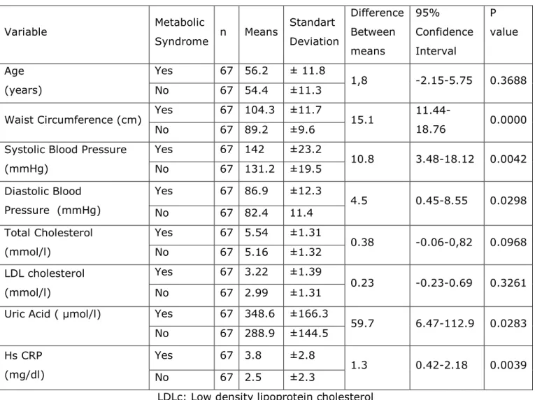
In the cases and controls groups there were 49 women (73.1%) and 18 men (26.9%). The results showed that in the studied population the waist circumference, systolic and diastolic blood pressure, level of high sensitivity c reactive protein and mean uric acid levels were significantly associated in both groups (table I)
Table I. Characteristic of the study population.
Variable Metabolic
Syndrome n Means
Standart Deviation Difference Between means 95% Confidence Interval P value Age (years)
Yes 67 56.2 ± 11.8
1,8 -2.15-5.75 0.3688
No 67 54.4 ±11.3
Waist Circumference (cm) Yes 67 104.3 ±11.7 15.1
11.44-18.76 0.0000
No 67 89.2 ±9.6
Systolic Blood Pressure (mmHg)
Yes 67 142 ±23.2
10.8 3.48-18.12 0.0042 No 67 131.2 ±19.5
Diastolic Blood Pressure (mmHg)
Yes 67 86.9 ±12.3
4.5 0.45-8.55 0.0298
No 67 82.4 11.4
Total Cholesterol (mmol/l)
Yes 67 5.54 ±1.31
0.38 -0.06-0,82 0.0968
No 67 5.16 ±1.32
LDL cholesterol (mmol/l)
Yes 67 3.22 ±1.39
0.23 -0.23-0.69 0.3261
No 67 2.99 ±1.31
Uric Acid ( μmol/l) Yes 67 348.6 ±166.3
59.7 6.47-112.9 0.0283 No 67 288.9 ±144.5
Hs CRP (mg/dl)
Yes 67 3.8 ±2.8
1.3 0.42-2.18 0.0039
No 67 2.5 ±2.3
LDLc: Low density lipoprotein cholesterol Hs CRP: High sensitivity c reactive protein
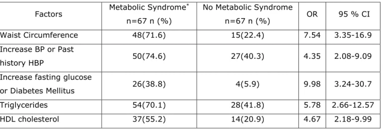
Table II. Factors considered as identifying abnormalities of metabolic syndrome according to ATP III definition.
Factors Metabolic Syndrome * n=67 n (%)
No Metabolic Syndrome
n=67 n (%) OR 95 % CI
Waist Circumference 48(71.6) 15(22.4) 7.54 3.35-16.9
Increase BP or Past
history HBP 50(74.6) 27(40.3) 4.35 2.08-9.09
Increase fasting glucose
or Diabetes Mellitus 26(38.8) 4(5.9) 9.98 3.24-30.7
Triglycerides 54(70.1) 28(41.8) 5.78 2.66-12.57
HDL cholesterol 37(55.2) 14(20.9) 4.67 2.18-9.99
Met S: Metabolic Syndrome HBP: High Blood Pressure
HDLc: High- density lipoprotein cholesterol
* Diagnosis of metabolic syndrome according to ATP III, 2005 (Adult Treatment Panel III)3
Elevated waist circumference: ≥ 102 cm in men and ≥ 88 cm in women
Elevated blood pressure: 130 mm Hg systolic BP or 85 mm Hg diastolic BP or, drug treatment for hypertension.
Elevated fasting glucose 100 mg/dl or drug treatment for elevated glucose
Elevated triglycerides:> 1.70 mmol/l
Reduced HDL cholesterol: <0.9 mmol/l in men and <1.1 mmol/l in women
Table III. Factors that increased probability of the metabolic syndrome.
Factors Metabolic Syndrome n=67 n (%)
No Metabolic Syndrome n=67
n (%)
OR 95 % CI
Smoking Yes 19(28.4) 20(29.8) 0.93 0.44-1.96
No 48(71.6) 47(70.1) 1.07 0.51-2.26
Body Mass Index (kg/m2)
< 24.9 9(13.4) 32(47.8) 0.16 0.07-0.39
25-29.9 20(29.8) 24(35.8) 0.76 0.36-1.57
> 30 40(59.7) 11(16.4) 7.54 3.35-16.9
Total Cholesterol (mmol/l)
< 6 44(65.7) 52(77.6) 0.55 0.65-1.18
6-7.2 14(20.9) 9(13.4) 1.70 0.68-4.25
> 7.2 9(13.4) 6(8.9) 1.57 0.52-4.70
LDL Cholesterol (mmol/l)
< 3.37 25(37.3) 40(59.7) 0.40 0.20-0.80
3.38-4.13 13(19.4) 15(22.4) 0.83 0.36-1.92
> 4.16 29(43.3) 12(17.9) 3.49 1.58-7.70
Uric acid (μmol/l) > 440(Male)
>350(Female) 27(40.3) 20(29.8) 1.58 0.77-3.24
Hs CRP (mg/dl) < 1 9(13.4) 24(35.8) 0.27 0.12-0.66
> 1 58(86.6) 43(64.2) 3.59 1.51-8.51
LDLc: Low-density lipoprotein cholesterol Hs CRP: High sensitivity c reactive protein
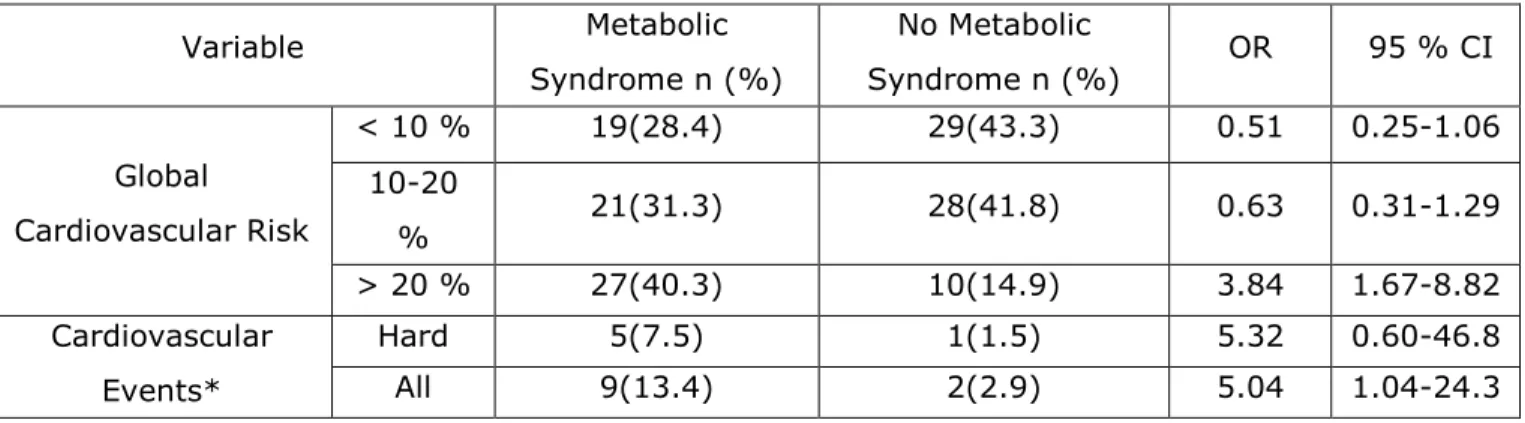
Total cardiovascular risk greater 20% was 3.84 times higher in the group with MetS (95 % CI: 1.67-8.82) and 13.4% of the patients with MetS developed a cardiovascular event during the period in comparison with 2.9% of patients in the group without MetS (OR= 5.4; 95% CI: 1.04-24.3) (table IV)
Table IV. Global cardiovascular risk and cardiovascular events according to study groups.
Variable Metabolic
Syndrome n (%)
No Metabolic
Syndrome n (%) OR 95 % CI
Global Cardiovascular Risk
< 10 % 19(28.4) 29(43.3) 0.51 0.25-1.06
10-20
% 21(31.3) 28(41.8) 0.63 0.31-1.29
> 20 % 27(40.3) 10(14.9) 3.84 1.67-8.82
Cardiovascular Events*
Hard 5(7.5) 1(1.5) 5.32 0.60-46.8
All 9(13.4) 2(2.9) 5.04 1.04-24.3
DISCUSSION
Among the contributing major cardiovascular risk factors, an increased waistline, elevated triglycerides, impaired fasting plasma glucose, high systolic blood pressure and low levels of HDL cholesterol are referred to as MetS3,9. Obesity is a growing burden worldwide; hence, forecasts predict an increase in prevalence from 35%, to 51% by 2030 in US Americans9.
In this research the waist circumference (p 0.0000) was significantly associated in both groups (table I) and the BMI > 30 kg/m2 showed significantly statistical differences (OR 7.54; 95 % CI: 3.35-16.9) (table III).
In spite of the fact that high BMI values are associated with adverse risk profiles for morbidity and mortality, particularly in relation to T2DM and atherothrombotic cardiovascular disease, various sub-types of obesity have been described which complement the apparent dose-response relationship between BMI and its impact on health. The most classic sub-types are android obesity (abdominal) and gynoid obesity (gluteal obesity) 10.
Less well-known sub-types are, however, also of interest. For example, a phenotype corresponding to metabolically obese normal-weight (MONW) individuals has been reported, these individuals have a normal BMI together with some of the characteristics of obese individuals such as insulin resistance, central adiposity, low levels of HDL and high levels of triglycerides, and high blood pressure. At the same time, metabolically healthy obese (MHO) individuals have also been identified. These individuals have BMI>30, but none of the metabolic alterations which are typical of obese individuals10.
The existence of these 2 "paradoxical" sub-types led to several studies over the last 2 decades which contributed to metabolically characterization and quantify visceral adipose tissue as opposed to subcutaneous adipose tissue. These studies showed that the quantity of visceral adipose tissue is directly correlated in both men and women with a seriously altered metabolic risk profile which leads the development of T2DM and cardiovascular disease. Therefore, although it is true that obesity increases the risk of chronic disease, it seems clear that patients with visceral obesity constitute a sub-group of individuals with the most serious metabolic alterations10.
tissue have consequently been identified to play a key role in systemic inflammation, atherosclerosis and cardiovascular death9,11.
It has been demonstrated that excess body fat, abdominal visceral fat, and larger waist circumference have been associated with accelerated arterial stiffening. Abdominal obesity has been associated with an adverse effect on blood vessels, independently of age, sex, blood pressure, fasting glucose and BMI. Combination of diabetes mellitus and abdominal obesity, but not overall obesity, has been associated with significant deterioration in terms of arterial stiffness parameters. Body fat distribution is one of the major determinants of metabolic health, and visceral adiposity has a stronger correlation with metabolic abnormalities and cardiovascular disease than subcutaneous adipose tissue11.
The mean level of uric acid was significantly associated in both groups (table I) but the level more than 440 and 350 μmol/l in male and female respectively were not significantly associated in the groups with and without MetS (table III).
Serum uric acid (UA) is a strong predictor of stroke, coronary artery disease, and MetS, however, the definite role of uric acid in these diseases is still the subject of much discussion and debate because it is always accompanied with other risk factors such as diet, obesity, and dyslipidemia. Specifically, disputation exists about whether serum uric acid is a causative risk factor or only a coexisting marker of those pathologic processes12.
However, few studies have investigated the predictive role of UA levels, and studies that investigate the relation between UA and MetS criteria in patients previously detected with cardiovascular risk are still scarce13. Abreu da Silva, et al found higher UA levels in subjects showing MetS when compared to those who did not13 , previous epidemiologic studies have also shown increased serum UA levels in adults with MetS, as well as an association between increased UA levels and oxidative stress, endothelial dysfunction, inflammation, atherosclerosis, and increased risk of cardiovascular events13-15.
Evidence from prospective and interventional studies of various populations suggests that gout and hyperuricaemia are independent risk factors for cardiovascular diseases, the mechanisms linking hyperuricaemia and gout with cardiovascular events are unclear but may include oxidative stress generated by xanthine oxidase (XO), the enzyme that catalyses the formation of urate, other explanations are a direct contribution to endothelial dysfunction and low-grade inflammation associated with increased urate levels and tophi16.
During the recent years, an increasing number of publications have supported that allopurinol might have a benefit effect in a variety of cardiovascular diseases, and may provide a survival benefit among patients with hyperuricaemia. Indeed, allopurinol, a free radical scavenger, was found to improve both endothelial dysfunction and levels of some markers of oxidative stress16.
The precise mechanism by which allopurinol exerts a beneficial effect on the cardiovascular system is poorly known, the drug is thought to act mainly through an increase in the bioavailability of endothelium derived nitric oxide, presumably by blocking the production of reactive oxygen species generated by the XO activity16.
Recent data have also suggested that the cardiovascular benefit of allopurinol in patients with heart failure might be due to its urate lowering property, although this point is a matter of debate. Indeed, post hoc analyses of clinical trials have suggested that the reduced vascular risk afforded by therapeutics, such as atorvastatin and losartan which are not XOs, was due to their urate lowering property16.
The pathophysiological mechanism by which the MetS increases cardiovascular risk remains under debate; earlier definitions by the World Health Organization and the EGIR emphasize the independent role of insulin resistance as the underlying component of the MetS. Insulin resistance progresses toward hyperinsulinemia and hyperglycemia, thus triggering peripheral vasoconstriction and sodium retention. Hepatic production of very low-density lipoprotein also increases, leading to hypertriglyceridemia, low HDL cholesterol, elevated apolipoprotein B, elevated small LDL cholesterol, and consequently, atherosclerosis, as a result of these lipid imbalances, individuals with the MetS typically exhibit a prothrombotic and proinflammatory state17, 18.
At least half of the attributable risk in patients who end up with a thrombus or atherosclerosis is caused by this inflammatory response, major meta-analyses have shown that if you measure inflammation using the high-sensitivity C-reactive protein (hs-CRP) test, the magnitude of risk associated with hs-CRP is larger than the magnitude of risk of high blood pressure or high cholesterol19, 20.
However, all of these parameters are associated with elevated levels of hs-CRP, an easily measured inflammatory biomarker that has proven to be a strong, independent predictor of both incident diabetes and incident cardiovascular disease, CRP levels also correlate with several other components such as fasting insulin, microalbuminuria, and impaired fibrinolysis that are not easily evaluated in usual clinical practice19- 21.
Rapidly evolving work now demonstrates that in addition to being a marker of innate immunity, CRP also has several direct effects at the level of the vessel wall, these observations, along with basic research into the inflammatory mechanisms of both diabetes and vascular dysfunction, provide strong evidence that insulin resistance and atherosclerosis share a common inflammatory basis, CRP, however, is also associated with several aspects of the MetS not easily ascertained in usual clinical practice, including fasting insulin, hypofibrinolysis, and microalbuminuria19
MetS is associated with increased oxidative stress, furthermore, it appears that some component pathologies of the MetS contribute to a higher percentage of total oxidative stress than others; however, additional studies are needed to determine the exact contribution of individual components to total oxidative stress4.
In this study no differences in the smoker in the groups were found, this can be explained by the prevalence of women in both groups, smoking and air pollution interact with the MetS which are not insufficiently assumed, but they clearly combined to deliver a cardiovascular risk factor which is greater than the sum of each of them.
Smokers and ex-smokers are more likely to have MetS than nonsmokers, both cigarette smoking and the MetS are strong independent risk factors for cardiovascular disease; however, smoking also potentiates the negative cardiovascular effects of the MetS. For example, MetS is associated with higher rates of cardiac events after acute myocardial infarction and smoking has an additive effect. These effects are largely mediated via Reactive Oxygen Species (ROS) generation4.
including lipid-related markers [i.e. apolipoprotein B, apo-lipoprotein A-I, lipoprotein (a), or lipoprotein-associated phospholipase A2] in prognostic models for CV disease risk prediction22-25.
MetS was commonly defined by the presence of obesity, diabetes, hypertension, and dyslipidemia, and these factors were involved in the mechanisms of insulin resistance, inflammation, and atherosclerosis, but does not include other novel risk factor, the tools for prediction of cardiovascular risk include other different risk factors and both (MetS and scores for cardiovascular risk prediction) do not include other risk factors, with the improving of knowledge on metabolic syndrome and the prediction of cardiovascular risk the investigators have opportunities for the certain identification of novel biomarkers or risk factors that could be considered for cardiovascular disease and the prevention of the cardiovascular diseases is based in the opportune identification in the primary health care with the biggest precision possible of the people with risk of developed cardiovascular disease26-28.
Specific strengths of the present study were the use of nested case control study in a dynamic cohort design tended to results in estimated with lower bias and greater precision, controls matched to cases on classical matching factors (eg, age, sex); a limitation of this study was its small sample due to lack of laboratory reagents and suitable technology that did not allow to investigate more people.
CONCLUSIONS
In conclusion, this research showed relation statistically significant between the MetS and the mean level of uric acid, the level of hs CRP higher than 1 mgl/dl, level of LDLc greater than 4.16 mmol/l, the global cardiovascular risk higher than 20% and the incidence of all cardiovascular events.
REFERENCES
1. Zhou H, Guo Zr, Hu Xs , Yu Lg, Xu Bx , Wu M, et al. An exploratory analysis of dynamic change of metabolic syndrome in relation to the risk of developing cardiovascular disease in a chinese cohort. Iranian J Publ Health. 2012[cited 2013 Jan 14]; 41(4):26-34. Available in:
http://www.ncbi.nlm.nih.gov/pmc/articles/PMC3481612/pdf/ijph-41-26.pdf
prediction: A longitudinal study. PLoS ONE. 2013[cited 2014 Jul 10]; 8(12): 1-10. Available in:
http://www.ncbi.nlm.nih.gov/pmc/articles/PMC3866125/pdf/pone.0084204.pdf
3. Zimmer P, George K, Alberti M, Serrano M. A new international diabetes federation worldwide definition of the metabolic syndrome: the rationale and the results. Rev Esp Cardiol. 2005[cited 6 Oct 2006]; 58(12):1371-1375. Available in:
http://www.revespcardiol.org/es/content/articulo/13082533/
4. Hutcheson R, Rocic P. The metabolic syndrome, oxidative stress, environment, and cardiovascular disease: The great exploration. Exp Diabetes Res. 2012[cited 27 Jun
2013];2012(2012):1-13. Available in: http://www.ncbi.nlm.nih.gov/pmc/articles/PMC3399393/
5. Simmons RK, Alberti KG, Gale EA, .Colagiuri S, Tuomilehto J, Qiao Q, et al. The metabolic syndrome: useful concept or clinical tool? Report of a WHO expert Consultation . Diabetologia. 2010. [cited 14 Jan 2011];53(4):600–605. Available in:
http://link.springer.com/article/10.1007/s00125-009-1620-4
6. Pineda CA. Metabolic syndrome: definition, history, criterion. Colombia Med.2013[cited 14 Jan 2014];39(1):96-106.Available in:
http://bibliotecadigital.univalle.edu.co/bitstream/10893/4753/1/Metabolic%20syndrome.pdf
7. D’Agostino RB, Pencina MJ, Massaro, JM, Coady S. Cardiovascular disease risk assessment: Insights from Framingham. Glob Heart. 2013. [cited 2014 Oct 7]; 8(1): 11–23. Available in:
http://www.ncbi.nlm.nih.gov/pmc/articles/PMC3673738/
8. D'Agostino RB, Vasan RS, Pencina MJ, Wolf PA, Cobain M, Massaro JM. General cardiovascular risk profile for use in primary care. The Framingham Heart Study. Circulation. 2008 [cited 15 Feb 2013]; 117(6):743-753.Available in: http://circ.ahajournals.org/cgi/content/full/117/6/743
9. Rohla M, Weiss TW. Adipose tissue, inflammation and atherosclerosis. Clin Lipidology. 2014[cited 30 Jun 2014]; 9(1):71-81.Available in:
http://www.futuremedicine.com/doi/pdf/10.2217/clp.13.80
11. Lukich A, Gavish D, Shargorodsky M. Normal weight diabetic patients versus obese diabetics: Relation of overall and abdominal adiposity to vascular health. Cardiovasc Diabetol. 2014[cited 14 Dec 2014]; 13(141).Available in: http://www.medscape.com/viewarticle/835945_1
12. Peng TC, Wang CC, Wei Kao T, Hsin Chan J, Yang YH, Wen ChangY, et al. Relationship between hyperuricemia and lipid profile in US adults. Biomed Research International.2015 [cited 2015 Jul 10 ]:1-7.Available in: http://www.ncbi.nlm.nih.gov/pmc/articles/PMC4299312/
13. Abreu da Silva H, Cardoso JC, Bressan J, Miranda H. Relation between uric acid and metabolic syndrome in subjects with cardiometabolic risk. Einstein. 2015 [cited 2015 May
19]13(2): 202-208. Available in: http://www.scielo.br/scielo.php?script=sci_arttext&pid=S1679-45082015005053194&lng=en&nrm=iso&tlng=en
14. Lee JK , Ryoo JH , Choi JM , Park SK. Serum uric acid level and the incidence of metabolic syndrome in middle-aged Korean men: A 5-year follow up study. J Prev Med Public Health 2014 [cited 2015 Jan 14];47:317-326. Available in:
http://www.ncbi.nlm.nih.gov/pmc/articles/PMC4263007/
15. Li LX, Dong XH, Li MF, Zhan R, Li TT, ShenJ,et al. Serum uric acid levels are associated with hypertension and metabolic syndrome but not atherosclerosis in Chinese inpatients with type 2 diabetes. Journal of Hypertension 2015 [cited 2015 Jul 10]; 33(3): 482-490. Available
in:http://www.ncbi.nlm.nih.gov/pmc/articles/PMC4309490/
16. Grimaldi-Bensouda L, Alperovitch A, Aubrum E, Danchin N, Rossigni M, Abenhaim L, et al. Impact of allopurinol on risk of myocardial infarction. Ann Rheum Dis. 2015.[cited 2015 Jul 10]; 74(5): 836-842. Available in: http://www.medscape.com/viewarticle/844859?src=wnl_edit_tpal
17. Miguel-Soca P. Predictores de riesgo cardiometabolico. Revista Finlay. 2015 [cited 2015 Jul 3]; 5(2):80-82. Disponible en: http://www.revfinlay.sld.cu/index.php/finlay/article/view/357
18. Mottillo F. Filion K, Genest J, Joseph J,Pilote L, Poirier P, et al. The metabolic syndrome and cardiovascular risk. A systematic review and meta-analysis.JACC.2010 [cited 2011 Jun 27]; 56(14):1113-32.Available in:
http://www.sciencedirect.com/science/article/pii/S0735109710026380
Circulation. 2003[cited 2005 Sep 19]; 107:391-397.Available in:
http://circ.ahajournals.org/content/107/3/391.full.pdf+html
20. The Emerging Risk Factors Collaboration. C - reactive protein, fibrinogen, and cardiovascular disease prediction. N Engl J Med. 2012[cited 2013 Jan 14]; 367(14): 1310–1320. Available in:
http://www.ncbi.nlm.nih.gov/pmc/articles/PMC3714101/pdf/nihms488711.pdf
21. Vega J, Guimara M, Garces Y, Garcia Y, Vega L. Proteina C reactiva de alta sensibilidad y riesgo de enfermedad cardiovascular. CCM. 2015[cited 2015 Jul 10]; 19(2):190-201. Disponible en: http://www.revcocmed.sld.cu/index.php/cocmed/article/view/978/621
22. Charakida M, Masi S, Deanfield JE. The Year in Cardiology 2012: focus on cardiovascular disease prevention. Eur Heart J. 2013 [cited 2014 Nov 21]; 34: 314–7. Available in
http://eurheartj.oxfordjournals.org/content/ehj/early/2013/01/02/eurheartj.ehs429.full.pdf
23. Nordet P, Mendis M, Dueñas A, de la Noval R, Armas N. Total cardiovascular risk assessment and management using two prediction tools, with and without blood cholesterol. MEDICC
Review.2013 [cited 2014 Nov 26]; 15(4). Available in:
http://www.scielosp.org/scielo.php?pid=S1555-79602013000400009&script=sci_arttext
24. Aljutaili M, Becker C, Witt S, Holle R, Leidl L. Should health insurers target prevention of cardiovascular disease?: A cost-effectiveness analysis of an individualized programme in Germany based on routine data. BMC Health Services Research.2014 [cited 2014 Nov 26];
14:263.Available in: http://www.biomedcentral.com/1472-6963/14/263
25. Vega J, Guimará M, Vega L. Riesgo cardiovascular, una herramienta útil para la prevención de las enfermedades cardiovasculares. Rev Cubana Med Gen Integr.2011 [cited 2013 Dec 12]; 27(1):91-97. Available in: http://scielo.sld.cu/pdf/mgi/v27n1/mgi10111.pdf
26. Vega J, Guimara M, Garces Y, Vega L, Rivas M. Predicción del riesgo coronario y
cardiovascular global en atención primaria de salud. CCM. 2015 [cited 2015 Nov 27] 19(2):202-211. Available in: http://www.revcocmed.sld.cu/index.php/cocmed/article/download/979/622
27. Tarragó E, Miguel Soca PE, Cruz L, Santiesteban Y. Factores de riesgo y prevención de la enfermedad isquémica. CCM. 2012 [cited 2014 Nov 20]; 16(2). Disponible en:
28. Rivas Vázquez D, Miguel Soca PE, Llorente Columbié Y, Marrero Ramírez GM. Comportamiento clínico y epidemiológico del síndrome metabólico en adultos. Rev Cubana Med Gen Integr. 2015 [citado 2016 Mar 09]; 31(3). Available in:
http://scielo.sld.cu/scielo.php?script=sci_arttext&pid=S0864-21252015000300001&lng=es
Recibido: 15 de julio de 2015 Aprobado: 14 de marzo de 2016