Prevalence and risk factors of isolated anti-HBc
antibody and occult hepatitis B infection in hemodialysis patients:
a nationwide study
Hossein Keyvani,* Shahram Agah,** Ali Kabir,*** Seyed-Moayed Alavian****
* Department of Virology, Tehran University of Medical Sciences, Tehran, Iran.
** Tehran University of Medical Sciences, Gastrointestinal and Liver Diseases Research Center, Tehran, Iran.
*** Department of Epidemiology, Faculty of Public Health, Shahid Beheshti University of Medical sciences; Center for Educational Research in Medical Sciences, Tehran University of Medical Sciences, Tehran, Iran.
**** Baqyiatallah University of Medical Sciences, Research Center for Gastroenterology and Liver Dieases; Rezvan Medical Research Institute (R-MRI), Tehran, Iran.
ABSTRACT
Background. Recent studies have demonstrated that prevention of hepatitis B virus (HBV) transmission re-mains a challenge in hemodialysis (HD) patients. The aim of this study is to explore factors which increase the possibility of occult HBV (OHB) infection in patients under HD in Iran. Material and methods. Among 2188 cases who spent an average of 60.2 ± 61.1 months under HD, we selected 103 cases with isolated anti-hepatitis B virus core antibody (anti-HBc) and 231 controls without any HBV, anti-hepatitis D virus (HDV) and hepatitis C virus (HCV) serologic markers. Socio-demographic data, past medical history, and clinical signs and symptoms were assessed. Results. The frequency of checking hepatitis B serologic markers was 15.3%. The rate of OHB infection in HD patients who were monitored for their HBV markers was 4.9%. Cases with isolated anti-HBc had significantly higher percentage of positive HBV DNA [odds ratio: OR (95% confidence interval: 95% CI) = 12.1 (1.4-105)], visual disturbances [OR (95% CI) = 1.8 (1.1-3.03)], history of diabetes mellitus [OR (95% CI) = 2.1 (1.3-3.5)], higher age, higher age when dialysis started and were mostly married, illiterate, disabled and retired. Diabetes mellitus was the only independent predictor of HBV DNA status in cases with isolated anti-HBc. Conclusion. In our region, OHB infection is prevalent among hemodialysis patients and displays a direct correlation with factors which are age related except diabetes mellitus. Thus, the presence of isolated anti-HBc should prompt the clinician to evaluate a possible OHB infection especially when it is detected in conjunction with a history of diabetes mellitus.
Key words. Renal dialysis. Hepatitis B antibodies. Occult HBV.
Correspondence and reprint request: Seyed-Moayed Alavian, M.D. Professor of Gastroenterology and Hepatology, Director of Baqiyatallah Re-search Center for Gastroenterology and Liver Disease
P.O. Box 14155/3651, Tehran, I.R. Iran
Tel. and Fax: +98 21 88945186-8, +98 21 81262072 E-mail: alavian@thc.ir, aikabir@yahoo.com
Manuscript received: June 16, 2012. Manuscript accepted: August 22, 2012. INTRODUCTION
Occult hepatitis B virus (HBV) infection (nega-tive hepatitis B surface antigen (HBsAg) with posi(nega-tive HBV DNA)1 is different from isolated anti-hepatitis
B virus core antibody [isolated anti-HBc: negative HBsAg, negative hepatitis B surface antibody (anti-HBs) and positive anti-HBc].2
HBV is a major problem especially in developing countries with limited facilities for hemodialysis (HD) patients and lower health standard precautions.3
HBV is the most important cause of transmitted infections by the parenteral route in patients on maintenance hemodialysis.4 The prevalence of HBV
infection in HD patients varies markedly around the world.5
The prevalence of HBV, anti-HBc and isolated anti-HBc in Iran is 2.14%,6 over 35% and 5.13%,7
respectively. The prevalence of HBsAg, anti-HBc and isolated anti-HBc is 2.6-6.72%,8-9 25.16%9 and
6.2%10 for HD patients in Iran, respectively.
The number of published studies available in the literature on occult hepatitis B (OHB) infection in hemodialysis patients is quite low.11
decades, the prevalence and possibility of infection is still considerably high.
Most other studies in Iran and even other coun-tries are single center studies. Studies which are from more than one dialysis unit are mainly from a single state or city. Our study is from 14 provinces of Iran. The strength of our study lies in our nation-wide sampling, better generalizability and larger anti-HBc positive samples.
This study evaluates the seroprevalence and risk factors of isolated anti-HBc and OHB infection in a large population of patients on HD.
MATERIAL AND METHODS
Since 1975, Ministry of Health (MOH) of Iran in-troduced a treatment program for end stage renal disease (ESRD) patients. As a routine, all hemo-dialysis patients in Iran give biannual blood samples for assessment of serum HBsAg, anti-HBs and hepa-titis C virus antibody (anti-HCV) [enzyme-linked im-munosorbant assay (ELISA)]. The national data is collected in the related office in MOH.13
In this cross sectional study, we have analyzed the data from 14 provinces (from total 31 centers) in Iran constituting 43.1% of a 74,733,230 person tar-get population in 2010.14 The first phase of this
work was done on 2,188 HD patients [mean age (standard deviation: SD): 51.2 (15.1) years, age ran-ge: 9-97 years, 1,299 (59.4%) males, mean (SD) du-ration of HD: 60.2 (61.1) months] who were assessed in December 2007 using a standardized questionnaire to collect data about sociodemographic, hemodialysis, hepatitis, other diseases, labora-tory and clinical status. In this study what is referred to as visual disturbances is pertaining to any abnormalities of sight like diplopia, blurred vision, reduced visual acuity, reduced visual field and partial or total loss of vision, halos, blind spots, floaters, and other symptoms expressed by the patients.
Our pilot study on 197 similar cases showed that our self administered questionnaire with 44 questions has a reliability of 0.715, according to the Cronbach’s alpha coefficient. All of the questions had two choices (Yes-No).
All HBsAg positive patients were put on separate dialysis machines.13 After identifying HBsAg positive
patients prior to HD, each patient was assigned to his or her own machine to decrease the risk of spreading the infection.
Monthly blood samples were taken from all HD patients for assessment of serum levels of different
elements in the blood prior to the HD session. All samples were tested for alanine aminotransferase (ALT) and aspartate aminotransferase (AST) levels by a colorimetric method. In addition to checking HBsAg and anti-HBs (Both by Hepanosticka Biome-rieux, Boxtel, The Netherlands), the patients were also screened for human immunodeficiency virus (HIV) 1 and 2 (ELISA, MP Biomedicals, Illkirch, France), and anti-HBc by enzyme-linked immuno-sorbent assays (ELISA, Abbott Laboratories, US) every three months. ELISA generation III is the as-say that is usually used to check for HCV antibody. Qualitative HBV DNA (Roche Diagnostics GmbH, Mannheim, Germany) was checked for cases with positive HBsAg or isolated anti-HBc. HCV RNA was ordered for all cases with positive anti-HCV (Biorad, Segrate, Italy).
To analyze the obtained data we used mean ± SD, t-test, one way ANOVA, Kruskal-Wallis and chi-square (and fisher’s exact) statistical methods. Odds ratio (OR) and its 95% confidence interval (CI) was used for assessing the strength of association of risk factors for positive anti-HBc or HBV DNA. Forward Wald logistic regression was used for determining the most important independent variables of being HBV DNA positive. Variables with p-value less than 0.2 (in univariate analysis) were entered into logis-tic regression analysis. Likelihood Ratio test was used to evaluate significant differences between lo-gistic models. Cronbach’s alpha was used for assess-ing internal consistency of the questionnaire. Authors considered differences and correlations with P < 0.05 statistically significant. In our analysis, SPSS 16 software (SPSS Inc. Chicago, Illinois, USA) and Stata 10 (STATA Corp. LP) were used.
RESULTS
Fifteen and three percent of the population is regularly checked for hepatitis B serologic markers. Of the 2188 HD cases, 103 cases were isolated anti-HBc, 231 cases were not. So, the rate of isolated anti- HBc in HD patients was 44.6%.
Percent of positive cases (among cases with deter-mined status) for HBsAg, HBs, HBc, anti-HDV, anti-HCV, HBV DNA and HCV RNA were 18.2, 13.9, 36.7, 2.9, 40.8, 22.8 and 58.7, respectively. Persons who had been monitored for HBV serolog-ical markers were significantly higher among high risk males who had more symptoms and a lower history of vaccination.
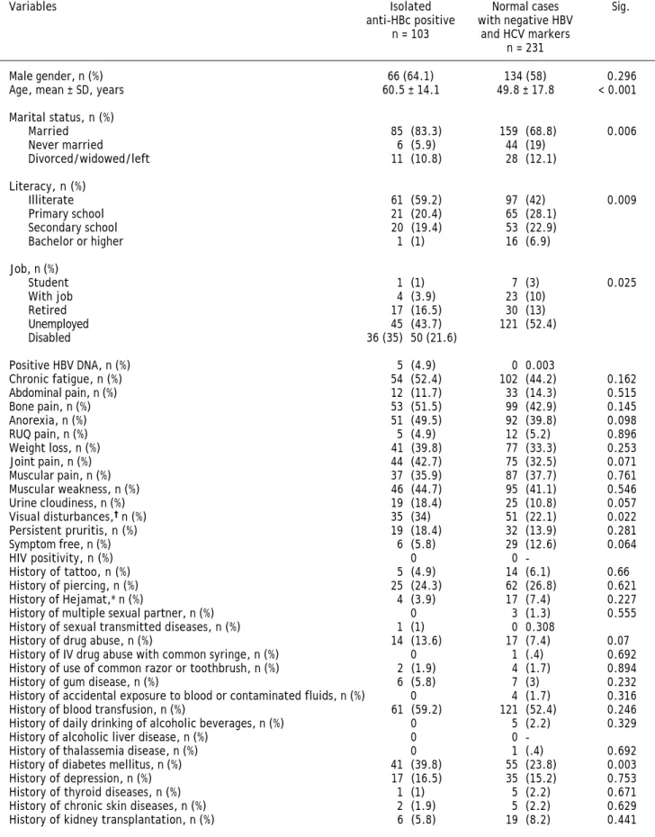
Table 1. Comparison of cases with isolated anti-HBc and cases with negative HBV and HCV markers.
Variables Isolated Normal cases Sig.
anti-HBc positive with negative HBV n = 103 and HCV markers
n = 231
Male gender, n (%) 66 (64.1) 134 (58) 0.296 Age, mean ± SD, years 60.5 ± 14.1 49.8 ± 17.8 < 0.001
Marital status, n (%)
Married 85 (83.3) 159 (68.8) 0.006
Never married 6 (5.9) 44 (19)
Divorced/widowed/left 11 (10.8) 28 (12.1)
Literacy, n (%)
Illiterate 61 (59.2) 97 (42) 0.009
Primary school 21 (20.4) 65 (28.1)
Secondary school 20 (19.4) 53 (22.9)
Bachelor or higher 1 (1) 16 (6.9)
Job, n (%)
Student 1 (1) 7 (3) 0.025
With job 4 (3.9) 23 (10)
Retired 17 (16.5) 30 (13)
Unemployed 45 (43.7) 121 (52.4)
Disabled 36 (35) 50 (21.6)
Positive HBV DNA, n (%) 5 (4.9) 0 0.003
Chronic fatigue, n (%) 54 (52.4) 102 (44.2) 0.162 Abdominal pain, n (%) 12 (11.7) 33 (14.3) 0.515 Bone pain, n (%) 53 (51.5) 99 (42.9) 0.145
Anorexia, n (%) 51 (49.5) 92 (39.8) 0.098
RUQ pain, n (%) 5 (4.9) 12 (5.2) 0.896
Weight loss, n (%) 41 (39.8) 77 (33.3) 0.253 Joint pain, n (%) 44 (42.7) 75 (32.5) 0.071 Muscular pain, n (%) 37 (35.9) 87 (37.7) 0.761 Muscular weakness, n (%) 46 (44.7) 95 (41.1) 0.546 Urine cloudiness, n (%) 19 (18.4) 25 (10.8) 0.057 Visual disturbances,† n (%) 35 (34) 51 (22.1) 0.022 Persistent pruritis, n (%) 19 (18.4) 32 (13.9) 0.281 Symptom free, n (%) 6 (5.8) 29 (12.6) 0.064
HIV positivity, n (%) 0 0
-History of tattoo, n (%) 5 (4.9) 14 (6.1) 0.66 History of piercing, n (%) 25 (24.3) 62 (26.8) 0.621 History of Hejamat,* n (%) 4 (3.9) 17 (7.4) 0.227 History of multiple sexual partner, n (%) 0 3 (1.3) 0.555 History of sexual transmitted diseases, n (%) 1 (1) 0 0.308
History of drug abuse, n (%) 14 (13.6) 17 (7.4) 0.07 History of IV drug abuse with common syringe, n (%) 0 1 (.4) 0.692 History of use of common razor or toothbrush, n (%) 2 (1.9) 4 (1.7) 0.894 History of gum disease, n (%) 6 (5.8) 7 (3) 0.232 History of accidental exposure to blood or contaminated fluids, n (%) 0 4 (1.7) 0.316 History of blood transfusion, n (%) 61 (59.2) 121 (52.4) 0.246 History of daily drinking of alcoholic beverages, n (%) 0 5 (2.2) 0.329 History of alcoholic liver disease, n (%) 0 0
64.1% (66 cases) male, 83.3% (85 cases) married at present, 59.2% (61 cases) illiterate, 43.7% (45 cases) unemployed, all supported by a health insurance, and 4.9% (5 cases) with HBV DNA positive test.
Considering all cases with negative HBsAg and anti-HCV tests, we analyzed cases with positive (103 cases) and negative anti-HBc (231 cases). Cases with isolated anti-HBc had significantly higher per-centage of positive HBV DNA [OR (95% CI) = 12.1 (1.4-105)], visual disturbances [OR (95% CI) = 1.8 (1.1-3.03)], history of diabetes mellitus [OR (95% CI) = 2.1 (1.3-3.5)], higher age, higher age when dialysis started and were more likely to be married, illiterate, disabled and retired (Table 1). They did not have significant differences according to other basic characteristics and risk factors.
Cases with isolated anti-HBc who had positive HBV DNA result, had significantly more diabetes, visual disturbances and history of depression (Table 2). All cases with isolated anti-HBc and positive HBV DNA vs. 75.8% (72 cases) with negative HBV DNA had vaccination history against HBV. This difference was not statistically significant.
Forward Wald Logistic regression model showed that only history of diabetes mellitus has a
signifi-cant correlation with HBV DNA positivity in cases with isolated anti-HBc [P = 0.005, OR (95% CI) = 25.2 (2.6-243.7)]. This analysis was done on 100 cases and eight variables (with P-value < 0.2) were entered and the model was significant (P = 0.001).
DISCUSSION
Our results verify the proposal given by earlier studies that while anti-HBc screening has little value in a low prevalence population, it can be useful in high prevalence populations.15-17
Routine blood donor screening for anti-HBc sta-tus has been implemented in some countries result-ing in a decrease in the risk of post-transfusion HBV infection.18 Hence, in our poor resource
set-ting, inclusion of anti-HBc testing for donor screen-ing will definitely remove possible HBV infected donations. Although this screening will result in the rejection of a large number of donations, it will constitute a valuable asset in reducing the risk of HBV transmission with its potential consequences, particularly among immunocompromised recipients. If our policy does not require that we reject all blood donated by anti-HBc infected people, we can at least
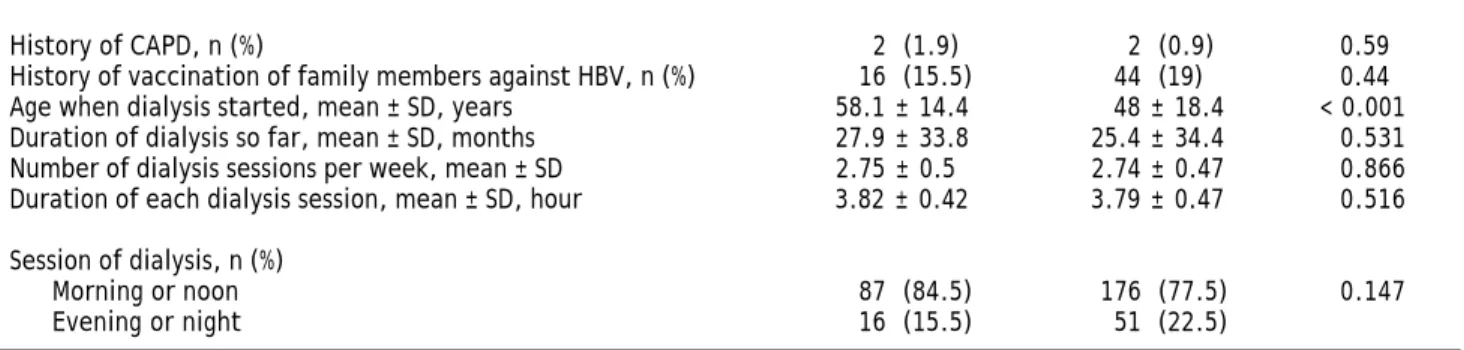
History of CAPD, n (%) 2 (1.9) 2 (0.9) 0.59 History of vaccination of family members against HBV, n (%) 16 (15.5) 44 (19) 0.44 Age when dialysis started, mean ± SD, years 58.1 ± 14.4 48 ± 18.4 < 0.001 Duration of dialysis so far, mean ± SD, months 27.9 ± 33.8 25.4 ± 34.4 0.531 Number of dialysis sessions per week, mean ± SD 2.75 ± 0.5 2.74 ± 0.47 0.866 Duration of each dialysis session, mean ± SD, hour 3.82 ± 0.42 3.79 ± 0.47 0.516
Session of dialysis, n (%)
Morning or noon 87 (84.5) 176 (77.5) 0.147 Evening or night 16 (15.5) 51 (22.5)
Since these numbers (percents) are calculated based on crosstabs; they are by considering that missing cases are deleted. Anti-HBs: hepatitis B surface antibo-dy. CAPD: continuous ambulatory peritoneal dialysis. HBV: hepatitis B virus. HCV: hepatitis C virus. HIV: human immunodeficiency virus. IV: intravenous. RUQ: right upper quadrant. †††††In this study what is referred to as visual disturbances is pertaining to any abnormalities of sight like diplopia, blurred vision,
redu-ced visual acuity, reduredu-ced visual field and partial or total loss of vision, halos, blind spots, floaters, and other symptoms expressed by the patients. *A proredu-cedu- procedu-re in Iranian traditional medicine done by making shallow cuts on the trunk (upper back) and producing a suction effect that procedu-results in drawing blood from cuts (< 100 cc). It is usually done by a non-physician, using non-standard instruments (done for healing or cure purposes).
Table 2. Comparison of cases with positive (occult HBV infection) and negative HBV DNA in patients with isolated anti-HBc.* Variables HBV DNA Crude OR (95% CI) Sig.
Positive, n = 5 Negative, n = 98
History of diabetes mellitus, n (%) 5 (100) 34 (35.8) 1.1 (1.02-1.3) 0.008 History of depression, n (%) 4 (80) 13 (13.7) 19.5 (2.3-164.1) 0.003 Visual disturbance,† n (%) 4 (80) 30 (31.6) 7.8 (0.9-66.8) 0.044
*The remainder of the variables was not significant. †††††In this study what is referred to as visual disturbances is pertaining to any abnormalities of sight like
exclude donors who are high risk for being HBV DNA positive (persons with history of diabetes mellitus, depression or visual disturbance).
A similar paper on 289 patients on chronic HD from five dialysis units in Tehran with similar char-acteristics showed that 18 subjects (6.2%) had iso-lated anti-HBc. Positive HBV-DNA was detectable in 9 out of 18 patients (50%) who had isolated anti-HBc. Only one of them was anti-HCV positive. None of them were HIV infected. There was no significant difference between HBV DNA positive and negative patients regarding age, sex, ALT and AST levels or length of time on dialysis, maybe due to low sample size.10
Most studies have shown low copies/mL in viral load of HBV DNA positive cases. Therefore, a low HBV viral load seems the most likely explanation for our HBsAg negative/DNA positive samples.
An Iranian study on 90 HD patients found no one with positive HBV DNA and 30 cases with positive HCV RNA, 10 cases of which were positive for anti-HBc among HCV RNA positive subjects.19
A Brazilian study on 1476 HD subjects showed that positive anti-HBc, HBsAg and HBV DNA were 34.1%, 15.4% and 8.1%, respectively, while the inci-dence of HBV was null. This study suggests that employing more than one HBV marker and repeated follow-up evaluations may improve HBV screening in HD units.20
One Turkish study found that 9.1% (2/22) of non HD subjects with isolated anti-HBc have OHB infec-tion.21 Another Turkish study on 188 subjects on
HD revealed that five (2.7%) cases have OHB. There was isolated anti-HBc in 12 (6.4%) patients, three (7.9%) of with anti-HCV and two (40%) with OHB.22
A study on one group of HBsAg negative patients (n = 213) from a large cohort (n = 585) of Italian chronic dialysis patients showed that OHB infection was absent. Persistent HBsAg carriers were less fre-quent than anti-HBc positive patients in this study group [1.88% (11 of 585) vs. 36% (216 of 585), P = 0.0001]. No significant association between abnormal biochemical liver tests and serum anti-HBc was noted in our population. Nominal logistic regression analy-sis demonstrated an independent and significant rela-tionship between anti-HCV and anti-HBc status in serum (P = 0.0001). The rate of patients seropositive for anti-HBc was higher among study patients than controls with normal renal function [36.9% (216 of 585) vs. 21.4% (59 of 275), P = 0.0001]; this difference partially persisted after correction for demographic parameters, and viral markers.23
A prospective study on 86 Spanish patients, prior to the start of a HD program in 2003 showed that 18.7% are anti-HBc positive. Logistic regression showed that only age (OR = 1.03), being born in the jungle area (OR = 13.1), and food consumption in restaurants (OR = 5.0) were related to total cases with positive anti-HBc.24
In the present study, there were only 5.3% who had checked their anti-HBs status. Among patients who had been tested for hepatitis B serologic mark-ers, 44.6% were isolated anti-HBc. So, it is logical to vaccinate isolated anti-HBc cases.
Our present study found that persons who had been tested for their serological markers of HBV were higher risk males with more symptoms but lower history of vaccination. So, we have overesti-mated the prevalence of isolated anti-HBc in our patients tested for hepatitis B serologic markers. Total HD cases have a lower percent of OHB in-fection from that shown in our selected study group.
Table 3 summarizes the results of some relatively similar studies showing a wide range of isolated anti-HBc in various populations from 2.5% to 55.7%. However, some of these results are not reli-able due to low sample size. One interesting finding is that different risk factors of isolated anti-HBc or OHB infection are not consistent among these pa-pers and the present study. Another important issue is the different comparison groups in these studies which makes it hard to draw definitive conclusions. On the other hand, the different control groups in these studies give us valuable information pertaining to all possible risk factors in different nationalities.
One limitation of this study is that we only explored the situation of HBV DNA in anti-HBc cases and not all cases; while, OHB infection can be detec-ted in anti-HBc negative cases as well. However, one similar study has shown that HBV DNA detection was more frequent when positive anti-HBc was de-tected in isolation (72%) than when associated with anti-HBsAg antibodies (31%).25 In addition, other
studies have found that isolated anti-HBc positivity was more frequent in patients with OHB than in those without (40% [2/5] vs. 5.5% [10/183], p = 0.002).22 So, we should check the status of HBV
Significant difference between persons who had checked hepatitis B serologic markers with others shows that we can only generalize our results to these patients.
In conclusion, our study underscores the high rate of OHB infection in hemodialysis patients in most parts of Iran. Except diabetes mellitus, factors correlated with anti-HBc status are age related (higher age, higher age when dialysis started, illit-erate, disabled and retired, visual disturbances, and even being married). Diabetes mellitus was the only independent predictor of HBV DNA status in cases with isolated anti-HBc.
ACKNOWLEDGEMENT
We would like to express our special thanks for all participants and other colleagues who helped make this study possible.
CONFLICTS OF INTEREST
There is no conflict of interest.
REFERENCES
1. Raimondo G, Pollicino T, Romano L, Zanetti AR. A 2010 up-date on occult hepatitis B infection. Pathol Biol (Paris)
2010; 58: 254-7.
2. Kabir A, Keshvari M, Kashani AH, Alavian SM. Predicting response to HBV vaccination in people with positive anti-HBc but negative HBsAg and anti-HBs. Hum Vaccin 2008; 4: 379-83.
3. Mahdavimazdeh M, Hosseini-Moghaddam SM, Alavian SM, Yahyazadeh H. Hepatitis B Infection in Hemodialysis Pa-tients in Tehran Province, Iran. Hepat Mon 2009; 9: 206-10.
4. Fabrizi F, Bunnapradist S, Lunghi G, Aucella F, Martin P. Epidemiology and clinical significance of hepatotropic in-fections in dialysis patients. Minerva Urol Nephrol 2004; 56: 249-57.
5. Fabrizi F, Lunghi G, Martin P. Hepatitis B virus infection in hemodialysis: Recent discoveries. J Nephrol 2002; 15: 463-8.
6. Alavian SM, Hajarizadeh B, Ahmadzad-Asl M, Kabir A, Bag-heri-Lankarani K. Hepatitis B Virus Infection in Iran: A Systematic Review. Hepat Mon 2008; 8: 281-94.
7. Merat S, Malekzadeh R, Rezvan H, Khatibian M. Hepatitis B in Iran. Arch Irn Med 2000; 3: 192-201.
8. Alavian SM, Bagheri-Lankarani K, Mahdavi-Mazdeh M, Nourozi S. Hepatitis B and C in dialysis units in Iran: chan-ging the epidemiology. Hemodial Int 2008; 12: 378-82.
Table 3. Characteristics and findings of similar studies about isolated anti-HBc in hemodialysis patients.
Study: Characteristics, sample size, Risk factors of Reference country, year percent of positive isolated anti-HBc isolated anti-HBc/occult
Case group Control group HBV infection
Egypt, 2009. HD patients, . Healthy blood donor, Multiple blood 26 143, 9 100, 8. transfusion.
Egypt, 1995. HD patients, Healthy persons, ND. 27 64, 51.8% 15, ND.* of anti-HCV
positive patients.
Brazil, 1995. HD patients, Peritoneal dialysis ND. 28 185, 55.7. patients, 124, ND.
Turkey, 2006. HD patients, No control group. ND. 22 188, 6.4.
Hong Kong, 1989. HD patients, Personnel working in the Higher incidence of 29 63, 19. dialysis unit, ND, ND. repeated liver dysfunction,
Healthy controls, ND, ND. elevated alanine transfusion requirement. transaminase levels,
and a higher
Italy, 2009. HD patients, No control group. HCV seropositivity and 25 128, 26.6 (occult the positive anti-HBs,
HBV infection). isolated anti-HBcAg.
Brazil, 2006. HD patients, No control group. ND. 30 1095, 2.5.
9. Mostaghni AA, Soltanian A, Mokhtari E, Japoni S, Mehraba-ni D. Seroprevalence of hepatitis B virus among hemodialy-sis patients in Bushehr province, southern Iran. Hepat Mon 2011; 11: 200-2.
10. Aghakhani A, Banifazl M, Kalantar E, Eslamifar A, Ahmadi F, Razeghi E, Atabak S, et al. Occult hepatitis B virus infec-tion in hemodialysis patients with isolated hepatitis B core antibody: a multicenter study. Ther Apher Dial 2010; 14: 349-53.
11. Hollinger FB, Habibollahi P, Daneshmand A, Alavian SM. Oc-cult Hepatitis B Infection in Chronic Hemodialysis Pa-tients: Current Cocepts and Strategy. Hepat Mon 2010; 10: 199-204.
12. Edey M, Barraclough K, Johnson DW. Review article: Hepati-tis B and dialysis. Nephrology (Carlton) 2010; 15: 137-45. 13. Mahdavi-Mazdeh M, Zamyadi M, Nafar M. Assessment of
management and treatment response in hemodialysis pa-tients in Tehran province, Iran. Nephrol Dial Transplant
2008; 23: 288-93.
14. Available from: http://www.amar.org.ir/default-2649.aspx [Accessed 31 Oct 2010].
15. Allain JP, Hewitt PE, Teddler RS, Williamson LM. Evidence that anti-HBc but not HBV DNA testing may prevent some HBV transmission by transfusion. Br J Haematol 1999; 107: 186-95.
16. Douglas DD, Taswell HF, Rakela J, Rabe D. Absence of he-patitis B virus DNA detected by polymerase chain reactio-nin blood donors who are hepatitis B surface antigen negative and antibody to hepatitis B core antigen positi-ve from a United States population with a low prevalence of hepatitis B serologic markers. Transfusion 1993; 33: 212-6.
17. Wang JT, Wang TH, Sheu JC, Shih LN, Lin JT, Chen DS. De-tection of hepatitis B virus DNA by polymerase chain reac-tion in plasma of volunteer blood donors negative for hepatitis B surface antigen. J Infect Dis 1991; 163: 397-9. 18. Kleinman SH, Kuhns MC, Todd DS, Glynn SA, McNamara A, Di
Marco A, Busch MP. Frequency of HBV DNA detection in US blood donors testing positive for the presence of anti-HBc: implications for transfusion transmission and donor screening. Transfusion 2003; 43: 696-704.
19. Arababadi MK, Hassanshahi G, Yousefi H. HBV-DNA in he-modialysis patients infected by HCV. Saudi J Kidney Dis Transpl 2009; 20: 398-401.
20. Moreira RC, Deguti MM, Lemos MF, Saraceni CP, Oba IT, Spina AM, Nascimento-Lima AS, et al. HBV markers in
hae-modialysis Brazilian patients: a prospective 12-month fo-llow-up. Mem Inst Oswaldo Cruz 2010; 105: 107-8.
21. Altindis M, Uslan I, Cetinkaya Z, Yüksel S, Ciftçi IH, Demir-türk N, Ozdemir M, et al. Investigation of hemodialysis pa-tients in terms of the presence of occult hepatitis B.
Mikrobiyol Bul 2007; 41: 227-33.
22. Yakaryilmaz F, Gurbuz OA, Guliter S, Mert A, Songur Y, Ka-rakan T, Keles H. Prevalence of occult hepatitis B and he-patitis C virus infections in Turkish hemodialysis patients.
Ren Fail 2006; 28: 729-35.
23. Fabrizi F, Messa PG, Lunghi G, Aucella F, Bisegna S, Man-gano S, Villa M, et al. Occult hepatitis B virus infection in dialysis patients: a multicentre survey. Aliment Pharma-col Ther 2005; 21: 1341-7.
24. Loza Munárriz C, Depaz Dolores M, Suarez Jara M, Loza Munárriz R, Valenzuela Córdova R, Bravo Tejada J, Valencia Rodriguez J, et al. Rate of serological mar-kers of hepatitis B and C viruses in first-time users of the hemodialysis program at Hospital Nacional Cayeta-no Heredia (HNCH). Rev Gastroenterol Peru 2005; 25: 320-7.
25. Di Stefano M, Volpe A, Stallone G, Tartaglia L, Prato R, Martinelli D, Pastore G, et al. Occult HBV infection in he-modialysis setting is marked by presence of isolated anti-bodies to HBcAg and HCV. J Nephrol 2009; 22: 381-6. 26. Elghannam DM, Aly RM, Goda EF, Eltoraby EE, Farag RE.
Cli-nical significance of antibody to hepatitis B core antigen in multitransfused hemodialysis patients. Asian J Transfus Sci 2009; 3: 14-7.
27. Gohar SA, Khalil RY, Elaish NM, Khedr EM, Ahmed MS. Pre-valence of antibodies to hepatitis C virus in hemodialysis patients and renal transplant recipients. J Egypt Public Health Assoc 1995; 70: 465-84.
28. Cendoroglo Neto M, Manzano SI, Canziani ME, Silva AE, Ci-renza LF, Sesso Rde C, Ajzen H, et al. Environmental transmission of hepatitis B and hepatitis C viruses within the hemodialysis unit. Artif Organs 1995; 19: 251-5. 29. Lai KN, Tam JS, Lai FM, Lin HJ. Isolated presence of
anti-body to hepatitis B core antigen in dialysis patients: occu-rrence of subclinical hepatitis? Am J Kidney Dis 1989; 13: 370-6.
30. Ferreira RC, Teles SA, Dias MA, Tavares VR, Silva SA, Go-mes SA, Yoshida CF, et al. Hepatitis B virus infection pro-file in hemodialysis patients in Central Brazil: prevalence, risk factors, and genotypes. Mem Inst Oswaldo Cruz