Liver fibrosis in young Egyptian beta-thalassemia
major patients: relation to hepatitis C virus and
compliance with chelation
Mohsen S. Elalfy,* Gamal Esmat,** Randa M. Matter,* Hesham E. Abdel Aziz,* Walid A. Massoud***
* Department of Pediatric Hematology, Ain Shams University, Cairo 11566, Egypt. ** Department of Tropical Medicine, Cairo University, Cairo 12613, Egypt.
*** Research Department, National Center for Examinations and Educational Evaluation (NCEEE), Cairo 11571, Egypt.
ABSTRACT
Background. The main causes of liver fibrosis in transfusion-dependent thalassemia major are hepatitis C vi-rus (HCV) infection and hepatic iron overload. The study aimed to assess liver fibrosis in Egyptian adoles-cents and young adult poly-transfused beta thalassemia patients infected with HCV using liver FibroScan in relation to iron overload and Liver iron concentration (LIC). Material and methods. Fifty-one regularly transfused beta thalassemia patients above 12 years old were subjected to measurement of serum alanine transaminase (ALT), serum ferritin (SF), HCV (antibody and RNA), LIC assessed by hepatic R2* and transient elastography (TE) (FibroScan). FibroTest and liver biopsy were done to 25 patients. Results. Eighty two% of studied thalassemia patients were HCV antibody positive; 21(49%) of them were viremic (HCV RNA positive); median LIC was 12 mg/gm dry weight. There were strong positive correlation between the degree of liver stiffness and Ishak fibrosis score assessed in liver biopsy specimens (P = 0.002) and between FibroScan and FibroTest results (P < 0.001). Patients with HCV viremia showed significantly higher ALT, γ-glutamyl trans-peptidase (GGT), SF, LIC and increased liver stiffness compared to patients with no viremia (P = 0.0001, 0.001, 0.012, 0.006 and 0.001) respectively. Liver cirrhosis (TE values > 12.5kPa) was encountered in 23.5% and variable degrees of liver fibrosis (TE values > 6-12.5 kPa) in 35% of studied thalassemic patients. Conclusion. Young beta thalassemia patients with active hepatitis C infection may have hepatic cirrhosis or fibrosis at young age when accompanied with hepatic siderosis. Non invasive Liver FibroScan and Fibro-Test were reliable methods to assess liver fibrosis in young thalassemic-patients.
Key words. Iron overload. Liver iron concentration. Cirrhosis. FibroScan. FibroTest.
Correspondence and reprint request: Mohsen S. Elalfy, MD, PhD, Professor of Pediatrics.
2 sayed Zakaria Khalil Street, Morabaa 1153, Masaken Sheraton Heliopolis, Cairo, Egypt.
Tel.: +2-01000-864343. Fax: +202 22694900 E-mail: [email protected]
Manuscript received: Jun 03, 2012. Manuscript accepted: Jul 30, 2012.
INTRODUCTION
Worldwide, no patients get more red cell products than those with thalassemia major. The life-long need for transfusion renders patients vulnerable to transfusion-transmitted viral infections. Hepatitis C virus infection has emerged as the major risk in the
last decades.1
Hepatitis virus C infection is the main risk fac-tor for liver fibrosis in transfusion-dependent tha-lassemics. Excess liver iron is now clearly recognized as a cofactor for the development of
ad-vanced fibrosis and cirrhosis in patients with
HCV infection.2-4
In adults, the non-invasive assessment of fibrosis in chronic hepatitis, especially of viral etiology, is more and more accepted, partially replacing liver biopsy in some countries. Guidelines from France recommend that the first-line test for untreated pa-tients with HCV chronic hepatitis, with no comorbi-dities, should be a non invasive procedure (either
FibroTest® or FibroScan®).5
The vast majority of studies assessing transient elastography (TE) as compared to liver biopsy were
performed in patients with HCV chronic hepatitis.6-9
In beta-thalassemia patients (especially HCV in-fected ones) at higher risk of liver biopsy related complications as compared to other chronically HCV-infected patients, the availability of a
noninva-sive method to measure hepatic fibrosis is crucial.10
The purpose of this study was to assess liver fi-brosis status in young polytransfused beta thalasse-mia patients infected with HCV using both FibroScan and FibroTest in relation to iron over-load and liver iron concentration (LIC).
MATERIAL AND METHODS
This cross sectional study included fifty one regu-larly transfused beta thalassemia patients (26 males and 25 females with age range 12-24 years from Thalassemia Center, Ain Shams University during the period from March 2009 to March 2010. Written informed consent was obtained from adult patients, patients’ parents or their legal guardians after approval of the study by the Local Ethical Committee, Ain Shams University. The study protocol confor-med to the ethical guidelines of Declaration of Helsinki 1975.
Inclusion criteria
Beta thalassemia major patients aged ≥ 12 years,
poly-transfused > 100 transfusion, with liver sidero-sis; LIC > 2 mg Fe/g liver dry weight [dw], HCV antibody positive patients have had their infection for at least 10 years as recorded from their files. All patients received iron chelation therapy either in the form of subcutaneous infusion of desferoxamine (Desferal; Sigma, Saint Louis, Missouri, US) with a dose of 40 mg/kg for 5 days/week. Alternatively, oral chelation therapy Deferiprone (Ferriprox®; Apo-Pharma, Toronto, Canada) was administered at 75 mg/kg/d for 7 days/week.
Compliance defined as the extent to which patients take medications as prescribed by their health care providers; was assessed by the following questions:
• How many doses were missed per day during the preceding 4 weeks, and
• What is the amount of medication remaining?
At each clinic visit, the old vials were brought and the remaining tablets were counted.
Exclusion criteria
Non applicability conditions of FT: diabetes melli-tus, patients with ALT > 10 times ULN (Upper Li-mit of Normal), serum bilirubin > 5 mg /dL.
Thalassemic patients were categorized on the ba-sis of risk to develop liver fibroba-sis i.e. both LIC va-lues and HCV viremia into 3 subgroups:
• Group I. Patients with both HCV-RNA positivi-ty and LIC > 14 mg/gm dw.
• Group II. Patients with either HCV-RNA positi-vity or LIC > 14 mg/gm dw, and
• Group III. Patients with negative viremia with LIC < 14 mg/gm dw.
Blood samples were taken 4 weeks from last blood transfusion; Complete blood count was performed using coulter B66 (Miami, Florida, USA), Liver function tests including AST, ALT using Synchron CX9 autoanalyzer (Brea, California, USA), serum fe-rritin on Immulite instrument (Diagnostic products corporation 5700 West 96 St. Los Angeles, USA), hepatitis B surface antigen, human immunodeficien-cy virus antibody using ELISA technique (R & D system, USA) and HCV antibody using ELISA tech-nique (R & D system, USA) and HCV RNA by poly-merase chain reaction (Amplicor HCV; Roche Molecular Systems, Basel, Switzerland) were done to all patients according to manufacturer’s instructions. Transfusional iron intake was calculated in
mg/kg/day.11
FibroTest (Biopredictive, France, [FT]): this test consists of an algorithm of five fibrosis markers (alfa2-macroglobulin, apolipoprotein A1,
haptoglo-bin, GGT, bilirubin).12-13 It has been evaluated in 25
patients suitable for FT who performed also liver biopsy. Gamma-glutamyltranspeptidase (GGT), total bilirubin, apolipoprotein A1, and haptoglobin were measured with a Cobas Integra 400 analyzer (Roche, Indianapolis, Indiana, USA) and Roche Diagnostics reagents (Roche, USA). Alpha2-macroglobulin was assayed with a Cobas Integra 400 Turbidimetry with Dako utility channel reagents (Glostrup, Den-mark). All tests were performed by personnel blin-ded to all patients’ data, including biopsy results.
Liver iron concentration (LIC) was assessed by Magnetic resonance imaging (MRI) measurements of the proton transverse relaxation parameter R2 using 5 mm axial slices. R2 scans were
perfor-med using FerriScan® technology (Resonance Health).14
Results are expressed as mg/gm liver dry weight.
Liver histology and quantification of liver fibrosis
aaaaa aaaaa aaaaa aaaaa aaaaa aaaaa aaaaa aaaaa aaaaa aaaaa aaaaa aaaaa aaaaa aaaaa aaaaa aaaaa aaaaa aaaaa aaaaa aaaaa aaaaa aaaaa aaaaa aaaaa aaaaa aaaaa aaaaa aaaaa aaaaa aaaaa aaaaa aaaaa aaaaa trained pathologist blinded to the results of
non-inva-sive methods. Fibrosis was scored by Ishak
clas-sification.15 None of the patients experienced biopsy
complications.
Transient elastography (TE)
All patients were examined by TE (FibroScan®; Echosens, Paris, France). The procedures were per-formed by the same investigator who was blind to clinical, serological, and histological data. Details of the technical background and examination
procedu-re have been pprocedu-reviously described.16 The results
were expressed in kilopascals (kPa). The median va-lue was considered representative of the elastic mo-dulus of the liver. Only procedures with at least 10 successful acquisitions and a success rate of at least 60% with interquartile range (IQR) of all validated measurements less than 30% of the median value were considered reliable. Twenty five patients who had the liver biopsy and FT were retested after 2 weeks to verify the results. Cirrhosis was defined according to the published cut-offs in patient with
hepatitis C: 12.5 kPa.17 The cut-off value of TE 6 kPa
for diagnosing F ≥ 1 (18) was used to exclude hepatic
fibrosis with smaller values. No liver stiffness measu-rement failure was observed in the present study.
Statistical analysis
Analysis of data was performed by using SPSS (version 15). Comparison between 2 groups of pa-tients was made using Student’s t-test for parame-tric measures and Wilcoxon signed-rank test (Z value) for non parametric measures. Spearman’s rank correlation coefficient was used to correlate between two quantitative variables. P value < 0.05 was considered the cut-off value for significance.
RESULTS
Fifty-one consecutive patients (26 males and 25 females; mean age 15.92 ± 3.11 years); 25 with a suitable liver biopsy entered the study. All beta tha-lassemia major patients were transfusion-dependent: they transfused packed red cells every 15-21 days with pretrasfusional Hb levels from 7.4 to 9.4 g/dL.
Median BMI was 19.5 (range 14.5-28.3 kg/m2).
Eighty-two percent of thalassemia patients were HCV antibody positive; 21 of them (49%) were viremic (HCV RNA positive), 4% were hepatitis B surface antigen positive and none of them was human immunodeficiency virus positive. Median LIC was
12 mg/gm liver dw. Ishak fibrosis score was 6 in 4 patients, 5 in 3 patients, 4 in 4 patients, 3 in 4 pa-tients and 2 in 10 papa-tients. Mean stiffness value was 10.75 ± 10.41 kPa (median 6.8, range 2.8-49.7 kPa).
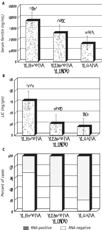
Figure 1. Serum ferritin (A), LIC (B) and RNA positive cases (C) among beta thalassemia major patients with different TE values. A B C 5,000 4,000 3,000 2,000 1,000 0
Serum ferritin (ng/mL)
TE > 12.5 kPa TE: 6-12.5 kPa TE < 6 kPa
TE values 50 40 30 20 10 0 LIC (mg/gm)
TE > 12.5 kPa TE: 6-12.5 kPa TE < 6 kPa
TE values 100 80 60 40 20 0
Percent of cases
TE > 12.5 kPa TE: 6-12.5 kPa TE < 6 kPa
TE values 3,712 2,598 1,626 31.31 10.59 7.81 aa
Using FibroScan; liver cirrhosis (TE values > 12.5 kPa) was encountered in 23.5% and variable degrees of liver fibrosis (TE values > 6-12.5 kPa) in 35% of studied thalassemic patients. There was strong posi-tive correlation between degree of liver stiffness and Ishak fibrosis score detected by liver biopsy (r = 0.40, P = 0.002). All patients with liver cirrhosis (TE values > 12.5 kPa) were HCV antibody positive and showed highest HCV RNA positivity (Figure 1). Patients with HCV viremia showed significantly hig-her ALT, GGT, SF, LIC and increased liver stiffness compared to patients with no viremia (P = 0.0001, 0.001, 0.012, 0.006 and 0.001) respectively (Table 1). Non viremic HCV antibody positive patients showed
significantly higher LIC (12.10 ± 6.56 vs. 6.56 ±
2.79 mg/gm liver dry weight, P = 0.006) and
increa-sed liver stiffness (8.26 ± 5.30 vs. 4.44 ± 1.175 kPa,
P = 0.006) compared to HCV antibody negative pa-tients. Highest serum ferritin (SF), LIC and RNA positivity were observed among beta thalassemia major patients with TE values in the cirrhotic range (above 12.5kPa) (Figure 1).
Lowest liver stiffness, fibrotest results and best compliance with chelation therapy were obtained in
thalassemic patients with negative viremia and LIC less than 14 mg/gm dw whereas highest TE values, worst compliance with chelation and highest fibro-test results were shown in patients with both HCV-RNA positivity and LIC above 14 mg/gm dw followed by patients with either HCV-RNA positivity or LIC above 14 mg/gm dw (Table 2). Details of compliance of each subgroup were shown in figure 2, where 57% of the patients compliant with Deferoxa-mine had LIC below 14 mg/gm dw and negative vire-mia. Most of the non-compliant patients to Deferoxamine were HCV RNA positive with LIC above 14 mg/gm dw and 83% of the patients com-pliant with Deferiprone had LIC below 14 mg/gm dw and negative viremia.
Relation between compliance with chelators and TE values showed that most non compliant patients with Deferoxamine had TE more than 12.5 kPa whe-reas most compliant patients with Deferiprone had TE less than 6 kPa (Figure 3). Strong positive co-rrelations were shown between TE values and both SF levels and LIC (Figure 4), between TE values and fibrotest (r = 0.935, p < 0.001) and also bet-ween SF levels and LIC (r = 0.538, P < 0.001).
Table 1. Demographic and biochemical markers of HCV viremic vs. non viremic Beta Thalassemia major patients.
HCV Viremia Non viremic HCV P
Age (years) 16.67±3.45 (N = 21) 15.53± 0.39 (N = 30) n.s.
T I (mg/kg) 0.45±0.10 (N = 21) 0.38± 0.08 (N = 30) n.s.
ALT (IU/L) 126.38±49.16 (N = 21) 53.60± 18.49 (N = 30) 0.0001*
AST (IU/L) 95.05±49.31 (N = 21) 38.00± 20.45 (N = 30) 0.0001*
GGT (IU/L) 48.83±19.47 (N = 12) 23.46± 8.24 (N = 13) 0.001*
S.F. (ng/mL) 3081±1329 (N = 21) 2063± 1317 (N = 30) 0.012*
LIC (mg/gm liver dw) 20.86±15.27 (N = 21) 10.43± 6.21 (N = 30) 0.006*
FT 0.71±0.21 (N = 12) 0.54± 0.22 (N = 13) n.s.
TE (kPa) 15.95±13.77 (N = 21) 7.11± 4.79 (N = 30) 0.001*
*Significant. TI: transfused iron. ALT: alanine aminotransferase. AST: aspartate aminotransferase. GGT: γ-glutamyl transpeptidase. SF: serum ferritin. LIC: liver iron concentration. dw: dry weight. FT: FibroTest. TE: transient elastography. kPa: kilopascal.
Table 2. Risk of liver fibrosis in relation to HCV RNA positivity and LIC in Beta Thalassemia major patients.
(Risk ) Liver fibrosis (HCV RNA & LIC values)
Group I (N = 9) Group II (N = 16) Group III (N = 26) P1 P2
TI (mg/kg) 0.50 ±0.12 (N = 9) 0.43±0.08 (N=16) 0.38± 0.08 (N = 26) n.s. 0.047* Compliance with chelator 0.11 ±0.33 (N = 9) 0.31±0.48 (N=16) 0.73± 0.45 (N = 26) n.s. 0.030* FT 0.81 ±0.22 (N = 6) 0.68±0.20 (N=7) 0.47± 0.13 (N = 12) 0.004* 0.002* TE (kPa) 28.19 ±13.23 (N = 9) 9.46±5.27 (N=16) 5.51± 2.28 (N = 26) 0.0001* 0.0001*
aaaaaa aaaaaa aaaaaa aaaaaa aaaaaa aaaaaa aaaaaa aaaaaa aaaaaa aaaaaa aaaaaa aaaaaa aaaaaa aaaaaa aaaaaa aaaaaa aaaaaa aaaaaa aaaaaa aaaaaa aaaaaa aaaaaa aaaaaa aaaaaa aaaaa aaaaaa aaaaaa aaaaaa aaaaaa aaaaaa aaaaaa aaaaaa aaaaaa aaaaaa aaaaaa aaaaaa aaaaaa aaaaaa aaaaaa aaaaaa aaaaaa aaaaaa aaaaa aaaaaa aaaaaa aaaaaa aaaaaa aaaaaa aaaaaa aaaaaa aaaaaa aaaaaa aaaaaa aaaaaa aaaaaa aaaaaa aaaaaa aaaaaa aaaaaa aaaaaa aaaaaa aaaaa aaaaaa aaaaaa aaaaaa aaaaaa aaaaaa aaaaaa aaaaaa aaaaaa aaaaaa aaaaaa aaaaaa aaaaa aaaaaa aaaaaa aaaaaa aaaaaa aaaaaa aaaaaa aaaaaa aaaaaa aaaaaa aaaaaa aaaaaa aaaaaa aaaaaa aaaaaa aaaaaa aaaaaa aaaaaa aaaaaa aaaaaa aaaaaa aaaaaa aaaaaa aaaaaa aaaaaa aaaaaa aaaaaaa a
DISCUSSION
The present study revealed that 82% of thalasse-mia patients were HCV antibody positive; 21(49%) of them were viremic (HCV RNA positive). Previous studies on the prevalence of hepatitis C in Egyptian
thalassemic children reported that 44%19 to 75.6%20
had hepatitis C antibodies. HCV-PCR was positive in
64% of studied Egyptian patients.21 While in Italian
multicenter study;22 anti-HCV antibodies were found
in 91% of thalassemic patients and 72% of them were viremic.
Figure 3. Compliance with iron chelation therapy among beta thalassemia major patients with different TE values.
Figure 4. Correlation between Fibro Scan and both serum Ferritin (A) (r = 0.555**, P < 0.001) and LIC (B) (r = 0.929, P < 0.0001).
Figure 2. Risk of Liver fibrosis/cirrhosis (RNA Positivity and LIC) in beta thalassemia major patients according to com-pliance with iron chelation therapy.
A high percentage of fibrosis/cirrhosis was en-countered in the studied young beta thalassemia major patients. The development and the severity of liver fibrosis were strongly related to the presence of chronic HCV infection and to the extent of liver iron overload. Patients with liver cirrhosis (TE values > 12.5kPa) had multiple risk factors; all were HCV
antibody positive; had high LIC (≥ 14 mg/gm) and
SF levels above 2,500 ng/mL. Several studies22-26
re-ported similar data. Patients with HCV viremia showed higher TE values and increased fibrosis. However; two non viremic patients showed TE
A B
Percent of cases
100 80 60 40 20 0
Non compliant Complaint Non compliant Complaint
Deferoxamine Deferiprone 58 25 17 0 43 57 7 57 36 6 11 83
Percent of cases
100 80 60 40 20 0
Non compliant Complaint Non compliant Complaint
Deferoxamine Deferiprone 5,000 4,000 3,000 2,000 1,000 0
0 10.0 20.0 30.0 40.0 50.0
Fibroscan 60 50 40 30 20 10 0
0 10.0 20.0 30.0 40.0 50.0
Fibroscan
S Ferritin
LIC
RNA pos & LIC ≥ 14
RNA pos & LIC < 14 or RNA neg & LIC ≥ 14 RNA neg & LIC < 14
aaaa aaaaa
TE > 12.5 kPa TE: 6-12.5 kPa TE < 6 kPA
reby avoid liver biopsy in a significant percentage
of patients.37
In the current study, cirrhosis was revealed by FibroScan and confirmed by biopsy in young thalas-semics as early as 12 years old. The relatively long duration of infection and poor compliance with che-lation may explain this observation.
TE values increased proportionally according to the Ishak stage. This goes in agreement with
previous reports.10,33-34 Highest TE values
obser-ved in patients with both HCV-RNA positivity and high LIC (> 14 mg/gm dw) may confirm that thalassemic patients with active HCV replication and severe iron overload develop severe fibrosis or cirrhosis more frequently. Their concomitant presence results in a striking increase in risk for
liver fibrosis progression.38 Difference in
com-pliance with chelators in our patient subgroups may help explain these findings. Moreover, strong correlation between TE and fibrosis stage detected by liver biopsy may denote that high LIC did not affect the usefulness of TE. Liver Iron, in association with HCV viremia, may lead to an increased rate of fibrosis detected by TE, but further studies are required on wider scale before this can be determined.
FibroScan should be verified on thalassemic pa-tients of younger age group as we had cirrhotic tha-lassemic patients who died early in the second decade of life with liver cell failure.
In the current study, most compliant patients with deferoxamine or deferiprone had the lowest TE values whereas most non compliant patients had higher TE values. It is noteworthy that most compliant patients with deferiprone showed TE va-lues within normal range. These results can
con-firm previous results39-41 which demonstrated no
evidence of hepatic fibrosis induced by deferiprone. Non compliance with chelation in the group with HCV viremia and high LIC was marked. Adherence to adequate chelation therapy can prevent the deve-lopment of liver fibrosis in thalassemics free of HCV-infection and may reduce the risks of develo-ping severe fibrosis in thalassemics with chronic
hepatitis C.42
CONCLUSIONS
Liver cirrhosis and/or fibrosis were commonly encountered in young beta thalassemia patients with chronic active hepatitis C infection and hea-vy iron overload. Thalassemic patients compliant with adequate chelation may have normal liver values in the cirrhotic range and some non viremic
patients had fibrosis; this might be explained by the undetectable persistent low level of HCV viremia for long duration with high liver iron due to poor com-pliance with chelation therapy. This observation is
in concordance with previous study27 who reported
fibrosis by liver biopsy in non viremic HCV antibo-dy-positive patients suggesting that HCV may per-sist in the liver in the majority of HCV RNA-negative cases.
Liver iron concentration is related to transfusio-nal iron intake, type and compliance with chelation therapy. In transfusion-dependent thalassemia major, hepatic iron overload is one of the major problems for the progression of the liver disease and is due to regular transfusion regimen that
leads to iron overload.28 Previous studies suggested
the role of iron loading as a factor in fibrosis
pro-gression in hepatitis C.22,29-31 The positive
correla-tions observed between TE values and both SF levels and LIC in our study are in agreement with
Fraquelli, et al.,10 who reported that in thalassemia
major patients with higher ferritin levels, TE in-creased progressively; viremic patients with higher ferritin levels showed a higher increase of TE as compared with non viremic ones suggesting a possi-ble synergistic effect of iron overload and ongoing HCV on hepatic fibrosis.
In this study; serum ferritin (SF) levels were assessed as a potential surrogate marker for LIC and showed positive correlation with LIC. There are controversies on the relation between SF and hepatic fibrosis. Data on a strong correla-tion between SF and the degree of hepatic fibrosis was observed in thalassemia major patients not infected with HCV; however, SF levels alone were not sufficient to assess the degree of fibrosis
in HCV positive thalassemia major patients.32
Meanwhile; others found no correlation between TE values and the degree of iron overload in
beta-thalassemia major patients.33-34
The positive correlation between Fibrotest re-sults and TE values in studied thalassemic pa-tients was in concordance with previous studies in
other hepatic disorders.35-36 In patients with
Fi-broScan and Fibrotest concordant results liver biopsy might be avoided. FibroScan and Fibrotest appear to be valuable methods for detecting early stages of fibrosis among patients with chronic HCV infection, allowing avoiding the progression
of liver damage.36 Combinations of two modalities
the-stiffness with reduced LIC. Liver FibroScan and FibroTest were reliable methods as surrogate for liver biopsy to assess fibrosis progression in tha-lassemic patients.
ABBREVIATIONS
• HCV: hepatitis C virus.
• LIC: liver iron concentration.
• ALT: alanine aminotransferase.
• SF: serum ferritin.
• RNA: ribonucleic acid.
• TE: transient elastography.
• FT: FibroTest.
• GGT:γ-glutamyl transpeptidase.
• kPa: kilopascal.
• dw: dry weight.
• ULN: upper limit of normal.
• MRI: magnetic resonance imaging.
• IQR: inter-quartile range.
• BMI: body mass index.
• PCR: polymerase chain reaction.
• AST: aspartate aminotransferase.
• TI: transfused iron.
GRANTS AND FINANCIAL SUPPORT
None.
REFERENCES
1. Cohen AR, Galanello R, Pennell DJ, Cunningham MJ, Vichins-ky E. Thalassemia. Hematology Am Soc Hematol Educ Pro-gram 2004: 14-34.
2. Fernandez-Rodriguez CM, Gutierrez ML, Serrano PL, Lledó JL, Santander C, Fernández TP, Tomás E, et al. Factors in-fluencing the rate of fibrosis progression in chronic hepa-titis C. Dig Dis Sci 2004; 49: 1971-6.
3. Lin TJ, Liao LY, Lin SY, Lin CL, Chang TA. Influence of iron on the severity of hepatic fibrosis in patients with chronic hepatitis C. World J Gastroenterol 2006; 12: 4897-901. 4. Guyader D, Thirouard AS, Erdtmann L, Rakba N,
Jacqueli-net S, Danielou H, Perrin M, et al. Liver iron is a surrogate marker of severe fibrosis in chronic hepatitis C. J Hepa-tol 2007; 46: 587-95.
5. Fontaine H, Petitprez K, Roudot-Thoraval F, Trinchet JC. Guidelines for the diagnosis of uncomplicated cirrhosis.
Gastroenterol Clin Biol 2007; 31: 504-9.
6. Ziol M, Handra-Luca A, Kettaneh A, Christidis C, Mal F, Ka-zemi F, de Lédinghen V, et al. Noninvasive assessment of liver fibrosis by measurement of stiffness in patients with chronic hepatitis C. Hepatology 2005; 41: 48-54.
7. Sporea I, Sirli R, Deleanu A, Tudora A, Curescu M, Cornianu M, Lazar D. Comparison of the liver stiffness measurement by transient elastography with the liver biopsy. World J Gastroenterol 2008; 14: 6513-7.
8. Lupsor M, Badea R, Stefãnescu H, Grigorescu M, Sparchez Z, Serban A, Branda H, et al. Analysis of histopathological changes that influence liver stiffness in chronic hepatitis
C. Results from a cohort of 324 patients. J Gastrointestin Liver Dis 2008; 17: 155-63.
9. Blanc PL, Gabbuti A, Marino N, Mecocci L, Mazzotta F. Li-ver stiffness in chronic hepatitis C: will it modify the as-sessment of patients? J Hepatol 2007; 46(Suppl. 1): S201-S202.
10. Fraquelli M, Cassinerio E, Roghi A, Rigamonti C, Casazza G, Colombo M, Massironi S, et al. Transient elastography in the assessment of liver fibrosis in adult thalassemia pa-tients. Am J Hematol 2010; 85: 564-8.
11. Cohen AR, Glimm E, Porter JB. Effect of transfusional iron intake on response to chelation therapy in beta-thalasse-mia major. Blood 2008; 111: 583-7.
12. Poynard T, Imbert-Bismut F, Munteanu M, Messous D, Myers RP, Thabut D, Ratziu V, et al. Overview of the diag-nostic value of biochemical markers of liver fibrosis (FibroTest, HCV FibroSure) and necrosis (ActiTest) in patients with chronic hepatitis C. Comp Hepatol 2004; 23: 8.
13. Shaheen AA, Wan AF, Myers RP. FibroTest and FibroScan for the Prediction of Hepatitis C-Related Fibrosis: A Syste-matic Review of Diagnostic Test Accuracy. Am J Gas-troenterol 2007; 102: 2589-600.
14. St. Pierre TG, Clark PR, Chua-anusorn W, Fleming AJ, Je-ffrey GP, Olynyk JK, Pootrakul P, et al. Noninvasive mea-surement and imaging of liver iron concentrations using proton magnetic resonance. Blood 2005; 105: 855-61. 15. Ishak K, Baptista A, Bianchi L, Callea F, De Groote J,
Gu-dat F, Denk H, et al Histological grading and staging of chronic hepatitis. J Hepatol 1995; 22: 696-9.
16. Sandrin L, Fourquet B, Hasquenoph JM, Yon S, Fournier C, Mal F, Christidis C, et al. Transient elastography: a new noninvasive method for assessment of hepatic fibrosis. Ul-trasound Med Biol 2003; 29: 1705-13.
17. Castera L, Vergniol J, Foucher J, Le Bail B, Chanteloup E, Haaser M, Darriet M, et al. Prospective comparison of transient elastography, Fibrotest, APRI, and liver biopsy for the assessment of fibrosis in chronic hepatitis C. Gas-troenterology 2005; 128: 343-50.
18. Wang JH, Changchien CS, Hung CH, Eng HL, Tung WC, Kee KM, Chen CH, et al. FibroScan and ultrasonography in the prediction of hepatic fibrosis in patients with chronic vi-ral hepatitis. J Gastroenterol 2009; 44: 439-46.
19. el-Gohary A, Hassan A, Nooman Z, Lavanchy D, Mayerat C, el-Ayat A, Fawaz N, et al. High prevalence of hepatitis C virus among urban and rural population groups in Egypt.
Acta Trop 1995; 59: 155-61.
20. el-Nanawy AA, el-Azzouni OF, Soliman AT, Amer AE, Demian RS, el-Sayed HM. Prevalence of hepatitis-C antibody sero-positivity in healthy Egyptian children and four high risk groups. J Trop Pediatr 1995; 41: 341-3.
21. Ragab L, Helal S, Zaghloul N, El-Raziky M, Afifi R, Musallam KM, Taher A. Clinic-virologic analysis of hepatitis C infec-tion in transfusion-dependent beta-thalassemia major chil-dren. Int J Lab Hematol 2010; 32: 184-90.
22. Prati D, Maggioni M, Milani S, Cerino M, Cianciulli P, Coggi G, Forni GL, et al.; Cooley care Cooperative Group. Clini-cal and histologiClini-cal characterization of liver disease in pa-tients with transfusion-dependent beta-thalassemia. A multicenter study of 117 cases. Haematologica 2004; 89: 1179-86.
23. Li CK, Chik KW, Lam CW, To KF, Yu SC, Lee V, Shing MM, et al. Liver disease in transfusion dependent thalassaemia major. Arch Dis Child 2002; 86: 344-7.
thalassemia major in North America. Blood 2004; 104: 34-9.
25. Ardalan FA, Osquei MR, Toosi MN, Irvanloo G. Synergic effect of chronic hepatitis C infection and beta thalasse-mia major with marked hepatic iron overload on liver fi-brosis: a retrospective cross-sectional study. BMC Gastroenterol 2004; 4: 17.
26. Perifanis V, Tziomalos K, Tsatra I, Karyda S, Patsiaoura K, Athanassiou-Metaxa M. Prevalence and severity of liver disease in patients with β thalassemia major. A single-ins-titution fifteen-year experience. Haematologica 2005; 90: 1136-8.
27. Hoare M, Gelson WT, Rushbrook SM, Curran MD, Woodall T, Coleman N, Davies SE, et al. Histological changes in HCV antibody-positive, HCV RNA-negative subjects suggest persistent virus infection. Hepatology 2008; 48: 1737-45. 28. Papastamataki M, Delaporta P, Premetis E, Kattamis A,
La-dis V, Papassotiriou I. Evaluation of liver fibrosis in pa-tients with thalassemia: the important role of hyaluronic acid. Blood Cells Mol Dis 2010; 45: 215-8.
29. Piperno A, D’Alba R, Fargion S, Roffi L, Sampietro M, Parma S, Arosio V, et al. Liver iron concentration in chronic viral hepatitis: a study of 98 patients. Eur J Gastroenterol He-patol 1995; 7: 1203-8.
30. Rigamonti C, Andorno S, Maduli E, Morelli S, Pittau S, Nico-sia G, Boldorini R, et al. Iron, hepatic stellate cells and fi-brosis in chronic hepatitis C. Eur J Clin Invest 2002; 32(Suppl 1): 28-35.
31. Beinker NK, Vogt MD, Arendse M, Smit J, Stander IA, Kirsch RE. Threshold effect of liver iron content on hepatic inflammation and fibrosis in hepatitis B and C. J Hepatol
1996; 25: 633-8.
32. Anwar M, Nadeem A, Jamal S, Dilawar M, Ali W, Aziz S, Ayyub M, et al. Effect of HCV infection on hepatic fibrosis in patients of thalassaemia major. J Coll Physicians Surg Pak 2006; 16: 200-3.
33. Mirault T, Lucidarme D, Turlin B, Vandevenne P, Gosset P, Ernst O, Rose C. Non-invasive assessment of liver fibrosis by transient elastography in post transfusional iron over-load. Eur J Haematol 2008; 80: 337-40.
34. Di Marco V, Bronte F, Cabibi D, Calvaruso V, Alaimo G, Borsellino Z, Gagliardotto F, et al. Noninvasive assessment of liver fibrosis in thalassaemia major patients by tran-sient elastography (TE)-lack of interference by iron depo-sition. Br J Haematol 2010; 148(3): 476-9.
35. de Lédinghen V, Le Bail B, Rebouissoux L, Fournier C, Foucher J, Miette V, Castéra L, et al. Liver stiffness mea-surement in children using FibroScan: feasibility study and comparison with Fibrotest, aspartate transaminase to platelets ratio index, and liver biopsy. J Pediatr Gastroen-terol Nutr 2007; 45: 443-50.
36. Dolmazashvili E, Zhamutashvili M, Svanidze M, Nizharadze N, Abutidze A. Fibroscan and FibroTest/FibroMax to assess li-ver fibrosis/cirrhosis in patients with chronic HBV and HCV infection in Georgia. Georgian Med News 2008; (165): 83-7. 37. Smith JO, Sterling RK. Systematic review: non-invasive
me-thods of fibrosis analysis in chronic hepatitis C. Aliment Pharmacol Ther 2009; 30(6): 557-76.
38. Angelucci E, Muretto P, Nicolucci A, Baronciani D, Erer B, Gaziev J, Ripalti M, et al. Effects of iron overload and he-patitis C virus positivity in determining progression of li-ver fibrosis in thalassemia following bone marrow transplantation. Blood 2002; 100(1): 17-21.
39. Wanless IR, Sweeney G, Dhillon AP, Guido M, Piga A, Gala-nello R, Gamberini MR, et al. Lack of progressive hepatic fibrosis during long-term therapy with deferiprone in sub-jects with transfusion-dependent beta-thalassemia. Blood
2002; 100: 1566-9.
40. Wu SF, Peng CT, Wu KH, Tsai CH. Liver fibrosis and iron le-vels during long-term deferiprone treatment of thalasse-mia major patients. Hemoglobin 2006; 30: 215-8.
41. Chen AC, Peng CT, Wu SF, Wu KH, Chiang IP, Tsai CH. Effect of deferiprone on liver iron overload and fibrosis in hepatitis-C-virus-infected thalassemia. Hemoglobin 2006; 30(2): 209-14.