Pathophysiological basis of albumin use in cirrhosis
Vicente Arroyo,* Javier Fernandez** Liver Unit, Institut de Malalties Digestives i Metaboliques (IMDiM), Hospital Clínic and Institut d’Investigacions Biomèdiques August Pi i Sunyer (IDIBAPS);
University of Barcelona and Centro de Investigación Biomédica en
Red en el Área temática de Enfermedades Hepáticas y Digestivas (CIBERehd); Barcelona, Spain.
ABSTRACT
During the course of cirrhosis, a progressive reduction of splanchnic vascular resistance takes place in pa-rallel with a deterioration of cardiac function manifested by the disappearance of the hyperdynamic circu-lation due to a fall in cardiac output. This compromises arterial pressure and determines a homeostatic activation of endogenous vasoconstrictor systems. Cirrhotic patients are prone to developing renal vaso-constriction, decreased renal perfusion and renal failure in response to insults that impairs the effective arterial blood volume such as severe bacterial infections or other clinical events that produce hypovole-mia. Although circulatory dysfunction in cirrhosis predominantly affects the kidney, it has also effects on other organs and systems: brain edema and encephalopathy, increased portal pressure and decreased in-testinal motility. Albumin infusion is effective in the prevention of circulatory dysfunction after therapeu-tic paracentesis or acute bacterial infections and in in the treatment of hepatorenal syndrome. This effectiveness may be related to the dual effect of albumin on the cardio-circulatory function, the increa-se in the cardiac output and in the systemic vascular resistance. The administration of intravenous albu-min not only expands the plasma volume and increases cardiac preload and cardiac output but also induces arterial vasoconstriction at the level of splanchnic microcirculation. Moreover, albumin is a powerful antioxidant as well as plays a crucial role in the transport of physiologic substances and disposal of toxic substances. Impairment of albumin function is one of the most characteristic traits of cirrhosis. Administration of exogenous albumin could be beneficial because of its positive effects on microcircula-tion.
Key words. Splanchnic microcirculation. Paracentesis. Bacterial infection. Liver failure.
Correspondence and reprint request: Dr. Vicente Arroyo Director del Centro de Investigación Biomédica Esther Koplowitz Hospital Clínic de Barcelona
C/ Villarroel No. 170. 08036 Barcelona, Spain Tel: +34 9322-71724, Fax: +34 9331-29403 E-mail: varroyo@clinic.ub.es
Manuscript received: December 16, 2010. Manuscript accepted: December 16, 2010. USE OF HUMAN
ALBUMIN IN CIRRHOSIS
For decades, intravenous administration of hu-man albumin has been one of the treatments most frequently used in patients with decompensate liver cirrhosis. Initially, its main indication was for the management of tense ascites, as it was considered that hypoalbuminemia was a key factor in the gene-sis of this disorder. Moreover, it is well known that
plasma-volume expansion enhances the effect of fu-rosemide and spironolactone. Currently, the main indication of albumin is in the treatment and pre-vention of severe circulatory dysfunction and hepa-torenal syndrome usually appearing in cirrhotic patients with bacterial infections, particularly spon-taneous bacterial peritonitis and the prevention of the circulatory dysfunction associated to therapeutic paracentesis.1
sinusoids. The hepatic sinusoids are freely permea-ble to albumin and other high molecular weight proteins, such as fibrinogen. Albumin is synthesi-zed by the liver cells, passes into circulation via the sinusoids and remains in the bloodstream for about 21 days.2
Albumin plays a crucial role in the transport of substances resulting from cellular catabolism from their place of production to the excretory organs, mainly the liver and kidney. Albumin also participa-tes in the transport and disposal of toxic substances that accumulate in the course of acute and chronic pathological conditions, such as sepsis, cancer, kid-ney failure and diabetes. Finally, albumin also takes part in transporting hormones and drugs to target cells.3-6 The presence of hypoalbuminemia,
therefo-re, can limit the body’s ability to eliminate toxic substances, to transport substances with essential physiological effects and it can alter drug pharmaco-kinetics. Albumin also has an important antioxidant capacity. Given the high concentration of albumin in blood, this protein is the body’s most powerful ex-tracellular antioxidant mechanism.7 Albumin is able
to bind free radicals and, once oxidized, is rapidly removed from circulation. Free radicals affect cell function, since they have harmful effects on cell membranes and intracellular organelles. It is, there-fore, not surprising that oxidative stress has impor-tant effects on the function of many organs and systems, including the antibacterial capacity of granulocytes and macrophages,8 and on
microcircu-lation homeostasis.9
Hypoalbuminemia is one of the most characteris-tic traits of chronic liver failure. Traditionally hypoalbuminemia was considered to play an impor-tant role in the pathophysiology of ascites. Portal hypertension and decreased plasma oncotic pressure would induce an altered Starling balance in the he-patic and splanchnic microcirculation favoring the escape of fluid into the peritoneal cavity. Intrave-nous administration of albumin was previously used to correct this process. Subsequent investigations, however, showed that the formation of ascites is a process linked to a decrease in splanchnic vascular resistance and not to hypoalbuminemia.10
Splanch-nic arterial vasodilation produces two different types of processes.
On the one hand, it induces a strong increase in the volume of blood flowing through the splanchnic circulation at high pressure, thus favoring the esca-pe of fluid into the esca-peritoneal cavity. On the other hand, it causes an effective arterial hypovolemia, the activation of systems that stimulate renal
reab-sorption of sodium and water (the renin-angioten-sin-aldosterone system, sympathetic nervous system and antidiuretic hormone system) and fluid reten-tion, which accumulates in the peritoneal cavity thus perpetuating the formation of ascites.10,11
The use of albumin in cirrhosis was reinforced af-ter the reintroduction of therapeutic paracentesis. If performed without plasma volume expansion, this treatment is associated with persistent circulatory dysfunction in 75% of patients, as well as with kid-ney failure in approximately 15-20%. Moreover, it may shorten survival.12,13 Plasma volume expansion
with albumin decreases the incidence of post-para-centesis circulatory dysfunction from 75 to 15% approximately.13,14
The effectiveness of albumin in the prevention of post-paracentesis circulatory dysfunction encoura-ged researchers to test other potential indications, and this leads to new indications on the use of albu-min into the management of decompensated cirrho-sis. First, it was demonstrated that albumin infusion at the time of diagnosis of the infection re-duces the incidence of type 1 hepatorenal syndrome and hospital mortality by more than 60% in pa-tients with spontaneous bacterial peritonitis.15
Se-condly, the combination of albumin and vasoconstrictors is able to reverse the circulatory dysfunction and renal failure in patients with type 1 hepatorenal syndrome.1,11,16-18
Investigations are currently being carried out on large series of patients in Spain and Italy in search of a new indication. They are trying to demonstrate that the continuous improvement in circulatory dys-function in cirrhosis, by weekly or fortnightly admi-nistration of albumin, reduces the incidence of other complications of cirrhosis, such us hepatic encepha-lopathy, gastrointestinal bleeding and bacterial in-fections.
SPONTANEOUS CIRCULATORY DYSFUNCTION IN CIRRHOSIS
Traditionally, the circulatory dysfunction in de-compensated cirrhosis was considered to be seconda-ry to arterial vasodilation10 (Figure 1). It was later
demonstrated that this vasodilation occurs in the splanchnic area, and there is evidence that it could be related to a massive release of vasodilators as a result of portal hypertension. Numerous studies have shown that nitric oxide might be of significan-ce.19,20 However, the most widespread theory is that
subs-tance P,22 carbon monoxide,23 calcitonin gene
rela-ted peptide24 and endocannabinoids25 might also be
involved.
Two recent findings have demonstrated that the mechanism of circulatory dysfunction in cirrhosis is much more complex than previously thought. First-ly, experimental studies in cirrhosis have demons-trated the existence of intense vascular neoformation in the liver and in the splanchnic area related to the presence of high levels of proangioge-nic substances.26,27 The total number of blood
ves-sels is much higher in cirrhotic rats than in healthy animals. The decrease in vascular resistance in the splanchnic area would therefore result not only from arteriolar vasodilation but also from an increase in the concentration of blood vessels. Evidence that the use of drugs with antiangiogenic activity improves circulatory function confirms the importance of the latter mechanism.28 Secondly, studies in patients
with liver cirrhosis found that in parallel to the pro-gressive reduction of splanchnic vascular resistance during the course of cirrhosis, there is also a pro-gressive deterioration of cardiac function manifested by the disappearance of the hyperdynamic circula-tion due to a fall in cardiac output.29,30 Spontaneous
circulatory dysfunction in cirrhosis is therefore the result of a fall in splanchnic vascular resistance and decreased cardiac function, both of which progresses during the course of the disease. Several factors con-tribute to the deterioration of cardiac function.
The-re may be a decThe-rease in venous The-return; furthermoThe-re, there is evidence of a cirrhotic cardiomyopathy with diastolic dysfunction that may compromise the ino-tropic function. Finally, chronoino-tropic heart function is severely impaired and patients do not increase heart rate, despite having an intense sympathetic nerve activity.31,32
One of the most significant aspects of the circula-tory function in cirrhosis is its extreme sensitivity to events that produce arterial hypovolemia. This is manifested primarily by the development of renal fa-ilure. The administration of diuretics, therapeutic paracentesis without albumin12,13 or severe bacterial
infections15,33 are associated with renal failure in
approximately 15-30% of these patients. Under phy-siological conditions the regulation of blood pressu-re and effective arterial blood volume is carried out mainly in the splanchnic and renal vascular compar-tments. When an effective hypovolemia takes place, whether as a result of loss of volume or vasodila-tion, vasoconstriction systems become activated to maintain blood pressure by acting in these vascular territories.34 In cirrhosis, the regulatory effect of
blood pressure, however, occurs preferentially in the kidney, since the splanchnic vasculature is highly resistant to the effect of endogenous vasoconstric-tors due to the massive release of vasodilator subs-tances occurring in the vascular compartment.19-25
It is also possible that newly formed vessels in the splanchnic area do not react with the required in-Figure 1. Periferal arterial vasodi-latation hypotesis of renal dysfunction in cirrhosis. The main mechanism is a progressive reduction in splanchnic vas-cular resistance due to arterial vasodi-latation. Initially this is compensated by a increase in cardiac output. Howe-ver, subsequently there is an impair-ment in cardiac function and decrease in cardiac output that contributes to the decrease in effective arterial blood volume. RAAS: Renin-angiotensin-al-dosterone system. SNS: Sympathetic nervous system. ADH: Antidiuretic hor-mone.
Normal effective
blood volume
Effective arterial hypovolemia
Compensated cirrhosis
Time (years) Ascites
Hyponatremia Type-2 HRS Cardiac output
Splanchnic arterial vasodilatation
Systemic vascular resistance Extra-splanchnic vasoconstriction
Degree of activation of RAAS, SNS, ADH
C
h
tensity. Cirrhotic patients are therefore prone to de-veloping renal vasoconstriction, decreased renal per-fusion and renal failure in response to insults that compromise the effective arterial blood volume.11
Type-2 hepatorenal syndrome is the most extre-me manifestation of the circulatory dysfunction that spontaneously develops in cirrhotic patients11
Type 1 hepatorenal syndrome is probably a diffe-rent process than type 2 (Figure 2). Although the pathophysiology is similar in both syndromes, a decreased peripheral resistance and cardiac func-tion, circulatory dysfunction in type-1 hepatore-nal syndrome is very fast, intense and frequently associated with failure in the function of other or-gans including the brain, adrenal glands, lungs, and coagulation. On the other hand it often oc-curs in close relationship to a precipitating factor, often an infection, a fact not common in type-2 hepatorenal syndrome.11
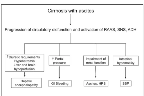
Although circulatory dysfunction in cirrhosis predominantly affects the kidney, it is also obser-ved in other organs and systems (Figure 3). Sym-pathetic overactivity secondary to circulatory dysfunction decreases intestinal motility, induces intestinal bacterial overgrowth, and may promote bacterial translocation from the intestinal lumen into the systemic circulation, causing severe infec-tions.35 The intrahepatic portal circulation is also
sensitive to the vasoconstrictor action of angio-tensin-II, norepinephrine and antidiuretic hormo-ne. The intense activation of these systems as a result of circulatory dysfunction may contribute
to the high portal pressure that is observed in pa-tients with advanced cirrhosis and ascites.29,36,37
Hyponatremia secondary to the hypersecretion of antidiuretic hormone may induce brain edema and encephalopathy.38 The use of diuretics for the
treatment of ascites that responds poorly to these drugs (refractory or recurrent ascites) is associa-ted to hepatic encephalopathy in 25% of cases.39
Studies have shown the existence of a close rela-tionship between renal blood flow and cerebral blood flow as well as between them and the degree of activation of endogenous vasoactive systems.40
Patients with hepatorenal syndrome are those with a lower cerebral blood flow. The role of this cerebral hypoperfusion in the predisposition of he-patic encephalopathy in these patients is not known.41,42
Finally, recent studies have found that critica-lly-ill patients with decompensated cirrhosis and bacterial infections present relative adrenal insu-fficiency with a frequency of 50-70%, with inappropriately low cortisol levels and poor res-ponse to cortocotropin.43-45 Cortisol is essential
for successful vascular response to endogenous vasoconstrictor systems. Relative adrenal insuffi-ciency, therefore, could contribute to circulatory dysfunction in these patients. The impairment in multiorgan function associated to circulatory dysfunction in cirrhosis constitutes the rationale for the prolonged use of albumin in the long-term studies that are being developed in Spain and Italy.
Figure 2. Clinical course of a pa-tient with type-2 HRS and refractory as-cites requiring frequent hospitalizations for repeated paracen-tesis that developed type-1 HRS follo-wing an episode or spontaneous bacterial peritonitis treated by cefo-taxime. Type-1 HRS occurred despite ra-pid resolution of the infection. The patient also developed aggravation of liver failure and hepatic encephalopaty and died 3 weeks after the onset of the infection.
Type-2 HRS Type-1 HRS
Cefotaxime
Encephalopathy Jaundice Therapeutic paracentesis
-6 -4 -2 0 1 2 3
Months Weeks
6
5
4
3
2
1
0
POST-PARACENTESIS CIRCULATORY DYSFUNCTION
It occurs in approximately 70% of patients trea-ted with total paracentesis without administration of plasma expanders.12,13 The incidence is very low
when the volume of ascites removed is less than 5 L, but progressively increases in parallel to the volu-men of paracentesis when the amount of fluid
remo-ved is above this limit.14 The mechanism of
circulatory dysfunction is dual. Patients develop a decrease in peripheral vascular resistance, indica-ting an arterial vasodilation. The site where this process occurs is unknown, although it is possible that it takes place in the splanchnic area. This di-sorder is accompanied by a reactive activation of en-dogenous vasoconstrictor systems with increased plasma renin activity and noradrenaline. Despite this sympathetic hyperactivity, the cardiac output and pulse rate do not increase, indicating that in addition to the decrease in systemic vascular resis-tance, inadequate cardiac response to vasodilation participates in the circulatory impairment.46
CIRCULATORY DYSFUNCTION AFTER BACTERIAL INFECTION
Bacterial infections are one of the most important causes of circulatory dysfunction and renal failure in cirrhosis; approximately 30-40% of patients with severe infections develop renal failure.33,47,48 In
many cases, renal failure is reversible, disappearing after resolution of infection; in others, however,
re-nal failure persists in spite of the resolution of infec-tion. In most of these patients renal failure follows a rapidly progressive course (type 1 hepatorenal syn-drome), and this is frequently associated to failure in the function of other organs. Finally, in other pa-tients renal insufficiency remains stable after reso-lution of the infection (type 2 hepatorenal syndrome).11,30,33 The infection that most often
cau-ses renal failure is spontaneous bacterial peritonitis, followed by symptomatic urinary tract infection (acute pyelonephritis) and cellulites.47,48
There are experimental studies showing that bac-terial infections in cirrhotic rats are associated with a much stronger inflammatory response than in healthy animals.49 Moreover, in cirrhotic patients
with spontaneous bacterial peritonitis, it has been shown that patients who develop renal failure are those with a greater inflammatory response, as esti-mated by the concentration of leukocytes in ascitic fluid and cytokine levels in ascites and blood.50
Studies performed in patients with spontaneous bacterial peritonitis have shown that the develop-ment of type-1 hepatorenal syndrome occurs as a re-sult of a dual mechanism. On the one hand, a rapid and intense decrease in vascular resistance that de-termines the activation of endogenous vasoconstric-tor systems. On the other hand, a failure in heart function with decreased cardiac output. This latter process is due to both a deterioration inotropic and chronotropic function. Patients do not develop ta-chycardia despite circulatory dysfunction and in-crease in the activity of the sympathetic nervous system.29
ROLE OF ALBUMIN IN LIVER FAILURE
Traditionally, the therapeutic role of albumin has been attributed solely to its oncotic properties. The use of albumin in the management of fluid retention and edema in diseases such as cirrhosis, nephrotic syndrome or protein-losing enteropathy is a clear example of this. However, the albumin molecule has many other functions that are important in physio-logy and pathophysiophysio-logy. The albumin molecule has sites with high and low affinity for binding fatty acids and other hydrophobic substances3-6,51 (Figure
4). Albumin, therefore, plays a major role in the transport of lipids and other hydrophobic molecules from their site of absorption or synthesis to periphe-ral tissues. Many drugs are transferred to the target organs through this process. Also many toxic subs-tances from cellular catabolism are also transported to their excretory organs (liver and kidney) by bin-ding to albumin. This phenomenon is especially rele-vant in diseases with high cellular catabolism (cancer, diabetes, sepsis). At the level of cis-34, the albumin molecule has a domain with ability to stabi-lize free-radicals, which gives a significant antioxi-dant capacity to the albumin molecule. Due to its high concentration in plasma and interstitial tissue, albumin is the most important antioxidant at extra-cellular level.7 Finally there are other domains,
par-ticularly at the N-terminal, with the ability to bind
metals which, if not removed, increase the oxidative stress.51
Functional proteomic studies using electron spec-troscopy techniques by magnetic resonance imaging (MRI) allow to assess many features of albumin func-tion, including the binding capacity, the efficiency in the transport of hydrophobic molecules (absorption, binding and release in target organs) and in the de-toxification of catabolic products (capacity of albu-min to bind, transport and finally release toxic substances produced through metabolism), the asses-sment of the conformation of the molecule at the bin-ding sites and the role of N-terminal chelation. Using these techniques it has been shown that albumin function in cirrhosis is deeply impaired.51 The
capaci-ty of binding fatcapaci-ty acids and other hydrophobic mole-cules and the ability for transport and detoxification is virtually absent (less than 20% as compared to healthy individuals). The mechanism that determines this decline in albumin function in cirrhosis is not known. It may be a reversible process. Liver failure determines the accumulation of large amounts of hydrophobic molecules. Sepsis, which also alters the function of albumin, is a frequent event in cirrhosis. Finally, both infections and liver failure are associa-ted with increased oxidative stress. All these proces-ses would saturate the binding sites of albumin and decrease its detoxification and transport functions. Albumin dialysis (MARS® system) is intended to
re-move these substances so as to make endogenous al-bumin functional again. However, a second possibility is that certain ligands irreversibly alter the molecular characteristics of albumin. Although traditionally it has been suggested that damaged al-bumin is rapidly eliminated, degraded and replaced by new molecules, this fact is probably not operating in cirrhosis, a condition in which albumin synthesis is profoundly impaired.
The clinical consequences of impaired albumin function in liver failure are not known, but could be significant. The combination of hypoalbumine-mia and functionally impaired endogenous albumin causes a profound alteration in the transport, me-tabolism and excretion of many endogenous and exogenous substances which, instead of being appropriately removed, will circulate as free com-pounds with the capacity to arbitrarily react. The pharmacokinetics and pharmacodynamics of many drugs and, therefore, effectiveness and side effects are severely affected. Finally, the existing oxidative stress caused by liver failure and other associated problems such as bacterial infections can not be co-rrected by the action of albumin. Increased oxidati-Figure 4. Molecular structure of albumin. BS Binding sites
ve stress can alter the microcirculation and cell function thus contributing to the multiple organ ilure that characterizes many patients with liver fa-ilure.52
EFFECT OF ALBUMIN
IN THE CIRCULATORY DYSFUNCTION OF CIRRHOSIS
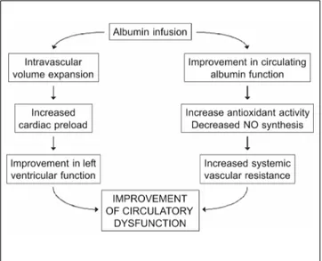
The administration of intravenous albumin in pa-tients with cirrhosis and ascites not only expands the plasma volume and increases cardiac preload and car-diac output but also increases peripheral vascular re-sistance, suggesting that arterial vasoconstriction occurs (Figure 5). This effect is not observed with synthetic expanders.53,54 The mechanism that
deter-mines the vasoconstrictor effect of albumin is not well studied. It may take place at the level of the mi-crocirculation and be secondary to a decrease in ni-tric oxide release. Plasma levels of von Willebrand Factor, whose synthesis is parallel to that of nitric oxide, are decreased in patients receiving albumin but not in those receiving plasma expanders.54 A decrease
in the levels of oxidative stress in the microcircula-tion after administramicrocircula-tion of exogenous albumin func-tionally active is another possibility.
The effectiveness of albumin in the prevention of post-paracentesis circulatory dysfunction, in the prevention of circulatory dysfunction after acute bacterial infections bacterial or in the treatment of hepatorenal syndrome may be related to this dual effect of albumin on the cardio-circulatory function, the increase in the cardiac output and in the syste-mic vascular resistance.
DISCLOSURE OF CONFLICT OF INTERESTS:
This article forms part of a supplement supported by an unrestricted grant from Grifols. The author received payment for the preparation of this article and attendance at the symposium in which it was presented.
REFERENCES
1. European Association for the Study of the Liver, Ginès P, Angeli P, Lenz K, Møller S, Moore K, Moreau R, Merkel C, et al. EASL clinical practice guidelines on the manage-ment of ascites, spontaneous bacterial peritonitis and hepatorenal syndrome in cirrhosis. J Hepatol 2010; 53: 397-417.
2. Rothschild MA, Oratz M, Schreiber SS. Albumin metabolism. Prog Liver Dis 1972; 4: 19-29.
3. Fasano M, Curry S, Terreno E, Galliano M, Fanali G, Narciso P, Notari S, et al. The extraordinary ligand binding proper-ties of human serum albumin. IUBMB Life 2005; 57: 787-96. 4. Varshney A, Sen P, Ahmad E, Rehan M, Subbarao N, Khan
RH. Ligand binding strategies of human serum albumin: how can the cargo be utilized? Chirality 2010; 22: 77-87. 5. Quinlan GJ, Martin GS, Evans TW. Albumin: biochemical
properties and therapeutic potential. Hepatol 2005; 41: 1211-9.
6. Oettl K, Stadlbauer V, Petter F, Greilberger J, Putz-Bankuti C, Hallstrom S, Lackner C, et al. Oxidative damage of albumin in advanced liver disease. Biochim Biophys Act 2008; 1782: 469-73.
7. Roche M, Rondeau P, Singh NR, Tarnus E, Bourdon E. The antioxidant properties of serum albumin. FEBS Lett 2008; 582: 1783-7.
8. Shawcross DL, Wright GA, Stadlbauer V, Hodges SJ, Davies NA, Wheeler-Jones C, Pitsillides AA, et al. Ammonia impairs neutrophil phagocytic function in liver disease. Hepatol 2008; 48: 1202-12.
9. Cicco G, Cicco S. The influence of oxygen supply, hemor-heology and microcirculation in the heart and vascular systems. Adv Exp Med Biol 2010; 662: 33-9.
10. Schrier RW, Arroyo V, Bernardi M, Epstein M, Henriksen JH, Rodés J. Peripheral arterial vasodilation hypothesis: a proposal for the initiation of renal sodium and water re-tention in cirrosis. Hepatol 1998; 8: 1151-7.
11. Arroyo V, Fernández J, Ginés P. Pathogenesis and treatment of hepatorenal syndrome. Semin Liver Dis 2008; 28-81-95.
12. Ruiz del Arbol L, Monescillo A, Jimenez W, Garcia Plaza A, Arroyo V, Rodés J. Paracentesis-induced circulatory dys-function: mechanism and effect on hepatic hemodynamics in cirrhosis. Gastroenterol 1997; 113: 579-86.
13. Ginès P, Titó L, Arroyo V, Planas R, Panés J, Viver J, To-rres M, et al. Randomized comparative study of therapeu-tic paracentesis with and without albumin in cirrhosis. Gastroenterol 1988; 94: 1494-502.
14. Ginès A, Fernández-Esparrach G, Monescillo A, Vila C, Do-mènech E, Abecasis R, Angeli P, et al. Randomized trial comparing albumin, dextran 70 and polygeline in cirrhotic patients with ascites treated by paracentesis. Gastroen-terol 1996; 114: 1002-10.
15. Sort P, Navasa M, Arroyo V, Aldeguer X, Planas R, Ruiz-del-Arbol L, Castells L, et al. Effect of intravenous albumin on Figure 5. Mechanism of the effect of I.V. albumin on
and spontaneous bacterial peritonitis. N Engl J Med 1999; 341: 403-9.
16. Salerno F, Gerbes A, Gines P, Wong F, Arroyo V. Diagnosis, prevention and treatment of hepatorenal syndrome. Gut 2007; 56: 1310-8.
17. Ortega R, Ginès P, Uriz J, Cárdenas A, Calahorra B, De Las Heras D, Guevara M, et al. Terlipressin therapy with and without albumin for patients with hepatorenal syndrome; results of a prospective non-randomized study. Hepatol 2002; 36: 941-8.
18. Gluud LL, Christensen K, Christensen E, Krag A. Systema-tic review of randomized trials on vasoconstrictor drugs for hepatorenal syndrome. Hepatol 2010; 51: 576-84. 19. Vallance P, Moncada S. Hyperdynamic circulation in
cirrho-sis: a role for nitric oxide? Lancet 1991; 337: 776-8. 20. Bomzon A, Blendis LM. The nitric oxide hypothesis and the
hyperdynamic circulation in cirrhosis. Hepatol 1994; 20: 1343-50.
21. Bruix J, Bosch J, Kravetz D, Mastai R, Rodés J. Effects of prostaglandin inhibition on systemic and hepatic hemody-namics in patients with cirrhosis of the liver. Gastroente-rol 1985; 88: 430-5.
22. Fernández-Rodriguez CM, Prieto J, Quiroga J, Zozoya JM, Andrade A, Núñez M, Sangro B, et al. Plasma levels of subs-tance P in liver cirrhosis: relationship to the activation of vasopressor systems and urinary sodium excretion. Hepa-tol 1995; 21: 35-40.
23. De las Heras D, Fernández J, Ginès P, Cárdenas A, Ortega R, Navasa M, Barberá JA, et al. Increased carbon monoxide production in patients with cirrhosis with and without spontaneous bacterial peritonitis.Hepatol 2003; 38: 452-9. 24. Bendtsen F, Schifter S, Henriksen JH. Increased circula-ting calcitonin gene-related peptide (CGRP) in cirrhosis. J Hepatol 1991; 12: 118-23.
25. Caraceni P, Domenicali M, Giannone F, Bernardi M. The role of the endocannabinoid system in liver diseases.Best Pract Res Clin Endocrinol Metab 2009; 23: 65-77.
26. Fernández M, Semela D, Bruix J, Colle I, Pinzani M, Bos-ch J. Angiogenesis in liver disease. J Hepatol 2009; 50: 604-20.
27. Paternostro C, David E, Novo E, Parola M. Hypoxia, angio-genesis and liver fibroangio-genesis in the progression of chro-nic liver disease. WJ Gastroenterol 2010; 16: 281-8. 28. Mejias M, Garcia-Pras E, Tiani C, Miquel R, Bosch J,
Fer-nandez M. Beneficial effects of sorafenib on splanchnic, intrahepatic, and portocollateral circulations in portal hy-pertensive and cirrhotic rats. Hepatol 2009; 49: 1245-5. 29. Ruiz-del-Arbol L, Urman J, Fernández J, González M,
Nava-sa M, Monescillo A, Albillos A, et al. Systemic, renal and he-patic hemodynamics derangement in cirrhotic patients with spontaneous bacterial peritonitis. Hepatology 2003; 38: 1210-8.
30. Ruiz-del-Arbol L, Monescillo A, Arocena C, Valer P, Ginès P, Moreira V, Milicua JM, et al. Circulatory function and hepatorenal syndrome in cirrhosis. Hepatol 2005; 42: 439-47.
31. Møller S, Henriksen JH. Cirrhotic cardiomyopathy. J Hepa-tol 2010; 53: 179-90.
32. Alqahtani SA, Fouad TR, Lee SS. Cirrhotic cardiomyopathy. Semin Liver Dis 2008; 28: 59-69.
33. Follo A, Llovet JM, Navasa M, Planas R, Forns X, Francito-rra A, Rimola A, et al. Renal impairment after spontaneous bacterial peritonitis in cirrhosis: incidence, clinical cour-se, predictive factors and prognosis. Hepatol 1994; 20: 1495-501.
Iwasaki K. Bell-shaped relationship between central blood volume and spontaneous baroreflex function. Auton Neu-rosci 2008; 143: 46-52.
35. Wiest R, Garcia-Tsao G. Bacterial translocation in cirrho-sis. Hepatol 2005; 41: 422-33.
36. Gatta A, Bolognesi M, Merkel C. Vasoactive factors and hemodynamic mechanisms in the pathophysiology of portal hypertension in cirrhosis. Mol Aspects Med 2008; 29: 119-29.
37. Cichoz-Lach H, Celinski K, Slomka M, Kasztelan-Szczer-binska B. Pathophysiology of portal hypertension. J Phy-siol Pharmacol 2008; 59(Suppl. 2): 231-8.
38. Guevara M, Baccaro ME, Torre A, Gómez-Ansón B, Ríos J, Torres F, Rami L, et al. Hyponatremia is a risk factor of hepatic encephalopathy in patients with cirrhosis: a pros-pective study with time-dependent analisis. Am J Gas-troenterol 2009; 104: 1382-9.
39. Arroyo V, Ginès P, Gerbes AL, Dudley FJ, Gentilini P, Laffi G, Reynolds TB, et al. Definition and diagnostic criteria of refractory ascites and hepatorenal syndrome in cirrhosis. Hepatol 1996; 23: 164-76.
40. Guevara M, Bru C, Ginès P, Fernández-Esparrach G, Sort P, Bataller R, Jiménez W, et al. Increased cerebrovascular resistance in cirrhotic patients with ascites. Hepatol 1998; 28: 39-44.
41. Ahl B, Weissenborn K, van den Hoff J, Fischer-Wasels D, Köstler H, Hecker H, Burchert W. Regional differences in cerebral blood flow and cerebral ammonia metabolism in patients with cirrhosis. Hepatol 2004; 40: 73-9.
42. Iversen P, Sørensen M, Bak LK, Waagepetersen HS, Va-faee MS, Borghammer P, Mouridsen K, et al. Low cerebral oxygen consumption and blood flow in patients with cirr-hosis and an acute episode of hepatic encephalopathy. Gastroenterol 2009; 136: 863-71.
43. Tsai MH, Peng YS, Chen YC, Liu NJ, Ho YP, Fang JT, Lien JM, et al. Adrenal insufficiency in patients with cirrhosis, severe sepsis and septic shock. Hepatol 2006; 43: 673-81. 44. Marik PE, Gayowski T, Starzl TE. The hepatoadrenal syn-drome: a common yet unrecognized clinical condition. Crit Care Med 2005; 33: 1254-59.
45. Fernández J, Escorsell A, Zabalza M, Felipe V, Navasa M, Mas A, Lacy AM, et al. Adrenal insufficiency in pa-tients with cirrhosis and septic shock: Effect of treat-ment with hydrocortisone on survival. Hepatol 2006; 44: 1288-95.
46. Luca A, García-Pagán JC, Bosch J, Feu F, Jiménez W, Ginés A, Fernández M, et al. Beneficial effects of intravenous al-bumin infusion on the hemodynamic and humoral changes af-ter total paracentesis.Hepatol 1995; 22: 753-8.
47. Terra C, Guevara M, Torre A, Gilabert R, Fernández J, Martín-Llahí M, Baccaro ME, et al. Renal failure in patients with cirrhosis and sepsis unrelated to spontaneous bacte-rial peritonitis: value of MELD score. Gastroenterol 2005; 129: 1944-5.
48. Fasolato S, Angeli P, Dallagnese L, Maresio G, Zola E, Maz-za E, Salines F, et al. A. Renal failure and bacterial infec-tions in patients with cirrhosis: epidemiology and clinical feature. Hepatol 2007; 45: 223-9.
49. Ramírez MJ, Ibáñez A, Navasa M, Casals E, Morales-Ruiz M, Jiménez W, Arroyo V, et al. High-density lipoproteins re-duce the effect of endotoxin on cytokine production and systemic hemodynamics in cirrhotic rats with ascites. J Hepatol 2004; 40: 424-30.
interleu-kin-6 in spontaneous bacterial peritonitis in cirrhosis: re-lationship with the development of renal impairment and mortality. Hepatol 1998; 27: 1227-32.
51. Jalan R, Schnurr K, Mookerjee RP, Sen S, Cheshire L, Hod-ges S, Muravsky V, et al. Alterations in the functional ca-pacity of albumin in patients with decompensated cirrhosis is associated with increased mortality. Hepatol 2009; 50: 555-64.
52. Arroyo V. Human serum albumin: not just a plasma volume expander. Hepatol 2009; 50: 355-7.
53. Fernández J, Navasa M, Garcia-Pagan JC, G-Abraldes J, Ji-ménez W, Bosch J, Arroyo V. Effect of intravenous albumin on systemic and hepatic hemodynamics and vasoactive neurohormonal systems in patients with cirrhosis and spon-taneous bacterial peritonitis. J Hepatol 2004; 41: 384-90. 54. Fernández J, Monteagudo J, Bargallo X, Jiménez W,