Acute liver failure in under two yearolds
-are there markers of metabolic disease on admission?
Ana Brett,* Carla Pinto,** Leonor Carvalho,** Paula Garcia,*** Luísa Diogo,*** Isabel Gonçalves**Unidade de Hepatologia, **Serviço de Cuidados Intensivos, ***Unidade de Doenças Metabólicas, Hospital Pediátrico de Coimbra, Centro Hospitalar e Universitário de Coimbra, Portugal.
ABSTRACT
Introduction. The early establishment of an etiology for acute liver failure (ALF) in infants is essential for the start of adequate treatment in the shortest timeframe possible. Aim. To identify markers of inherited metabolic disease on admission in children under two years of age with ALF. Material and methods. A re-trospective review of the medical records of all children (< 2 years old) with ALF admitted to the pediatric hepatology or intensive care units of a tertiary center over a twenty-three year period (January 1989 to December 2011) was done. Patients were divided into two groups: with (group A) or without (group B) a metabolic etiology. Clinical and laboratory parameters on admission were compared. Results. Twenty-three children met inclusion criteria. Twelve had ALF of metabolic origin (group A). The median age in this group was 2.25 (Q1-Q3: 0.63-4.65) months and in group B 8.0 (Q1-Q3: 1.5-15) months. History of failure to thrive and/or vomiting was more frequent in group A (p = 0.022). Age, gender, encephalopathy and left ventricular hypertrophy were similar in both groups (p = 0.147, p = 1.000, p = 0.637, p = 1.000, respectively). Laboratory tests on admission (plasma lactate, ammonia, cholesterol, phosphate, INR, glucose, bilirubin, ALT, base excess and the presence of reducing substances in urine) showed no statistically significant differences between groups. Conclusion. This study showed that although infants with inborn errors of metabolism showed a trend towards lower age at presentation, the only marker of inherited metabolic disease found on admission was history of vomiting and/or failure to thrive.
Key words. Liver disease. Pediatric. Inborn errors of metabolism. Failure to thrive.
Correspondence and reprint request: Carla Pinto, M.D.
Serviço de Cuidados Intensivos Pediátricos, Hospital Pediátrico de Coimbra, Centro Hospitalar e Universitário de Coimbra
Av. Afonso Romão-Alto da Baleia, 3000-602 Coimbra, Portugal Ph.: +351 239488700
E- mail: [email protected]
Manuscript received: November 12, 2012. Manuscript accepted: February 25, 2013.
INTRODUCTION
Acute liver failure (ALF) in children is a rare mul-tisystemic disorder with high morbidity and mortali-ty. It is defined as severe impairment of liver function, with or without encephalopathy, in associa-tion with hepatocellular necrosis, in a patient with no recognized underlying chronic liver disease.1
In young children, the identification of encephalo-pathy is not mandatory for the definition of ALF, since its signs are difficult to identify and encepha-lopathy can be a late event in the course of the
disease.2 INR, which expresses prothrombin time, is
very helpful in this age group and can be used as an indicator of the severity of liver damage, thus helping decide whether or not to list for transplantation.2
In infants, ALF has several etiologies, the most common of which are liver based inborn errors of metabolism (IEM) and perinatally acquired in-fections. The former can be responsible for up to 40% of cases.3 Management is directed at treating
and monitoring ALF, whilst anticipating its multisystemic complications.4 The diagnosis of the
underlying disorder may lead to clinical im-provement when appropriate treatment is initiated promptly.5
Galactosemia, tyrosinemia type 1, hereditary fruc-tose intolerance and mitochondrial respiratory chain disorders are the most common IEM associa-ted with ALF.6-8 Patients affected by the first three
conditions can improve dramatically with appro-priate therapeutic intervention.1 When ALF is
multisystemic disorders and this can be considered a contraindication for liver transplant.4,9
The number of patients presenting with ALF as a first manifestation of metabolic disease has decli-ned in the last few years, due to earlier diagnosis of liver metabolic diseases. Neonatal screening, imple-mented in Portugal in 1979 for phenylketonuria, was recently extended, allowing earlier recognition of liver involvement in neonates.
The aim of this study is to identify markers of metabolic disease on admission in children under two years of age with ALF.
MATERIAL AND METHODS
A descriptive retrospective review of the medical records of all children (< 2 years old) with ALF ad-mitted to the pediatric hepatology or intensive care units of a tertiary center over a 23-year period (January 1989 to December 2011) was made.
ALF was defined as coagulopathy (INR > 2) un-responsive to vitamin K, regardless of clinical hepa-tic encephalopathy or neurological abnormalities. Infants with evidence of chronic liver disease at presentation were excluded, as well as those without a final etiological diagnosis.
The initial baseline investigation in a child with ALF included: full blood count and blood smear, creatinine, urea and electrolytes, ammonia, lactacte, glucose, markers of liver synthesis (prothrombin time, INR, factors V and VII, fibrinogen, albumin, cholesterol), markers of necrosis (AST, ALT, LDH), markers of bile duct injury (ALP, GGT, bilirubin), venous blood pH and gases, base excess and presence of reducing substances in urine, abdominal ultrasound, echocardiogram and electro-encephalogram.
A diagnostic algorithm was used to establish the etiology of ALF, including the following exams in children < 2 years of age: routine metabolic investi-gation (neonatal screening for metabolic disease, plasma and urine amino acid chromatography), serum ferritin levels, transferrin saturation, triglyce-rides, viral markers (herpes simplex virus, enterovirus, adenovirus, herpes virus type 6, varicella zoster, parvovirus B19, Epstein-Barr virus) and DNA extraction. When family history was present, autoim-mune markers (ANA, ASMA, LKM, immunoglobu-lins, direct antiglobulin test) were also included. According to clinical history and exam results, further tests were requested on a child-to-child basis. Each patient included in the study group was screened with respect to initial presentation and
laboratory results. Several items were analyzed: age, gender, history of vomiting or failure to thrive, ence-phalopathy and family history of liver disease with onset in childhood. The presence of left ventricular hypertrophy was assessed by echocardiogram. Initial laboratory results were collected: lactate, ammonia, cholesterol, phosphate, INR, glucose, bilirubin, ALT, base excess and presence of reducing substances in urine. Patient’s follow-up was briefly described regarding orthotopic liver transplantation and whether death occurred due to ALF and its complications.
The standard classification of hepatic encepha-lopathy adapted to infants under 4 years of age was used,10 although the different grades of
seve-rity are not discriminated in this study. Since this classification cannot be applied to newborns, neurological distress was defined by irritability, poor sucking, inappropriate agitation or crying or a disturbed sleep-wake cycle in this age group.11
Left ventricular hypertrophy was defined as an increase in left ventricular mass (e.g.: ventricu-lar septum and left ventricuventricu-lar posterior wall thickness, according to the child’s age).
According to the etiology of ALF, two groups were defined: Group A with metabolic disease and Group B with other etiologies for ALF. These two groups were compared regarding initial presenta-tion, laboratory data and imaging results.
Statistical analysis was performed using the Sta-tistical Package for the Social Sciences version 19.0 (SPSS Inc., Chicago, IL). The description of the po-pulation was made by calculating measures of cen-tral tendency and dispersion for quantitative variables and by determination of absolute and rela-tive frequencies for qualitarela-tive variables. The Mann-Whitney U test was used to compare quantitative variables without normal distribution. The Fisher exact test was used for the association between two qualitative variables, as the lowest expected frequency was below 5. The Kaplan-Meier method was used to estimate in both groups the requirement for liver transplantation and the survival curve. The statis-tical significance level was set at p < 0.050.
RESULTS
half (n = 12) were included in group A (with metabo-lic disease) and 11 in group B (without meta-bolic disease).
The etiologies of ALF in the study group are shown in table 1. In the inherited metabolic diseases group (A) the most common disorders were mito-chondrial DNA depletion syndromes (3 cases, 2 with deoxyguanosine kinase - DGUOK - deficiency), follo-wed by tyrosinemia type 1, galactosemia and conge-nital disorders of glycosylation type X (2 each). In group B, which includes all other etiologies, viral in-fection was the most common diagnosis (4 patients), followed by mushroom poisoning and hemophagocy-tic lymphohistiocytosis (2 each).
Male/female ratio was similar in both groups (Table 2). Median age of presentation was 2.25 (Q1-Q3: 0.63-4.65) months in group A, and 8.0 (Q1-Q3: 1.5-15) months in group B, with no statistical significance (p = 0.147) (Table 2). The youngest infant presen-ted at 2 days of life and was diagnosed with neonatal hemochromatosis.
Family history was irrelevant in all cases except in one DGUOK patient whose brother had died in another hospital with the diagnosis of neonatal he-mochromatosis, later proved to be also a DGUOK deficiency.
Regarding past medical history, 9 patients in group A (82%) and 2 patients in group B (18%)
pre-Table 2. Clinical and diagnostic features.
Group A, n = 12 Group B, n = 11 p value
Female gender (n) 7 7 1.000*
Age, [median (Q1-Q3)] 2.25 (0.63-4.65) 8.0 (1.5-15) 0.147†
History of failure to thrive/vomiting (n) 9 2 0.022*
Encephalopathy (n) 3 5 0.637*
Left ventricular hypertrophy (n) 3 4 1.000*
n: number of patients. Q1: quartile 1. Q3: quartile 3. *Fisher’s exact test. †Mann-Whitney U test. Table 1. Etiology groups.
Group A n = 12 Group B n = 11
mtDNA depletion syndromes 3 Viral infections (adenovirus, EBV, varicella virus, HSV1) 4
Galactosemia 2 Mushroom poisoning 2
CDG 2 HLH 1
Tyrosinemia 2 Porto-caval fistula 1
LCHAD deficiency 1 Autoimmune hepatitis 1
Hereditary fructose intolerance 1 Neonatal hemochromatosis 1
Urea cycle disorder (OTC) 1
CDG: congenital disorders of glycosylation. EBV: Epstein-Barr virus. HLH: hemophagocytic lymphohistiocytosis. HSV1: herpes simplex virus 1. LCHAD: long-chain acyl CoA dehydrogenase. mtDNA: mitochondrial DNA. OTC: ornithine transcarbamylase.
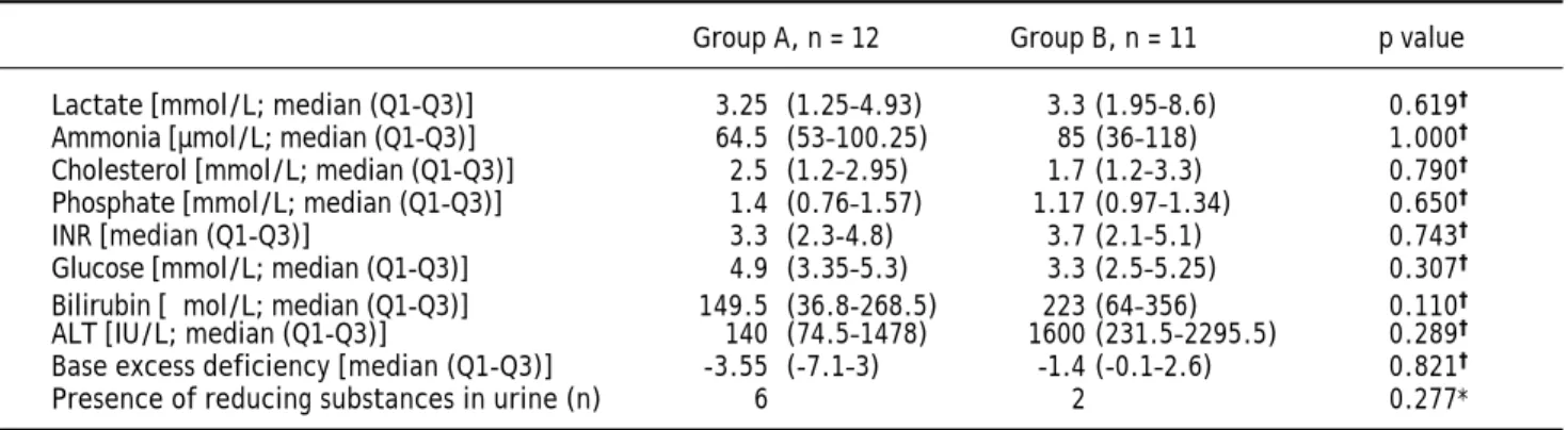
Table 3. Laboratory data evaluated on admission.
Group A, n = 12 Group B, n = 11 p value
Lactate [mmol/L; median (Q1-Q3)] 3.25 (1.25–4.93) 3.3 (1.95–8.6) 0.619†
Ammonia [µmol/L; median (Q1-Q3)] 64.5 (53–100.25) 85 (36–118) 1.000†
Cholesterol [mmol/L; median (Q1-Q3)] 2.5 (1.2–2.95) 1.7 (1.2–3.3) 0.790†
Phosphate [mmol/L; median (Q1-Q3)] 1.4 (0.76–1.57) 1.17 (0.97–1.34) 0.650†
INR [median (Q1-Q3)] 3.3 (2.3-4.8) 3.7 (2.1–5.1) 0.743†
Glucose [mmol/L; median (Q1-Q3)] 4.9 (3.35–5.3) 3.3 (2.5–5.25) 0.307†
Bilirubin [μmol/L; median (Q1-Q3)] 149.5 (36.8-268.5) 223 (64–356) 0.110†
ALT [IU/L; median (Q1-Q3)] 140 (74.5–1478) 1600 (231.5–2295.5) 0.289†
Base excess deficiency [median (Q1-Q3)] -3.55 (-7.1–3) -1.4 (-0.1–2.6) 0.821†
Presence of reducing substances in urine (n) 6 2 0.277*
sented with vomiting episodes and/or failure to thrive (p = 0.022). The presence of encephalopathy was also compared (3 in group A and 5 in group B), with no statistical significance (p = 0.637). A simi-lar number of patients presented with left ventricusimi-lar hypertrophy in both groups (p = 1.00) (Table 2).
The mean or median levels of each laboratory value analyzed (lactate, ammonia, cholesterol, phosphate, INR, glucose, bilirubin, ALT and base excess) and the number of patients with presence of reducing substances in urine in both groups are shown in ta-ble 3. No statistically significant differences were found in any of these parameters.
In the study group, 4 children, with the follo-wing disorders, were submitted to an orthotropic liver transplant: mtDNA depletion syndrome, vari-cella virus infection, mushroom poisoning and au-toimmune hepatitis. Figure 1A shows the proportion of patients without liver transplanta-tion in both groups.
A total of 9 children died due to ALF and its com-plications, 3 in group A (2 mtDNA depletion syndro-mes, 1 LCHAD deficiency) and 6 in group B (2 viral infections, 2 hemophagocytic lymphohistiocytosis, 1 porto-caval fistula, 1 autoimmune hepatitis) (p = 0.214). Out of these, 4 had contraindications for liver transplantation. Most children (n = 7) died from multiple organ failure, 1 from severe brain hemorrhage and 1 shortly after liver transplantation (autoimmune hepatitis). Figure 1B shows the survival curve in both groups.
DISCUSSION
In the present study the etiology for ALF was de-termined in 23 of 28 patients under 2 years of age, with only 18% remaining without a diagnosis. Another study, which reviewed patients with ALF younger than 1 year of age, had similar results, with 16% of cases with unknown diagnosis.11 This is in contrast
with two series of ALF, one in infants aged ≤ 90 days12 and another in children from birth to 18
years of age,13 that presented respectively with 38
and 49% of cases with indeterminate diagnosis. Liver based IEM present frequently in the neona-tal period with jaundice, severe hypoglycemia and sometimes with ALF.6 They represent an important
cause of ALF in < 2 year olds. Therefore, IEM must always be considered in an infant presenting with ALF, as an appropriate diet or specific treatment might be lifesaving.2 In several pediatric case series
of ALF the percentage of IEM as an etiology varied between 10% and 42.5%.11-13 This wide spectrum
may be explained not only by the different investiga-tion protocols, but also by the diverse age groups in-cluded in each study.
In the present study 43% of the 28 patients < 2 years of age were diagnosed with an inherited me-tabolic etiology for ALF. A similar percentage (42.5%) was found in a group of ALF patients un-der one year of age.11 In older children, IEM
present more rarely and secondary causes of ALF, namely viral and toxic, became more important.14 Figure 1. A. Proportion of patients without liver transplantation in both groups. B. Survival curve in both groups.
1.0
0.8
0.6
0.4
0.2
0.0
0 50 100 150 200 250 300
Months of follow up
Group A
Group B Group AGroup B
Proportion without liver transplantation
1.0
0.8
0.6
0.4
0.2
0.0
0 50 100 150 200 250 300
Months of follow up
Survival
The only exception might be neonatal hemochro-matosis, an alloimune disease and the most fre-quently recognized cause of neonatal ALF, which presents typically very early in life.15,16 In this
se-ries, which included one patient with neonatal hemochromatosis, children with IEM showed a trend towards lower age on admission [group A 2.25 (Q1-Q3: 0.63-4.65) vs. group B 8.0 (Q1-Q3: 1.5-15) months], although the age difference was not statistically significant.
A specific treatment is available in some IEM with significant liver involvement. Tyrosinemia type 1, galactosemia and fructosemia, three of the most commonly described etiologies, are IEM due to in-toxication, amenable to effective dietary/ pharmaco-logical treatment.6,7 In recent years, mitochondrial
respiratory chain defects, which are a broad group of diseases, frequently multisystemic, with no speci-fic treatment, have been implicated as an etiological factor for ALF in infants.17
The etiology of group A of the series in discus-sion was varied, with mitochondrial disorders, tyro-sinemia type 1, galactosemia and congenital disorders of glycosylation representing the most fre-quent diagnosis. The two cases with congenital di-sorders of glycosylation have been ascribed an X type, since the diagnosis was based on transferrin isoelectric focusing pattern abnormalities, not ex-plained by liver failure, with no enzyme or genetic diagnosis yet.
In group B, the most frequent diagnosis was a vi-ral infection, as expected.11-13 However, even if an
infectious etiology is found, an IEM should be consi-dered in this age group.18
Failure to thrive and/or vomiting have been descri-bed as striking features in the clinical presentation of IEM.19 In this study, only this presenting feature
proved to be statistically significant between both groups, with a larger number of cases in group A. Thus, its presence should point in the direction of an inherited metabolic disorder, particularly in infants.
Many IEM produce dysfunction in other organs such as the central nervous system and heart, which can be very helpful indicators in their differential diagnosis.20 However, ALF is an important cause of
encephalopathy. In the clinical setting, the distinc-tion between the role of liver failure or that of a pro-bable underlying central nervous system disease (e.g.: viral infection, multisystemic IEM) in a child with encephalopathy is very hard. As would be expec-ted in this age group, in this study only a small num-ber of children were described as having encephalopathy. The slightly larger number of
pa-tients with encephalopathy in group B (Table 2) might be explained by the older age of children in this group and, therefore, with easier to diagnose mental status changes. Although without statistical significance, this may be a possible study bias.
Heart involvement, namely hypertrophic cardiom-yopathy, is a rather common feature in IEM.21 In this
study, the number of patients with left ventricular hypertrophy was similar in both groups, contrarily to what was anticipated.
Very little data was found regarding biological markers to aid in differentiating metabolic from non-metabolic disease in children presenting with ALF. A recent study has showed that in a popula-tion of 148 infants ≤ 3months of age with ALF there was a moderate association between the cumulative biochemical profile and the presence of ALF in in-fants with IEM, when compared with neonatal he-mochromatosis: patients with IEM had moderately elevated aminotransferase levels, moderate cholesta-sis, and minimal coagulopathy.12
One of the routine tests for diagnosis of liver IEM, namely galactosemia and fructosemia, is to ascertain the presence of reducing substances in urine,22 and this test is frequently used to guide
in identifying the etiology of ALF. In this series, the number of infants with reducing substances in urine was greater in group A, although without statistical significance. Children with hepatic failure, regardless of their etiology, may have reducing substances in urine as liver meta-bolic functions are secondarily impaired. There-fore, it is important not to allow this test to supersede the search for other possible etiologies for ALF other than IEM.
In this study, bilirubin and ALT showed a trend towards higher levels in group B. Unexpec-tedly, plasma glucose levels were lower in group B than in group A. None of these differences or those of the other markers also mentioned in ta-ble 3 were statistically significant, thus not per-mitting a more direct approach in diagnosing the etiology of ALF. As a result, specific laboratory markers for each disease are still the mainstay for diagnosis.
In conclusion, ALF is an important cause of mor-bidity and mortality particularly in under two year-old children, and early establishment of an etiology is warranted. In this series, history of vomiting and/ or failure to thrive was shown to be a possible mar-ker of IEM on admission in this age group.
make definitive conclusions. Hence, more studies on this issue should be undertaken, with larger cohorts of patients, to understand if the infants’ clinical and laboratory profile may help to distin-guish between a probable IEM and a non-metabolic cause for ALF. This would be helpful in the manage-ment of these patients, namely in the decision ma-king of listing for liver transplantation.
ABBREVIATIONS
• ALF: acute liver failure. • ALP: alkaline phosphatase. • ALT: alanine aminotransferase. • ANA: antinuclear antibodies.
• ASMA: anti-smooth muscle antibodies. • AST: aspartate aminotransferase. • DGUOK: deoxyguanosine kinase. • GGT: gamma-glutamyl transpeptidase. • INR: international Normalized Ratio. • IEM: inborn errors of metabolism. • LDH: lactate dehydrogenase.
• LKM: liver/kidney microsomal antibody.
GRANTS AND FINANCIAL SUPPORT
None.
REFERENCES
1. Bhaduri BR, Mieli-Vergani G. Fulminant hepatic failure: pe-diatric aspects. Semin Liver Dis 1996; 16: 349-55.
2. Dhawan A, Mieli-Vergani G. Acute liver failure in neonates.
Early Hum Dev 2005; 81: 1005-10.
3. Krogstad P, Martin MG. Evaluation of acute liver failure.
Pediatr Infect Dis J 2003; 22: 831-2.
4. Squires RH Jr. Acute liver failure in children. Semin Liver Dis 2008; 28: 153-66.
5. Saenz MS, Van Hove J, Scharer G. Neonatal liver failure: a genetic and metabolic perspective. Curr Opin Pediatr
2010; 22: 241-5.
6. Saudubray JM, Nassogne MC, de Lonlay P, Touati G. Clini-cal approach to inherited metabolic disorders in neona-tes: an overview. Semin Neonatol 2002; 7: 3-15.
7. Kelly DA, McKiernan PJ. Metabolic liver disease in the pe-diatric patient. Clin Liver Dis 1998; 2: 1-30.
8. Nobre S, Grazina M, Silva F, Pinto C, Goncalves I, Diogo L. Neonatal liver failure due to deoxyguanosine kinase defi-ciency. BMJ Case Rep 2012; 2012.
9. Dimmock DP, Dunn JK, Feigenbaum A, Rupar A, Horvath R, Freisinger P, Mousson de Camaret B, et al. Abnormal neu-rological features predict poor survival and should preclu-de liver transplantation in patients with preclu-deoxyguanosine kinase deficiency. Liver Transpl 2008; 14: 1480-5. 10. Whitington PF, Alonso AE. Fulminant hepatitis and acute
li-ver failure. In: Kelly DA (ed). Diseases of the Lili-ver and Bi-liary System in Children. Oxford: Blackwell; 2003, p. 107-26.
11. Durand P, Debray D, Mandel R, Baujard C, Branchereau S, Gauthier F, Jacquemin E, et al. Acute liver failure in infan-cy: a 14-year experience of a pediatric liver transplanta-tion center. J Pediat 2001; 139: 871-6.
12. Sundaram SS, Alonso EM, Narkewicz MR, Zhang S, Squires RH, Pediatric Acute Liver Failure Study G. Characteriza-tion and outcomes of young infants with acute liver failu-re. J Pediat 2011; 159: 813-8 e1.
13. Squires RH Jr, Shneider BL, Bucuvalas J, Alonso E, Sokol RJ, Narkewicz MR, Dhawan A, et al. Acute liver failure in chil-dren: the first 348 patients in the pediatric acute liver fa-ilure study group. J Pediat 2006; 148: 652-8.
14. Dhawan A. Acute liver failure in children and adolescents.
Clin Res Hepatol Gastroenterol 2012; 36: 278-83.
15. Vohra P, Haller C, Emre S, Magid M, Holzman I, Ye MQ, Iofel E, et al. Neonatal hemochromatosis: the importance of ear-ly recognition of liver failure. J Pediat 2000; 136: 537-41. 16. Whitington PF, Kelly S, Ekong UD. Neonatal
hemochroma-tosis: fetal liver disease leading to liver failure in the fetus and newborn. Pediatr Transplant 2005; 9: 640-5.
17. Lee WS, Sokol RJ. Mitochondrial hepatopathies: advances in genetics and pathogenesis. Hepatology 2007; 45: 1555-65.
18. Dimmock DP, Zhang Q, Dionisi-Vici C, Carrozzo R, Shieh J, Tang LY, Truong C, et al. Clinical and molecular features of mitochondrial DNA depletion due to mutations in deoxyguanosine kinase. Hum Mutat 2008; 29: 330-1. 19. Burton BK. Inborn errors of metabolism in infancy: a guide
to diagnosis. Pediatrics 1998; 102: e69.
20. Clayton PT. Inborn errors presenting with liver dysfunc-tion. Semin Neonatol 2002; 7: 49-63.
21. Kreuder J, Kahler SG. Approach to the patient with car-diovascular disease. In: Hoffmann GF, Zschocke J, Nyhan WL (eds.). Inherited Metabolic Diseases: A Clini-cal Approach: Springer-Verlag Berlin Heidelberg; 2010, p. 69-88.