Otras secciones de este sitio:
☞ ☞ ☞ ☞
☞ Índice de este número
☞ ☞ ☞ ☞
☞ Más revistas
☞ ☞ ☞ ☞
☞ Búsqueda
Others sections in this web site:
☞ ☞ ☞ ☞
☞ Contents of this number ☞
☞ ☞ ☞
☞ More journals ☞
☞ ☞ ☞ ☞ Search Artículo:
Alcoholic liver disease. An update
Copyright © 2005: Mexican Association of Hepatology
Number 1 January-March 2 0 0 5
Volume 4
Annals of Hepatology 4(1) 2005: 32-42
MG
32
edigraphic.com
Annals of Hepatology 2005; 4(1): January-March: 32-42
Annals of Hepatology
Abstract
The prevalence and incidence of alcoholic liver disease are constantly evolving. Alcoholic liver disease has a wide clinical spectrum. It may progress to cirrhosis and to end-stage liver disease requiring liver transplantation. The histological manifestations range from steatosis without inflammation to liver cell injury and ultimately to fibrosis and cirrhosis. In some cases, the histological manifestation is steatohepatitis, morphologically charac-terized by inflammation and necrosis. Currently, al-though there are no specific tests to establish a diagnosis of steatohepatitis, some serological, radiological, or labo-ratory tests may be useful. Liver biopsy is useful in con-firming a suspected diagnosis and in assessing the extent of parenchymal damage. This review synthesizes the main aspects of the epidemiology, pathogenesis, morpho-logical characteristics, diagnosis, treatment, and progno-sis of alcoholic liver disease.
Key words: Alcoholic liver disease, fatty liver, steato-hepatitis, cirrhosis.
Epidemiology
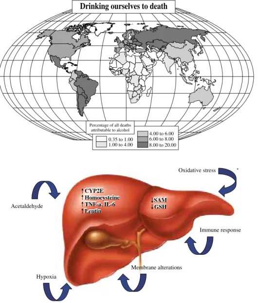
Cirrhosis causes nearly 150,000 deaths each year worldwide, and alcoholic cirrhosis accounts for approxi-mately 38–50% of all cirrhosis-related deaths (Figure
1).1,2 Alcohol liver disease (ALD) occurs in patients who
consume excessive amounts of alcohol. Approximately 7.4% of the U.S. population meets the diagnostic criteria for alcohol abuse or alcoholism; in Europe, 20–30% of the population consumes excessive amounts of alcohol; and in many developing nations, the increase in alcohol consumption is alarming. In Mexico, cirrhosis is the third leading cause of death in adults between 45 and 60 years of age, and alcohol is related to more than 50% of these
Concise Review
Alcoholic liver disease. An update
Nahum Méndez-Sánchez;1 Paloma Almeda-Valdés;1 Misael Uribe1
1Departments of Biomedical Research, Gastroenterology & Liver
Unit. Medica Sur Clinic & Foundation.
Address for correspondence: Nahum Méndez-Sánchez, M.D., PhD.
Departments of Biomedical Research, Gastroenterology & Liver Unit. Medica Sur Clinic & Foundation, Puente de Piedra 150, Col. Toriello Guerra, Tlalpan 14050, Mexico City, Mexico. Phone: (+525) 55 424-7200 ext. 4215; Fax: (+525) 55666-4031; E-mail: [email protected]
Manuscript received and accepted: 05 February, 2005
deaths. It is estimated that by the year 2050, there will be more than one million cases of ALD in Mexico.3-10
Usually, alcohol-related problems are not detected until a decompensated state occurs.11 Up to 90% of alcoholics have
fatty liver, and over a 20-year follow-up period, 5–15% of these patients will develop cirrhosis.12,13 In 1995, Teli et al.
published data demonstrating that patients with fatty liver can progress to cirrhosis. In that study, patients with alcohol-ic fatty liver had a 10% risk of progressing to cirrhosis over a period of 10.5 years and an 18% risk of cirrhosis or fibro-sis. The risk of developing cirrhosis increased to 30% and the risk of developing of cirrhosis or fibrosis to 37% in those who continued to drink alcohol.14 About 10–35% of
alcohol-ics exhibit changes on liver biopsy consistent with alcoholic hepatitis.15 The probability of developing cirrhosis in this
group of patients is approximately 10–20% per year, and ap-proximately 70% will finally develop cirrhosis. In 10% of patients, the changes associated with alcoholic hepatitis can be reversed and the liver function normalized with complete cessation of alcohol intake.16
Alcohol abuse and alcoholic liver disease are found predominately in men. However, 13–33% of Americans who either abuse or depend on alcohol are women. Alco-hol consumption by women is increasing in the United States, as well as in Europe and Asia. Women consume less alcohol on average than men and are less likely to be heavy users, but the duration of drinking is similar in both sexes.17,18 Drinking often starts at a relatively young age.
The age group at highest risk for hospitalization due to al-cohol-related liver disease is between 45 and 64 years, with a prevalence of 94.8 per 10,000. The overall preva-lence decreases with increasing age, but the prevapreva-lence remains higher for men than for women.19
The death rate among blacks with ALD exceeds that of whites. Stinson et al. analyzed alcoholic cirrhosis mortality from 1991 to 1997, and the rank order of mortality rates for men, from highest to lowest, was white Hispanic, black non-Hispanic, white non-Hispanic, and black Hispanic.20 It
is not clear whether ethnic differences in the rates of alco-holic cirrhosis and alcoalco-holic liver disease are due to genet-ic factors or to differences in the amounts and types of al-cohol consumed, or whether they are related to differences in socioeconomic status and access to medical care.21
33
edigraphic.com
:rop odarobale FDP
VC ed AS, cidemihparG
arap
acidémoiB arutaretiL :cihpargideM sustraídode-m.e.d.i.g.r.a.p.h.i.c
sustraídode-m.e.d.i.g.r.a.p.h.i.c cihpargidemedodabor
have higher levels of virus, are more likely to develop fi-brosis and cirrhosis, and develop cirrhosis more rapidly than do alcoholic patients without hepatitis C.22,23 The
in-teraction of alcohol and hepatitis B is incompletely un-derstood.
Nutrition and ALD
Malnutrition is not an important risk factor for the de-velopment of ALD. However, there are some nutritional factors that promote liver disease. Alcohol provides 7.1 kcal/g and constitutes 5% of all caloric energy in the American diet. Malnutrition is always present to varying degrees when chronic alcoholism progresses to ALD.24
Alcohol is metabolized rapidly in the liver with no net en-ergy storage. The substitution of alcohol for normal calo-ries results in weight loss, as seen in chronic addicted al-coholics. The effects of alcohol consumption on dimin-ishing fat oxidation can contribute to weight gain in a setting in which large amounts of alcohol are combined with a typical high-fat diet.25 Weight loss in ALD is
caused by three factors: anorexia with decreased intake of energy and protein-rich food, intestinal maldigestion of fat and protein, and a catabolic state that promotes gluco-neogenesis from endogenous skeletal and visceral pro-teins.
Among alcoholics, folate deficiency is manifest clini-cally as macrocytic anemia and by morphological changes in enterocytes, and it may contribute to elevated levels of circulating homocysteine. The causes of folate deficiency include dietary deficiency, intestinal malabsorption, re-duced liver uptake, increased renal loss, and rere-duced liver storage of folate.26 Thiamine deficiency is common in
chronic alcoholics and is clinically apparent as Wernicke– Korsakoff disease with ophthalmoplegia, polyneuropathy, loss of short-term memory and cognition, confusion, and disordered gait. The causes of thiamine deficiency in alco-holism include poor diet and reduced intestinal absorp-tion.27 Pyridoxine or vitamin B6 deficiency is expressed
clinically as peripheral neuropathy, sideroblastic anemia, and a disproportionate ratio of aspartate transaminase (AST) to alanine transaminase (ALT). Pyridoxine deficien-cy in alcoholics is related to ethanol metabolism in the liv-er because the production of acetaldehyde results in the displacement of pyridoxal phosphate from albumin, fol-lowed by urinary excretion of the unbound vitamin. Serum vitamin A is usually maintained at normal levels in chronic alcoholics, but vitamin A levels are universally depleted in liver biopsies from ALD patients. This may be due to mal-digestion secondary to a decrease in pancreatic esterase and reduced micelle incorporation.28
Pathogenesis
The enzymes involved in the transformation of ethanol to acetaldehyde are gastric alcohol dehydrogenase
(ADH), hepatic ADH, and the microsomal oxidative sys-tem, particularly cytochrome P-450 2E1 (CYP2E1), after which acetaldehyde is transformed to acetate via the ace-taldehyde dehydrogenase pathway.8 The expression and
activity of CYP2E are induced after ethanol consumption, indicating their role in the progression of ALD.
The development of ALD is influenced by nutritional factors, female sex, hepatitis viral infection, genetic pre-disposition, age, and intake of other drugs that induce CYP2E1. The nature of ALD is multifactorial, with com-plex interactions. The primary factors involved in the de-velopment of ALD are acetaldehyde, oxidative stress, hy-poxia, membrane changes, and the immune response
(Figure 2).29
The liver is composed of hepatocytes and nonparen-chymal liver cell (endothelial cells, Kupffer cells/hepatic macrophages, hepatic stellate cells, bile duct epithelial cells, and pit cells/liver NK cells). Hepatocytes are the site of ethanol oxidation and ethanol-induced injury. Nonparenchymal liver cells represent only one-third of total liver cells and have important cellular functions, supporting liver homeostasis and actively participating in pathological processes. For example, Kupffer cells have a direct regulatory role in hepatocyte injury caused by eth-anol by expressing tumor necrosis factor (TNF)-α.
ALD affects many of the enzymatic steps in methion-ine metabolism. The first step is the formation of S-ade-nosylmethionine (SAM), catalyzed by methionine adeno-syltransferase (MAT). Levels of methionine are elevated in patients with alcoholic cirrhosis, which is attributed to a 50–60% decrease in MAT activity. This reduction in MAT activity may be the result of a change in the equilib-rium of the liver-specific MAT isoform or covalent mod-ification of the enzyme. The decline in MAT activity re-sults in depletion of hepatic SAM and glutathione (GSH), and decreased transmethylation. Important consequences include the impairment of antioxidant defenses and changes in phospholipid composition, membrane fluidity, gene expression, and DNA stability. Impaired methionine resynthesis contributes to liver injury by decreasing the availability of SAM, with the consequences already de-scribed.
Changes in homocysteine metabolism result in in-creased homocysteine released by hepatocytes, which con-tributes to fibrogenesis. Mitochondria produce more reac-tive oxygen species (ROS) with ethanol consumption, a de-fect that is caused by impaired transport of GSH into the mitochondria. Reduced SAM levels can also affect mito-chondrial GSH transport, and cells are sensitized to oxida-tive-stress-induced injury by this mechanism.30 Chronic
MG
34
edigraphic.com
Drinking ourselves to death
0.35 to 1.00
1.00 to 4.00 8.00 to 20.006.00 to 8.00 4.00 to 6.00
Percentage of all deaths attributable to alcohol
Table I. Different forms or alcoholic liver disease.
• Steatosis • Alcoholic hepatitis • Cirrhosis
Modified from Méndez-Sánchez N, Uribe M. Conceptos actuales en hepatología Masson Doyma México. 2003: 259-264.
Another pathogenic mechanism relates to nuclear fac-tor kappa-beta (NF-κβ). In experimental animal models of ALD, there is increased expression of NF-κβ, with in-hibition of DNA synthesis and impaired liver regenera-tion.
Recent studies have shown that leptin, which normally regulates appetite, metabolic rate, and fat deposition, is in-creased in ALD and may have a role modulating the phe-notype of hepatic macrophages.32 Ethanol induces changes
in sinusoidal endothelial cells, causing reduced fenestra-tion and hyaluronan uptake and impairment of
receptor-Acetaldehyde
Hypoxia
Membrane alterations
Immune response Oxidative stress
SAM GSH SAM GSH CYP2E
Homocysteine TNF-a, IL-6 Leptin CYP2E Homocysteine TNF-a, IL-6 Leptin
Figure 2. Primary factors involved
in the development of ALD.
mediated endocytosis. Intracellular adhesion molecule 1 (ICAM-1) expression by endothelial cells is up-regulated in experimental ALD and correlates with plasma endotox-in, hepatic TNF-α mRNA, and liver inflammation and in-jury.33,34
Hepatic stellate cells are involved in liver homeostasis including vitamin A storage, regulation of sinusoidal blood flow, communication between hepatocytes, and the maintenance of the hepatocyte phenotype. They also reg-ulate matrix remodeling and the regulation of local in-flammation. Oxidative stress is particularly relevant to the mechanisms of hepatic stellate cell stimulation in al-coholic liver fibrogenesis. Collagen synthesis is induced by 4-hydroxynonenal (a lipid peroxidation product), and oxidative stress may contribute to the activation of hepat-ic stellate cells. Hyperhomocystenemia may also contrib-ute to the activation of hepatic stellate cells. Increased homocysteine released by hepatocytes in ethanol-induced liver injury due to abnormal methionine metabolism can
Figure 1. Alcohol mortality rates in
35
edigraphic.com
exert paracrine effects on stellate cells to induce fibrogen-esis.35
Alcoholism is believed to result from interactions be-tween genetic predisposition and environmental factors. The genes encoding the enzymes involved in alcohol metabolism (alcohol dehydrogenase and aldehyde dehy-drogenase) display functional polymorphisms. Because ADH2*2, ADH3*1, and ALDH2*2 are thought to pro-tect individuals from developing alcoholism, these
poly-morphisms should also protect them from developing ALD.36
Pathology
There are three main forms of ALD (see Table I). The lesions and microscopic findings of ALD are variable, and they include liver steatosis, ballooning degeneration, apoptosis, Mallory’s hyaline, megamitochondria, neutro-philic infiltrates, lipogranulomas in the lobules, perisinu-soidal collagen, perivenular fibrosis or veno-occlusive le-sions, lymphocytic or neutrophilic inflammation, bile ductular proliferation, and periportal fibrosis.37
Liver cell ballooning, Mallory’s hyaline, and perive-nular and perisinusoidal fibrosis predominate in acinar zone 3. In alcoholic hepatitis, the zone 3 group of lesions is known as “sclerosing hyaline necrosis”. With progres-sion, cirrhosis may occur with iron accumulation in the hepatocytes and Kupffer cells.38,39
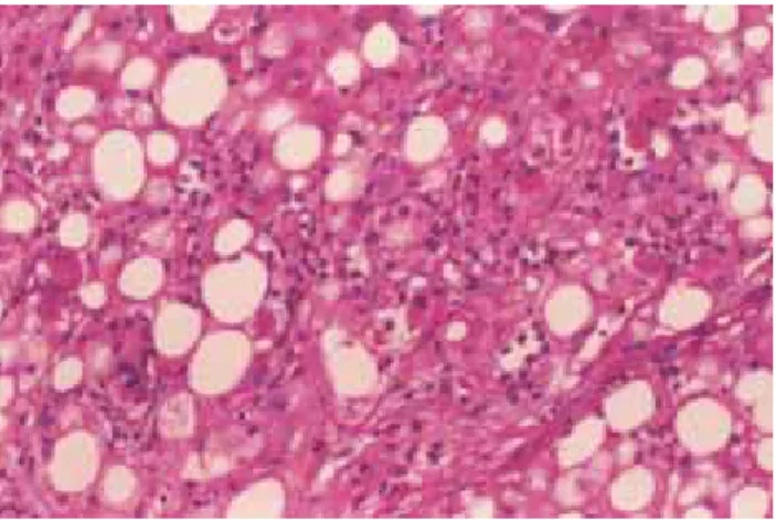
Steatosis, described as macrovesicular, microvesicu-lar, or mixed, is the earliest histopathological manifesta-tion of ALD. Macrosteatosis, more common in alcohol-ic steatohepatitis, is characterized by single large vacu-oles within hepatocytes that displace the cytoplasm and nucleus. It occurs as a result of metabolic imbalances in lipid delivery or synthesis within the liver, intracellular oxidation, and export from hepatocytes resulting in trig-lyceride accumulation. A mild degree of macrovesicular steatosis is a common finding in most liver biopsies
(Figure 3).40,41
Lipogranulomas are comprised of single or multiple fat globules surrounded by chronic inflammatory cells and Kupffer cells, sometimes with a few eosinophils.42
The presence of Mallory’s hyaline in noncirrhotic zone 3 liver cells is characteristic of ALD. The presence of megamitochondria in a liver with fat may indicate chron-ic alcohol abuse. They are observed by light mchron-icroscopy as round or cigar-shaped (Figure 4).43
Table II. CAGE Questionnaire.
• Have you ever felt the need to cut down on drinking? • Have you ever felt annoyed by criticism of your drinking? • Have you ever felt guilty about your drinking?
• Have you ever taken a morning eye opener?
Interpretation: Two “yes” answers are considered a positive screen. One “yes” answer should arouse a suspicion of alcohol abuse.
Modified from Mayfield D et al. The CAGE questionnaire: validation of a new alcoholism screening instrument. Am J Psychiatry 1974;131:1121.
Figure 4. Numerous neutrophils surround hepatocytes in various stages
of injury. Many cells are ballooned and contain Mallory bodies: Mega-mitochondria, wich are generally round and smaller than Mallory bo-dies, can be found in some of the ballooned cells.
Figure 3. The hepatocellular fat is largely macrovesicular. The edge of
the nodule appears irregular due to infiltration by inflammatory cells, fibrous tissue and ductular metaplasia.
Figure 5. High power view showing hepatocellular degeneration, fat,
MG
36
edigraphic.com
For a diagnosis of steatohepatitis, features of liver cell injury should be identified in a liver biopsy. The most com-mon form of injury is ballooning degeneration,
character-ized by swollen hepatocytes with rarefied cytoplasm, typi-cally found in zone 3. Other common findings include sep-ta of bridging necrosis and acidophil bodies, which are
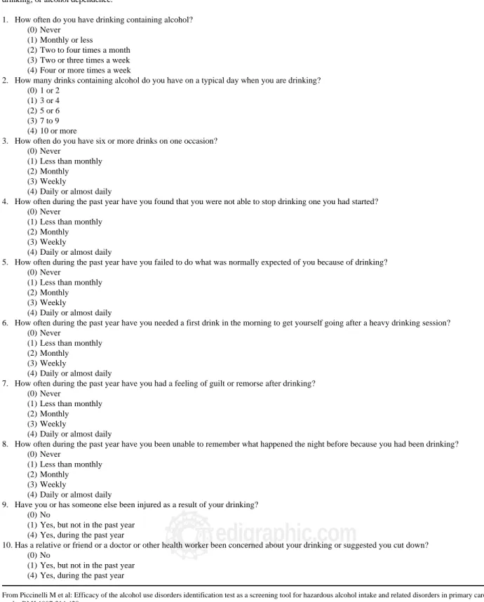
his-Table III. The alcohol use disorder identification TEST (AUDIT).
Scores for response categories are given in parentheses. Scores range from 0 to 40, with a cutoff score of > 5 indicating hazardous drinking, harmful drinking, or alcohol dependence.
1. How often do you have drinking containing alcohol? (0) Never
(1) Monthly or less
(2) Two to four times a month (3) Two or three times a week (4) Four or more times a week
2. How many drinks containing alcohol do you have on a typical day when you are drinking? (0) 1 or 2
(1) 3 or 4 (2) 5 or 6 (3) 7 to 9 (4) 10 or more
3. How often do you have six or more drinks on one occasion? (0) Never
(1) Less than monthly (2) Monthly (3) Weekly
(4) Daily or almost daily
4. How often during the past year have you found that you were not able to stop drinking one you had started? (0) Never
(1) Less than monthly (2) Monthly (3) Weekly
(4) Daily or almost daily
5. How often during the past year have you failed to do what was normally expected of you because of drinking? (0) Never
(1) Less than monthly (2) Monthly (3) Weekly
(4) Daily or almost daily
6. How often during the past year have you needed a first drink in the morning to get yourself going after a heavy drinking session? (0) Never
(1) Less than monthly (2) Monthly (3) Weekly
(4) Daily or almost daily
7. How often during the past year have you had a feeling of guilt or remorse after drinking? (0) Never
(1) Less than monthly (2) Monthly (3) Weekly
(4) Daily or almost daily
8. How often during the past year have you been unable to remember what happened the night before because you had been drinking? (0) Never
(1) Less than monthly (2) Monthly (3) Weekly
(4) Daily or almost daily
9. Have you or has someone else been injured as a result of your drinking? (0) No
(1) Yes, but not in the past year (4) Yes, during the past year
10. Has a relative or friend or a doctor or other health worker been concerned about your drinking or suggested you cut down? (0) No
(1) Yes, but not in the past year (4) Yes, during the past year
37
edigraphic.com
tological markers of apoptotic necrosis. There may be lob-ular infiltrates of polymorphonuclear leukocytes within the sinusoids and adjacent to injured liver cells. These are con-sidered to be an immune-mediated feature causing direct cytopathic damage to hepatocytes by the release of neutro-phil lysosomal granules (Figure 5). Mononuclear cell in-flammation of a mild degree may also be noted and some-times represents a resolution. Somesome-times there is a deposi-tion of collagen in the space of Disse (perisinusoidal fibrosis), initially located in acinar zone 3; with progres-sion to fibrosis, septum formation, and linkage between the central and portal regions, eventual cirrhosis may occur (micronodular, macronodular, or mixed). The presence of activated hepatic stellate cells in alcoholic steatohepatitis has been documented. Lesions of the hepatic veins are characteristic features of ALD. These include thickened veins and perisinusoidal fibrosis.44
Diagnosis
The revised hepatotoxic threshold in which ALD de-velops is 40 g (four drinks) and 20 g (two drinks) daily for men and women, respectively.14,45
All physicians should ask their patients about alcohol use. Standard validated questionnaires are more effective in the detection and diagnosis of ALD than routine clini-cal or laboratory tests. The usual questionnaire is the CAGE questionnaire (Table II). A positive answer for two or more questions indicates a positive result. A more recent test is the Alcohol Use Disorders Identification Test (AUDIT) published by the World Health Organiza-tion (Table III). Patients who admit to drinking over the toxic threshold or who have positive responses to ques-tionnaires should undergo a more complete evaluation for alcohol use and dependence.46 According to the American
Psychiatric Association’s Diagnostic and Statistical Man-ual of Mental Disorders (4th edition) (DSM-IV), at least
one of the following criteria is required for a diagnosis of alcohol abuse: failure to fulfill social obligations; recur-rent substance use in physically hazardous situations; re-current legal problems; and continued use despite alco-hol-related interpersonal or social problems. For alcohol dependence, at least three of the following criteria must be present: tolerance; withdrawal symptoms; use of alco-hol in larger quantities than intended; continued desire to control use or cut down; significant time lost because of obtaining, using, or recovering from alcohol; sacrifice of social, occupational, or recreational tasks; and continued use despite physical and psychological problems. These conditions should alert clinicians to look for clinical and laboratory evidence of ALD.47
The presentation of ALD correlates with the degree of liver injury. Alcoholic fatty liver has a milder clinical pre-sentation than alcoholic hepatitis or cirrhosis. Fatty liver occurs after short-term binge drinking and is the hallmark of acute ingestion. Patients are usually asymptomatic and have normal to mildly abnormal liver tests. Once alcohol ingestion ceases, the steatosis typically resolves. In ap-proximately 20% of patients, long-term alcohol consump-tion in those with alcoholic fatty liver may result in liver fibrosis and cirrhosis.13 Physical examination may reveal
jaundice, parotid hypertrophy, spider angiomata, gyneco-mastia, palmar erythema, Dupuytren’s contracture, and testicular atrophy. Table IV shows some of the physical signs associated with cirrhosis.
Patients with alcoholic hepatitis may be asymptomatic or may present with fever, jaundice, hepatomegaly, and occasionally ascites, portal hypertensive bleeding, and hepatic encephalopathy. On laboratory examination, leu-kocytosis is often present. A serum AST/ALT ratio great-er than 2.0 helps to diffgreat-erentiate ALD from othgreat-er livgreat-er diseases. Low ALT elevation is the result of a hepatic de-ficiency of pyridoxal-6-phosphate, necessary for ALT en-zymatic activity. Approximately 70% of patients with al-coholic hepatitis have cirrhosis.48,49 The clinical and
labo-ratory features (AST/ALT ratio, gamma glutamyl transferase [GGT], mean corpuscular volume [MCV]) are approximately 79% sensitive and 98% specific for the di-agnosis of ALD.
Table VI. Treatment for alcoholic liver disease.
• Abstinence • Nutritional support • Steroids if DF > 32
• S-adenosylmethionine (SAM) • Orthotopic liver transplant
Modified from Méndez-Sánchez N, Uribe M. Conceptos actuales en hepatología Masson Doyma México. 2003:259-264.
Table V. Diagnostic tests for ALD.
• Relation AST/ALT = 1.5 to 2 • Elevation of GGT
• Albumin diminished • Thrombin time prolongation
• Elevation of medium corpuscular volume • Thrombocytopenia
Modified from Méndez-Sánchez N, Uribe M. Conceptos actuales en hepatología Masson Doyma México. 2003:259-264.
Table IV. Physical signs in cirrhosis.
• Ascites • Splenomegaly • Spider angiomata • Dupuytren’s contractures • Palmar erythema • Hipocratic fingers • Periferic neuropathy • Asterixis
• Gynecomastia • Testicular atrophy
MG
38
edigraphic.com
Ultrasonography or computed tomography may be use-ful for the evaluation of cirrhosis and portal hypertension. On ultrasound, the features of cirrhosis and portal hyper-tension include liver nodules, sluggish or reversed portal vein flow, splenomegaly, and intraabdominal varices.
Patients with alcoholic cirrhosis have clinical features similar to patients with cirrhosis from other causes. Labo-ratory abnormalities are suggestive of liver synthetic dys-function, such as coagulopathy, hypoalbuminemia, and hyperbilirubinemia. In decompensated patients, the pre-sentation includes ascites, encephalopathy, and portal hy-pertensive bleeding.
In some cases, liver biopsy is needed prior to treatment to confirm the diagnosis of alcoholic hepatitis when alter-native diagnoses are suspected. It is also useful before a decision is taken to treat with corticosteroids or other ther-apies, and it provides prognostic information on the severi-ty of liver pathology.50 In patients with ALD and viral
hep-atitis, a liver biopsy is useful to determine the degree of necroinflammatory features, to make treatment decisions, and to obtain prognostic information: patients with ALD and hepatitis C have more significant liver disease, higher mortality, and a higher risk of hepatocellular carcinoma.51
Chronic alcohol consumption reduces the number of carbohydrate moieties attached to transferrin, leading to carbohydrate-deficient transferrin (CDT). Compared with other tests for alcohol use, the CDT test is more sensitive and specific for alcohol abuse. The limitations of CDT testing include wide ranges in sensitivity and specificity, and levels are affected by changes in serum transferrin or by hypertension, primary biliary cirrhosis, hepatic malig-nancy, chronic viral hepatitis, or decompensated liver dis-ease.52 Compounds of alcohol metabolism, such as
eryth-rocyte acetaldehyde and acetaldehyde adducts, have been shown to be effective markers for alcohol consumption but are not specific enough for clinical use.53
Treatment
Abstinence is the fundamental therapy for alcohol abuse. There is substantial evidence for the role of absti-nence in the improvement of the end-organ effects of al-cohol abuse in the heart, brain, pancreas, gastrointestinal tract, and reproductive organs. Continuing alcohol con-sumption is the best predictive factor of poor outcome. The diverse presentations of ALD (fatty liver, alcoholic hepatitis, and cirrhosis) are likely to improve or stabilize with the cessation of alcohol use and concurrent attention to malnutrition.54 Malnutrition in ALD patients should be
corrected with a diet of protein equivalent to 25–35 kcal and 1.25 g per kilogram of ideal body weight.8 Studies
have demonstrated that patients who consumed at least 2,500 kcal per day had improvements in six-month sur-vival, liver function, and nutritional status compared with a group that had a lower voluntary intake of calories.55
Enteral nutrition feeding is safe, maximizes digestion,
and may improve long-term survival in patients with ALD.56 An analysis of seven studies involving 239 ALD
patients concluded that parenteral nutrition treatment for periods of up to 30 days may improve liver function and nitrogen balance while normalizing the composition of plasma amino acids, but the long-term metabolic effects, risks, and benefits remain uncertain.57
Amino acid formulas enriched in branched-chain ami-no acids (BCAA; leucine, valine, and isoleucine) appear to confer an advantage by competing with aromatic ami-no acids for blood–brain barrier transport, improving he-patic encephalopathy, and stimulating protein synthesis, which could benefit the resolution of ascites. To date, there have been no studies comparing the potential bene-fits of BCAA with standard amino acid formulations in parenteral or enteral formulas.58
Obesity is a risk factor for the progression to ALD. However, aggressive weight reduction may not help in the treatment of alcoholic fatty liver and can be associat-ed with a worsening of liver injury.59
Treatment with S-adenosylmethionine (SAM) prevents the depletion of SAM and GSH levels and ameliorates liv-er injury, including fibrosis. The protective effects are me-diated via increased GSH levels. The administration of SAM prevents GSH uptake into the mitochondria. SAM also reduces liver injury by inhibiting TNF-α release from macrophages, either by itself or via its nucleoside metabo-lite 5-methylthioadenosine (MTA).60,61 SAM also attenu-ates the production of inducible nitric oxide synthase and the activation of NF-κβ. It also induces the expression and production of cytoprotective interleukin-10 (IL-10). SAM can also reduce alcoholic liver injury by lowering levels of homocysteine, which may be implicated directly in hepato-cellular apoptosis. Depletion of SAM may lead to a reduc-tion in phosphatidylcholine. The administrareduc-tion of phos-phatidylcholine has been advocated for the prevention and treatment of ALD because of its antifibrotic effects, which include a decrease in the transformation of intrahepatic li-pocytes to collagen-producing stellate cells, the induction of collagenase activity, the suppression of platelet-derived growth factor (PDGF), induced proliferation of hepatic stellate cells, protection against oxidant stress, and sup-pression of CYP2E1 induction.62,63
Betaine also facilitates phosphatidylcholine generation and very low-density lipoprotein export from the liver, and it offers the additional benefit of decreasing serum homocysteine levels.64 Antioxidants exert beneficial
ef-fects on hepatocytes by desensitizing them to oxidant stress, while inhibiting the priming mechanism for the ex-pression of proinflammatory and cytotoxic mediators via the suppression of NF-κβ.65 General measures
recom-mended for patients with ALD include the management of insulin resistance and the treatment of alcohol with-drawal symptoms with benzodiazepines.
hyperbiliru-39
edigraphic.com
binemia above 5 mg/dL, and coagulopathy) have a poor prognosis, with mortality rates as high as 50% in one year. The use of corticosteroids in selected patients with severe alcoholic hepatitis has been supported. The useful-ness of a Maddrey Discriminant Factor (MDF) score (4.6 × [prolongation of the prothrombin time in seconds] + to-tal serum bilirubin in mg/dL) greater than 32 in predicting increased mortality in alcoholic hepatitis has been vali-dated.66 A recent meta-analysis concluded that
corticos-teroids improved the short-term survival of patients with severe alcoholic hepatitis by approximately 20%. Corti-costeroids are given as oral prednisolone (32 mg) or pred-nisone and methylprednisolone (40 mg) daily for four weeks, followed by a four-week taper. The improvement in survival, lack of serious immediate side effects or com-plications, and the low relative cost support the use of corticosteroids.67,68
Pentoxifylline (400 mg orally three times daily) is an inhibitor of TNF-α and other cytokines. It offers an portant option for the protection of renal function and im-provement in short-term survival in severe alcoholic hep-atitis.69 Pilot studies have shown that neutralizing
anti-TNF-α agents (infliximab and etanercept) potentially interfere with an important mechanistic pathway of ALD. Excessive consumption of alcohol leads to the genera-tion of oxidative cellular stress and injury from increased endotoxemia, cytokine release, and membrane-bound lip-id peroxlip-idation. There is a broad spectrum of orally ad-ministered agents with antioxidant properties. Small pilot studies using N-acetylcysteine or vitamin E have shown no effect of these antioxidants in the context of severe al-coholic hepatitis. The long-term use of antioxidants may have a greater role in the treatment of cirrhotic disease, with the potential to prevent a decompensated state and the development of hepatocellular carcinoma.29
The cellular physiology and blood flow of the hepatic acinus leads to a relatively oxygen-poor microenviron-ment within the pericentral hepatocytes (zone 3) and an area with greater sensitivity to drug-induced, ischemic, and alcohol-induced injury. A “hypermetabolic” state is implicated in the pathogenesis of zone 3 alcoholic injury. Suppression of thyroid activity with propylthiouracil (PTU) has been proposed as a treatment for alcoholic in-jury. A systematic review combining the results of six randomized clinical trials demonstrated that PTU had no significant effect on mortality or liver-related mortality relative to the effects of placebos. Accordingly, there is no evidence to support the use of PTU for ALD.70
Some recent reports have demonstrated the regression or loss of hepatic fibrosis. Recognition of the importance of hepatic stellate cells and Kupffer cells as keys to the pro-gression and repro-gression of liver scarring offers new oppor-tunities for cirrhosis therapies.71 Recently, clarification of
the roles of leptin and adiponectin in insulin resistance and in the progression to chronic liver disease make these hor-mones candidates for future trials in patients with ALD.
Orthotopic liver transplantation (OLT) is the only ef-fective treatment for patients with terminal ALD. Cur-rently, ALD is the second leading indication for trans-plantation in the United States.72 Any patient with
decom-pensated liver disease without contraindications for transplantation can be listed for OLT. Current or recent substance abuse is a contraindication for OLT. Protein-calorie malnutrition is a common comorbidity in alcohol-ic patients and patients with end-stage liver disease, and it presents a significant risk of a poor outcome. Psychiatric diseases commonly coexist with alcohol dependence. Personality disorders, depression, anxiety, or psychoses require psychiatric involvement.73 Significant
cardiomy-opathy is another contraindication for OLT. Current rec-ommendations suggest pre-OLT cardiovascular assess-ment for symptomatic high-risk individuals with risk fac-tors for coronary artery disease.74 Drinking relapse after
OLT is common. Short-term recidivism rates range from 10–15% and cumulative relapse rates of 30–50% have been reported. The best predictor of continued sobriety after transplantation is a documented period of abstinence of six months or longer. Patient and graft survival rates after transplantation are similar to those of non-ALD pa-tients, with 66–82% five-year survival.75 Table VI shows
the actual and available treatments for ALD.
Prognosis
As mentioned above, evidence suggests that with the ingestion of more than 60–80 g/d of alcohol in men and more than 20 g/d in women, there is an increased risk of cirrhosis. However, Bellentani et al. showed that only 13.5% of patients with extremely high daily alcohol in-take (120 g/d) developed alcohol-induced liver dam-age.2,18 Alcohol consumption is necessary but not
suffi-cient for the development of fibrosis in alcoholic patients. Cirrhosis occurs in only 8–20% and alcoholic hepatitis in 6–30% of alcoholic patients. Older, overweight, female, hyperglycemic, or iron-overloaded patients exhibit the risk factors for progression to fibrosis and cirrhosis. Age and female sex are independent risk factors for the devel-opment of alcohol-induced cirrhosis.
The long-term prognosis for patients with cirrhosis im-proves with abstinence. The five-year survival in com-pensated cirrhosis patients who continue to drink is less than 70% compared with a survival as high as 90% if they abstain from further alcohol intake. In patients with dec-ompensated cirrhosis, the five-year survival rate is 60% in abstinent individuals but drops to less than 30% in those who continue to drink. Overall five-year survival in cirrhosis patients who continue drinking is approximately 35%.76
se-MG
40
edigraphic.com
vere liver disease is mainly dependent on the cessation or persistence of drinking, but abstinence from ethanol does not completely prevent the development of cirrhosis.
Women have an increased susceptibility to the detri-mental effects of alcohol. They are considered to be at in-creased risk of cirrhosis with a daily alcohol intake great-er than 20–60 g. Progression to cirrhosis in women who abstain may not necessarily be related to more severe or more extended liver damage. Some unrecognized factors related to sex might be responsible for the unfavorable course of alcoholic hepatitis in some women even if they stop drinking. Gastric ADH is less active in women than in men, and its activity seems to be depressed in both al-coholic women and men.7,8,77,78 Other mechanisms
under-lying the increased susceptibility to alcohol-related liver injury in women include differences in endotoxin levels and gut permeability, the effect of estrogens and andro-gens on endotoxin–alcohol-mediated liver injury, differ-ences in alcohol elimination rates either because of varia-tions in first-pass metabolism or enzymatic activity, and differences in the volumes of distribution and peak blood alcohol levels.79,80
The role of obesity as a risk factor in the development of alcoholic cirrhosis is unclear. Evidence indicates that in al-coholic patients, being overweight for at least 10 years cor-relates independently with the occurrence of cirrhosis. Obesity is associated with steatohepatitis, and alcohol in-take may increase liver damage in obese individuals.81
In addition to the directly toxic effects of alcohol and genetically based differences in alcohol-metabolizing en-zymes, there are other genetically and environmentally determined factors that may predispose individuals to the development of ALD: obesity, genetically determined polymorphisms in cytokines and their receptors, immune mechanisms, patterns of alcohol consumption, concurrent viral infections, and the use of drugs.82
The long-term survival of alcoholic patients is ad-versely affected by the lesions that characterize alcohol steatosis and steatohepatitis. Histological lesions premon-itory for the progression to cirrhosis include widespread liver cell necrosis and pericellular fibrosis, the formation of fibrous septa, widespread hepatic venular obstruction, and diffuse Mallory’s hyaline. Some lesions are revers-ible with abstinence from alcohol. The risk of developing cirrhosis is increased when steatohepatitis is present. Ste-atohepatitis may be considered the rate-limiting step in the progression to cirrhosis, and mortality due to alcohol-ic cirrhosis is adversely affected by its presence.83
The MDF (= 4.6 × [prothrombin time (PT) in seconds – control PT)] + serum bilirubin in mg/dL) was intro-duced in 1978 as a tool for predicting the risk of mortali-ty from alcoholic hepatitis. Patients with a MDF score of more than 32 have a mortality rate exceeding 50% and corticosteroid treatment is suggested for this group of pa-tients.65 The model for end-stage liver disease (MELD) is
a survival model based on three laboratory variables:
se-rum creatinine (Cr), sese-rum bilirubin, and the international normalized ratio (INR) using the following formula: MELD = 9.57 × loge(Cr mg/dL) + 3.78 × loge(bilirubin mg/dL) + 11.20 × loge(INR) + 6.43.84 In a study
undertak-en to compare the prognostic validity of MELD and MDF in alcoholic hepatitis, both were similarly valid for 30-day and 90-30-day mortality, but MELD emerged as the only independent predictor of 90-day mortality. That study identified a MELD score of 21 as having the highest sen-sitivity and specificity in predicting mortality, with an es-timated 90-day mortality of 20% for patients with this score.85
Physical examination for signs of encephalopathy or ascites may be useful in screening for mortality in alco-holic hepatitis. Alcohol can be considered both a primary cause of and a cofactor in the development of hepatocel-lular carcinoma (HCC). The risk of HCC increases when ethanol intake exceeds 60 g/d for more than 10 years. The rate of HCC increases significantly in patients with alco-hol use and hepatitis C. Alcoalco-hol and hepatitis C interact in the development of HCC.86
References
1. Corrao G, Ferrari P, Zambon A, Torchio P. Are the recent trends in liver cirrhosis mortality affected by the changes in alcohol consump-tion? Analysis of latency period in European countries. J Stud
Alco-hol 1997; 58: 486-494.
2. Bellentani S, Saccoccio G, Costa G, Tiribelli C, Manenti F, Sodde M, Saveria Croce L, Sasso F, Pozzato G, Cristianini G, Brandi G. Drinking habits as cofactors of risk for alcohol induced liver damage. The Dionysos Study Group. Gut 1997; 41: 845-850.
3. Dawson DA, Grant BF, Chou SP, Pickering RP. Subgroup variation in U.S. drinking patterns: results of the 1992 national longitudinal alcohol epidemiologic study. J Subst Abuse 1995; 7: 331-344. 4. Robertson I. Safe drinking. BMJ 1994; 308: 346.
5. Campollo O, Martínez MD, Valencia JJ, Segura-Ortega J. Drinking patterns and beverage preferences of liver cirrhosis patients in Mexico.
Subst Use Misuse 2001; 36: 387-398.
6. Rehm J, Rehn N, Room R, Monteiro M, Gmel G, Jernigan D, Frick U. The global distribution of average volume of alcohol consumption and patterns of drinking. Eur Addict Res 2003; 9: 147-156. 7. Méndez-Sánchez N, Uribe M. Conceptos actuales en hepatología.
Masson Doyma México. 2003: 259-264.
8. Saunders JB, Aasland OG, Amundsen A, Grant M. Alcohol consump-tion and related problems among primary health care patients: WHO collaborative project on early detection of persons with harmful alco-hol consumption—I. Addiction 1993; 88: 349-362.
9. Mendez-Sanchez N, Aguilar-Ramirez JR, Reyes A, Dehesa M, Juarez A, Castaneda B, Sanchez-Avila F, Poo JL, Guevara Gonzalez L, Lizardi J, Valdovinos MA, Uribe M, Contreras AM, Tirado P, Aguirre J, Rivera-Benitez C, Santiago-Santiago R, Bosques-Padilla F, Munoz L, Guerrero A, Ramos M, Rodriguez-Hernandez H, Jacobo-Karam J. Grupo de Estudio, Asociación Mexicana de Hepatologia. Etiology of liver cirrhosis in Mexico. Ann Hepatol 2004; 3: 30-33.
41
edigraphic.com
12. Leevy CM. Fatty liver: a study of 270 patients with biopsy provenfatty liver and review of the literature. Medicine 1962; 41: 249-276. 13. Sorensen TI, Orholm M, Bentsen KD, Hoybye G, Eghoje K, Christoffersen P. Prospective evaluation of alcohol abuse and alco-holic liver injury in men as predictors of development of cirrhosis.
Lancet 1984; 2: 241-244.
14. Teli MR, Day CP, Burt AD, Bennett MK, James OF. Determinants of progression to cirrhosis or fibrosis in pure alcoholic fatty liver.
Lancet 1995; 346: 987-990.
15. McCullough AJ, O´Connor JF. Alcoholic liver disease: proposed rec-ommendations for the American College of Gastroenterology. Am J
Gastroenterol 1998; 93: 2022-2036.
16. Brady KT, Randall CL. Gender differences in substance use disor-ders. Psychiatr Clin North Am 1999; 22: 241-252.
17. Blume SB. Women and alcohol. A review. JAMA 1986; 256: 1467-1470. 18. Norton R, Batey R, Dwyer T, MacMahon S. Alcohol consumption and the risk of alcohol related cirrhosis in women. Br Med J (Clin
Res Ed) 1987; 295: 80-82.
19. Adams WL, Yuan Z, Barboriak JJ, Rimm AA. Alcohol-related hos-pitalizations of elderly people. Prevalence and geographic variation in the United States. JAMA 1993; 270: 1222-1225.
20. Stinson FS, Grant BF, Dufour MC. The critical dimension of ethnicity in liver cirrhosis mortality statistics. Alcohol Clin Exp Res 2001; 25: 1181-1187.
21. Caetano R, Kaskutas LA. Changes in drinking patterns among whites, blacks and Hispanics, 1984-1992. J Stud Alcohol 1995; 56: 558-565. 22. Degos F. Hepatitis C and alcohol. J Hepatol 1999; 31 Suppl 1: 113-118. 23. Ostapowicz G, Watson KJ, Locarnini SA, Desmond PV. Role of al-cohol in the progression of liver disease caused by hepatitis C virus infection. Hepatology 1998; 27: 1730-1735.
24. Mendenhall CL, Anderson S, Weesner RE, Goldberg SJ, Crolic KA. Protein-calorie malnutrition associated with alcoholic hepatitis. Vet-erans Administration Cooperative Study Group on Alcoholic Hepati-tis. Am J Med 1984; 76: 211-222.
25. Suter PM, Schutz Y, Jequier E. The effect of ethanol on fat storage in healthy subjects. N Engl J Med 1992; 326: 983-987.
26. Cravo ML, Gloria LM, Selhub J, Nadeau MR, Camilo ME, Resende MP, Cardoso JN, Leitao CN, Mira FC. Hyperhomocysteinemia in chronic alcoholism: correlation with folate, vitamin B-12, and vita-min B-6 status. Am J Clin Nutr 1996; 63: 220-224.
27. Reuler JB, Girard DE, Cooney TG. Current concepts. Wernicke´s encephalopathy. N Engl J Med 1985; 312: 1035-1039.
28. Leo MA, Lieber CS. Hepatic vitamin A depletion in alcoholic liver injury. N Engl J Med 1982; 307: 597-601.
29. Tsukamoto H, Lu SC. Current concepts in the pathogenesis of alco-holic liver injury. FASEB J 2001; 15: 1335-1349.
30. Colell A, García-Ruiz C, Miranda M, Ardite E, Mari M, Morales A, Corrales F, Kaplowitz N, Fernandez-Checa JC. Selective glutathione depletion of mitochondria by ethanol sensitizes hepatocytes to tumor necrosis factor. Gastroenterology 1998; 115: 1541-1551.
31. Cahill A, Stabley GJ, Wang X, Hoek JB. Chronic ethanol consump-tion causes alteraconsump-tions in the structural integrity of mitochondrial DNA in aged rats. Hepatology 1999; 30: 881-888.
32. McCullough AJ, Bugianesi E, Marchesini G, Kalhan SC. Gender-dependent alterations in serum leptin in alcoholic cirrhosis.
Gastro-enterology 1998; 115: 947-953.
33. Sarphie TG, D´Souza NB, Spitzer JJ, Deaciuc IV. Chronic alcohol feeding in liquid diet or in drinking water has similar effects on elec-tron microscopic appearance of the hepatic sinusoid in the rat. Alchol.
Clin Exp Res 1996; 20: 973-979.
34. Nanji AA, Griniuviene B; Yacoub LK, Fogt F, Tahan SR. Intercellu-lar adhesion molecule-1 expression in experimental alcoholic liver disease: relationship to endotoxemia and TNF alpha messenger RNA.
Exp Mol Pathol 1995; 62: 45-51.
35. Nieto N, Friedman SL, Greenwel P, Cederbaum AI. CYP2E1-medi-ated oxidative stress induces collagen type I expression in rat hepatic stellate cells. Hepatology 1999; 30: 987-996.
36. Higuchi S, Matsushita S, Muramatsu T, Murayama M, Hayashida M. Alcohol and aldehyde dehydrogenase genotypes and drinking behav-ior in Japanese. Alcohol Clin Exp Res 1996; 20: 493-497.
37. MacSween RN, Burt AD. Histologic spectrum of alcoholic liver dis-ease. Semin Liver Dis 1986; 6: 221-232.
38. Baptista A, Bianchi L, de Groole. Alcoholic liver disease: morpho-logical manifestations. Review by an international group. Lancet 1981; 1: 707-711.
39. Brunt EM. Nonalcoholic steatohepatitis: definition and pathology.
Semin Liver Dis 2001: 21: 3-16.
40. MacSween RN, Burt AD. Histologic spectrum of alcoholic liver dis-ease. Semin Liver Dis 1986; 6: 221-232.
41. Brunt EM, Janney CG, Di Bisceglie AM, Neuschwander-Tetri BA, Bacon Br. Nonalcoholic steatohepatitis: a proposal for grading and staging the histological lesions. Am J Gastroenterol 1999; 94: 2467-2474.
42. Takahashi T, Kamimura T, Ichida F. Ultrastructural findings on poly-morphonuclear leucocyte infiltration and acute hepatocellular dam-age in alcoholic hepatitis. Liver 1987; 7: 347-358.
43. French SW, Nash J, Shitabata P, Kachi K, Hara C, Chedid A, Mendenhall CL. Pathology of alcoholic liver disease. VA Coopera-tive Study Group. Semin Liver Dis 1993; 13: 154-169.
44. Goodman ZD, Ishak KG. Occlusive venous lesions in alcoholic liver disease. A study of 200 cases. Gastroenterology 1982; 83: 786-796. 45. Becker U, Deis A, Sorensen TI, Gronbaek M, Borch-Johnsen K, Muller CF, Schnohr P, Jensen G. Prediction of risk of liver disease by alco-hol, intake, sex, and age: a prospective population study. Hepatology 1996; 23: 1025-1029.
46. Hoeksema HL, de Bock GH. The value of laboratory tests for the screening and recognition of alcohol abuse in primary care patients. J
Fam Pract 1993; 37: 268-276.
47. American Psychiatric Association. Diagnostic and Statistical Manual
of Mental Disorders (DSM-IV-TR), 4th ed. Washington, D. C.:
Ameri-can Psychiatric Association; 2000.
48. Mendenhall CL. Alcoholic hepatitis. Clin Gastroenterol 1981; 10: 417-441.
49. Williams AL, Hoofnagle JH. Ratio of serum aspartate to alanine aminotransferase in chronic hepatitis. Relationship to cirrhosis.
Gas-troenterology 1988; 95: 734-739.
50. Orrego H, Blake JE, Blendis LM, Medline A. Prognosis of alcoholic cirrhosis in the presence and absence of alcoholic hepatitis.
Gastro-enterology 1987; 92: 208-214.
51. Tanaka T, Yabusako T, Yamashita T, Kondo K, Nishiguchi S, Kuroki T, Monna T. Contribution of hepatitis C virus to the progression of alco-holic liver disease. Alcohol Clin Exp Res 2000; 24(4 Suppl): 112S-116S. 52. DiMartini A, Day N, Lane T, Beisler AT, Dew MA, Anton R. Carbo-hydrate deficient transferrin in abstaining patients with end-stage liver disease. Alcohol Clin Exp Res 2001; 25: 1729-1733.
53. Uppal R, Rosman A, Hernandez R, Baraona E, Lieber CS. Effects of liver disease on red blood cell acetaldehyde in alcoholics and non-alcoholics. Alcohol Alcohol Suppl 1991; 1: 323-326.
54. Pessione F, Ramond MJ, Peters L, Pham BN, Batel P, Rueff B, Valla DC. Five-year survival predictive factors in patients with excessive alcohol intake and cirrhosis. Effect of alcoholic hepatitis, smoking and abstinence. Liver Int 2003; 23: 45-53.
55. Mendenhall CL, Moritz TE, Roselle GA, Morgan TR, Nemchausky BA, Tamburro CH, Schiff ER, McClain CJ, Marsano LS, Allen JI. A study of oral nutritional support with oxandrolone in malnourished patients with alcoholic hepatitis: results of a Department of Veterans Affairs cooperative study. Hepatology 1993; 17: 564-576. 56. Soberon S, Pauley MP, Duplantier R, Fan A, Halsted CH. Metabolic
effects of enteral formula feeding in alcoholic hepatitis. Hepatology 1987; 7: 1204-1209.
57. Nompleggi DJ, Bonkovsky HL. Nutritional supplementation in chronic liver disease: an analytical review. Hepatology 1994; 19: 518-533. 58. Marchesini G, Bianchi G, Merli M, Amodio P, Panella C, Loguercio
C, Rossi Fanelli F, Abbiati R. Italian BCAA Study Group. Nutri-tional supplementation with branched-chain amino acids in advanced cirrhosis: a double-blind, randomized trial. Gastroenterology 2003; 124: 1792-1801.
MG
42
edigraphic.com
60. Colell A, Garcia-Ruiz C, Morales A, Ballesta A, Ookhtens M, RodesJ, Kaplowitz N, Fernandez-Checa JC. Transport of reduced glutathione in hepatic mitochondria and mitoplasts from ethanol-treated rats: ef-fect of membrane physical properties and S-adenosyl-L-methionine. Hepatology. 1997;26:699-708.
61. Watson WH, Zhao Y, Chawla RK. S-adenosyl-methionine attenu-ates the lipopolysaccharide-induced expression of the gene for tu-mour necrosis factor alpha. Biochem J 1999; 342(Pt 1): 21-25. 62. Brady LM, Fox ES, Fimmel CJ. Polyenylphosphatidylcholine
inhib-its PDGF-induced proliferation in rat hepatic stellate cells. Biochem
Biophys Res Commun 1998; 248: 174-179.
63. Aleynik MK, Leo MA, Aleynik SI, Lieber CS. Polyenylphosp hatidylcholine opposes the increase of cytochrome P-4502E1 by etha-nol and corrects its iron-induced decrease. Alcohol Clin Exp Res 1999; 23: 96-100.
64. Rai RM, Loffreda S, Karp CL, Yang SQ, Lin HZ, Diehl AM. Kupffer cell depletion abolishes induction of interleukin-10 and permits sus-tained overexpression of tumor necrosis factor alpha messenger RNA in the regenerating rat liver. Hepatology 1997; 25: 889-895. 65. Maddrey WC, Boitnott JK, Bedine MS, Weber FJ Jr, Mezey E, White
RI Jr. Corticosteroid therapy of alcoholic hepatitis. Gastroenterology 1978; 75: 193-199.
66. McClain CJ, Barve S, Deaciuc I, Hill DB. Tumor necrosis factor and alcoholic liver disease. Alcohol Clin Exp Res 1998; 22: 248S-252S. 67. Imperiale TF, McCullough AJ. Do corticosteroids reduce mortality
from alcoholic hepatitis? A meta-analysis of the randomized trials.
Ann Intern Med 1990; 113: 299-307.
68. Ramond MJ, Poynard T, Rueff B, Mathurin P, Theodore C, Chaput JC, Benhamou JP. A randomized trial of prednisolone in patients with severe alcoholic hepatitis. N Engl J Med 1992; 326: 507-512. 69. Akriviadis E, Botla R, Briggs W, Han S, Reynolds T, Shakil O.
Pentoxifylline improves short-term survival in severe acute alcoholic hepatitis: a double-blind, placebo-controlled trial. Gastroenterology 2000; 119: 1637-1648.
70. Rambaldi A, Gluud C. Propylthiouracil for alcoholic liver disease.
Cochrane Database Syst Rev 2002; CD002800.
71. Murphy F, Arthur M, Iredale J. Developing strategies for liver fibro-sis treatment. Expert Opin Investig Drugs 2002; 11: 1575-1585. 72. Neuberger J, Schulz KH, Day C, Fleig W, Berlakovich GA, Berenguer
M, Pageaux GP, Lucey M, Horsmans Y, Burroughts A, Hockerstedt K. Transplantation for alcoholic liver disease. J Hepatol 2002; 36: 130-137.
73. Howard L, Fahy T, Wong P. Sherman D, Gane E, Williams R. Psy-chiatric outcome in alcoholic liver transplant patients. QJM 1994; 87: 731-736.
74. Keeffe BG, Valantine H, Keeffe EB. Detection and treatment of coro-nary artery disease in liver transplant candidates. Liver Transpl 2001; 7: 755-761.
75. Fabrega E, Crespo J, Casafont F, De las Heras G, de la Pena J, Pons-Romero F. Alcoholic recidivism after liver transplantation for alco-holic cirrhosis. J Clin Gastroenterol 1998; 26: 204-206.
76. Diehl AM. Alcoholic liver disease: natural history. Liver Transpl Surg 1997; 3: 206-211.
77. Sato N, Lindros KO, Baraona E, Ikejima K, Mezey E, Jarvelainen HA, Ramchandani VA. Sex difference in alcohol-related organ in-jury. Alcohol Clin Exp Res 2001; 25 (5 Suppl ISBRA): 40S-45S. 78. Schenker S. Medical consequences of alcohol abuse: is gender a
fac-tor? Alcohol Clin Exp Res 1997; 21: 179-181.
79. Li TK, Beard JD, Orr WE, Kwo PY, Ramchandani VA, Thomasson HR. Variation in ethanol pharmacokinetics and perceived gender and ethnic differences in alcohol elimination. Alcohol Clin Exp Res 2000; 24: 415-416.
80. Naveau S, Giraud V, Borotto E, Aubert A, Capron F, Chaput JC. Excess weight risk factor for alcoholic liver disease. Hepatology 1997; 25: 108-111.
81. Yang SQ, Lin HZ, Lane MD, Clemens M, Diehl AM. Obesity in-creases sensitivity to endotoxin liver injury: implications for the pathogenesis of steatohepatitis. Proc Natl Acad Sci USA 1997; 94: 2557-2562.
82. Jarvelainen HA, Orpana A, Perola M, Savolainen VT; Karhunen PJ, Lindros KO. Promoter polymorphism of the CD14 endotoxin recep-tor gene as risk facrecep-tor for alcoholic liver disease. Hepatology 2001; 33: 1148-1153.
83. Marbet UA, Bianchi L, Meury U, Stalder, GA. Long-term histologi-cal evaluation of the natural history and prognostic factors of alco-holic liver disease. J Hepatol 1987; 4: 364-372.
84. Wiesner R, Edwards E, Freeman R, Harper A, Kim R, Kamath P, Kremers W, Lake J, Howard T, Merion RM, Wolfe RA, Krom R, United Network for Organ Sharing Liver Disease Severity Score Com-mittee. Model for end-stage liver disease (MELD) and allocation of donor livers. Gastroenterology 2003; 124: 91-96.
85. Dunn W, Jamil LH, Brown LS, Wiesner RH, Kim WR, Menon KV, Malinchoc M, Kamath PS, Shah V. MELD accurately pre-dicts mortality in patients with alcoholic hepatitis. Hepatology 2005; 41: 353-358.