Circulating levels of pentraxin-3
(PTX3) in patients with liver cirrhosis
Jéssica G. Pereira,* Telma Erotides Silva,† Emília T. O. Bansho,* Edelton F. Morato,‡ José T. Pinheiro,‡ Letícia Muraro-Wildner,§ Maria Luiza Bazzo,§ Esther Buzaglo Dantas-Corrêa,* Leonardo L. Schiavon,*,† Janaína L. Narciso-Schiavon*
* Núcleo de Estudos em Gastroenterologia e Hepatologia, Department of Internal Medicine, Federal University of Santa Catarina, Florianópolis, Santa Catarina, Brazil. † Postgraduate Program in Medical Sciences, Health Sciences Center, Federal University of Santa Catarina, Florianópolis, Santa Catarina, Brazil. ‡ Center for Assessment of Allergic Type Reactions to Drugs, University Hospital Polydoro Ernani de São Thiago - Federal University of Santa Catarina, Florianópolis, Santa Catarina, Brazil. § Clinical Analysis Laboratory, University Hospital Polydoro Ernani de São Thiago - Federal University of Santa Catarina, Florianópolis, Santa Catarina, Brazil.
September-October, Vol. 16 No. 5, 2017: 780-787
INTRODUCTION
Liver cirrhosis is the most advanced stage of chronic liver disease. It is characterized histologically by the pres-ence of regenerative nodules. Its prevalpres-ence is estimated at 0.27% in the United States,1 and it is associated with
mul-tiple etiologies, most commonly ethanol consumption, chronic viral hepatitis B and C and diabetes mellitus.1,2
In-hospital mortality for disease decompensation is 9.1% in South Korea3 and is 25.0% after 30 days of admission in
Brazil.4 Identifying patients with worse prognosis would
facilitate early management of potentially severe cases.
Several prognostic markers have been studied to identify mortality associated with decompensated cirrhosis, in-cluding the Model for End-Stage Liver Disease (MELD) score and its derivatives, Acute-on-Chronic Liver Failure (ACLF) score, Interleukins 2, 6 and 8 (2, 6 and IL-8, respectively), C-reactive protein (CRP) and even total leukocyte count.
Pentraxins are proteins formed by 5 monomers that form a ring in radial symmetry. They are a class of pattern recognition receptors. Among pentraxins, the main ones are pentraxin-3, CRP and serum amyloid P component. PTX3 is a long-chain pentraxin considered an acute phase
The Official Journal of the Mexican Association of Hepatology, the Latin-American Association for Study of the Liver and
the Canadian Association for the Study of the Liver
Manuscript received: Manuscript received: Manuscript received: Manuscript received:
Manuscript received: October 27, 2016. Manuscript accepted:Manuscript accepted:Manuscript accepted:Manuscript accepted:Manuscript accepted: March 20, 2017.
DOI:10.5604/01.3001.0010.2789.
A B S T R A C T A B S T R A C T A B S T R A C T A B S T R A C T A B S T R A C T
Background: Background:Background: Background:
Background: Despite the circulating levels of PTX3 were related to the severity of various diseases, there are no studies investi-gating its role in patients with liver cirrhosis. We aimed to study PTX3 levels in patients with liver cirrhosis. Material and meth-Material and meth-Material and meth-Material and meth-Material and meth-ods.
ods.ods. ods.
ods. A prospective cohort study included 130 patients hospitalized for acute decompensation of liver cirrhosis, 29 stable cirrhotic outpatients and 32 healthy controls evaluated in a tertiary hospital in Southern Brasil. Results.Results.Results.Results.Results. The median PTX3 level was signifi-cantly higher in stable cirrhotic patients compared to controls (2.6 vs. 1.1 ng/mL; p < 0.001), hospitalized cirrhotic patients compared to controls (3.8 vs. 1.1 ng/mL; p < 0.001), and hospitalized cirrhotic patients compared to stable cirrhotic patients (3.8 vs. 2.6 ng/ mL; p = 0.001). A positive correlation was found between PTX3 and serum creatinine (r = 0.220; p = 0.012), Chronic Liver Failure -Sequential Organ Failure Assessment score (CLIF-SOFA) (r = 0.220; p = 0.010), MELD (r = 0.279; p = 0.001) and Child-Pugh score (r = 0.224; p = 0.010). Significantly higher levels of PTX3 were observed in patients on admission with ACLF (8.9 vs. 3.1 ng/mL; p < 0.001) and MELD score ≥ 20 (6.6 vs. 3.4 ng/mL; p = 0.002). Death within 90 days occurred in 30.8% of patients and was associated with higher levels of PTX3 (5.3 vs. 3.4 ng/mL; p = 0.009). The probability of Kaplan-Meier survival was 77.0% in patients with PTX-3 < 5.3 ng mL (upper tercile) and 53.5% in those with PTX3 ≥ 5.3 ng/mL (p = 0.002). Conclusion.Conclusion.Conclusion.Conclusion.Conclusion. These re-sults indicate the potential for use of PTX3 as an inflammatory biomarker for the prognosis of patients with hepatic cirrhosis. Key words.
Key words.Key words. Key words.
marker produced mainly by endothelial and vascular smooth muscle cells at the site of inflammation. It is also produced by macrophages, fibroblasts, neutrophils, epi-thelial cells, dendritic cells and other cell types both near and far from the inflammation site,5,6 Pentraxin
produc-tion is influenced by certain inflammatory stimuli such as interleukin 1 beta (IL-1β) and tumor necrosis factor alfa (TNF-α).7 It differs considerably from CRP in terms of
expression patterns by affected organs. In particular, this is a short pentraxin mainly produced in the liver in response to IL-6.8
PTX3 has been recognized as an independent marker of inflammation associated with various disorders8,9 such as
atherosclerosis, cancer, respiratory diseases and central nervous system diseases in which increased levels are re-lated to the risk of the disease or its progression.10
Howev-er, according to our knowledge there are no studies that analyze its role in liver cirrhosis.
The aim of this study is to describe PTX3 levels in am-bulatory patients and hospitalized patients with liver cir-rhosis and their association with disease prognosis.
MATERIAL AND METHODS
Sample
This is a prospective cohort study with consecutive in-clusion of patients with liver cirrhosis treated at the hepa-tology ambulatory department and admitted to the emergency service of a tertiary hospital in southern Brazil between January 2011 and January 2014 because of disease decompensation. Patients were excluded from the study because of insufficient clinical and laboratory data in the medical records, hepatocellular carcinoma that did not meet Milan criteria and refusal to participate in the study.
During routine outpatient or emergency admission, subjects were asked to participate and sign the free and in-formed consent form. A family member or guardian would authorize data collection if the patient had encepha-lopathy grades III or IV. Clinical and laboratory variables were collected from interviews and from the medical records. The following clinical variables were studied: age, sex, skin color, etiology of cirrhosis and presence of ascites. Laboratory variables collected on admission in-cluded serum creatinine, MELD, Child-Pugh and Chron-ic Liver Failure - Sequential Organ Failure Assessment (CLIF-SOFA) scores, total leukocyte count, serum sodi-um, platelet count, international normalized ratio (INR), albumin, CRP and total bilirubin.
The diagnosis of cirrhosis was established either histo-logically when liver biopsy was available or by a combina-tion of clinical, imaging and laboratory data in patients with evidence of portal hypertension.
PTX3 assays were performed by Enzyme-Linked Im-munosorbent Assay with serum samples collected on ad-mission or at an outpatient visit and stored in a freezer at -80°C (ELISA; R&D Systems - Minneapolis, MN).
M e t h o d s
Patients were followed during hospitalization and 90-day mortality was assessed by telephone in case of dis-charge. The 90-day mortality rates did not include patients who underwent liver transplantation (because they were excluded from the study).
Individuals with suspected bacterial infection at ad-mission underwent clinical examination to confirm the diagnosis and establish the primary source of infection. The diagnosis of infection was performed according to the criteria of the Center for Disease Control.11
Diag-nostic paracentesis was performed for all patients with ascites present at admission. Hepatic encephalopathy was graded according to the criteria of West-Haven.12
If present, the precipitating factor was investigated and lactulose was administered with dose adjustment as needed.
The severity of liver disease was estimated by Child-Pugh13 and MELD scores14 calculated based on laboratory
tests performed on admission. ACLF and CLIF-SOFA were defined as proposed by the European Association for the Study of the Liver-Chronic Liver Failure (EASL-CLIF).15
Statistical analysis
All tests were performed using the Statistical Package for the Social Sciences, version 22.0 (IBM SPSS statistics, Chicago, Illinois, USA). P values lower than 0.05 were considered statistically significant.
The study protocol was in accordance with the ethical principles of the Declaration of Helsinki and was ap-proved by the local research ethics committee under the number 252709.
RESULTS
Casuistry analysis
Between January 2011 and January 2014, studied patients included 32 healthy controls, 29 subjects with liver cirrho-sis treated at the hepatology ambulatory department and 130 cirrhotic patients admitted to the hospital for disease decompensation. Table 1 shows the demographic and epi-demiological characteristics of patients included in the study. The mean age of the controls was 41.8 ± 15.4 years and 78.0% were male. Individuals with cirrhosis had a mean age of 54.1 ± 11.7 years, 73.8% were male, 35.6% were Child-Pugh class C and the mean MELD score was 15.0 ± 6.2. Only seven patients underwent needle biopsy of the liver to diagnose cirrhosis. The etiology of cirrhosis was related to alcohol in 35.0%, alcohol and hepatitis C vi-rus (HCV) in 22.5% and HCV only in 16.9%.
Comparative analysis of outpatient and hospitalized patients
with liver cirrhosis
Outpatients and hospitalized patients with liver cirrho-sis had similar clinical characteristics except for skin
color, in which there was a higher proportion of individu-als with white skin color among outpatients (50.0% vs.
71.3%; p = 0.025). These characteristics are shown in table 1. When comparing PTX3 levels between groups, it was observed that the cirrhotic outpatients had higher means compared to healthy controls (2.6 vs. 1.1 ng/mL; p < 0.001). Hospitalized cirrhotic patients had higher means compared both to healthy controls (3.8 vs. 1.1 ng/mL; p < 0.001) and to cirrhotic outpatients (3.8 vs. 2.6 ng/mL; p < 0.001) as can be seen in figure 1.
Correlation analysis of PTX3 with variables of interest
There was a positive correlation between serum levels of PTX3 and creatinine (r = 0.220; p = 0.012), MELD (r = 0.279; p = 0.001), Child-Pugh score (r = 0.224; p = 0.010) and CLIF-SOFA score (r = 0.225; p = 0.010). There was no correlation between PTX3 levels and total leukocyte count, serum sodium, platelet count, INR, al-bumin, CRP and total bilirubin.
Comparative analysis of serum PTX3 levels according to
complications on admission
When comparing PTX3 levels in terms of the presence or absence of ACLF (Figure 2), it was observed that pa-tients with ACLF showed higher PTX3 medians than those without ACLF (8.0 vs. 3.1 ng/mL; p < 0.001). When comparing PTX3 levels according to MELD score (Fig-ure 3), it was observed that patients with MELD higher than 20 had higher median PTX3 levels compared to oth-ers (6.7 vs. 3.4 ng/mL; p = 0.002).
Table 1. Demographic and epidemiological characteristics of the sample, according to the origin of the patients.
Characteristic Outpatients Emergency p
Male 86.7% 70.8% 0.074 χ
White skin color 50.0% 71.3% 0.025 χ
Diabetes mellitus 23.3% 23.4% 0.990 χ
Systemic arterial hypertension 30.0% 21.7% 0.333 χ Human immunodeficiency virus 3.3% 3.1% 1.000 f
Previous alcoholism 76.7% 69.0% 0.411 t
Current alcoholism 36.7% 36.9% 0.979 t
Previous decompensation 50.0% 64.3% 0.148 χ
Child-Pugh A 10.0% 15.4% 0.572 f
Child-Pugh B 63.3% 46.9% 0.105 χ
Child-Pugh C 26.7% 37.7% 0.256 χ
Model for End-Stage Liver Disease 10.8 ± 2.4 15.9 ± 6.4 <0.001t
Etiology = hepatitis B virus 13.3% 19.2% 0.450 χ Etiology = hepatitis C virus 36.7% 40.0% 0.736 χ Etiology = alcoholism 66.7% 56.5% 0.328 χ
When comparing PTX3 levels according to Child clas-sification, it was observed that individuals in Child class C had similar levels of PTX3 to the others (3.9 vs. 3.6 ng/mL; p = 0.559). When comparing PTX3 levels in terms of the presence of complications of cirrhosis, similar levels of PTX3 were observed in subjects presenting decompensa-tion in infecdecompensa-tion (4.5 vs. 3.5 ng/mL; p = 0.197), ascites (4.0
vs. 3.6 ng/mL; p = 0.384) and grades III or IV encephalopa-thy (4.2 vs. 3.0 ng/mL; p = 0.247).
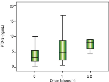
It was observed that the number of organ failures was associated with higher mean serum levels of PTX3 (Fig-ure 4) as follows: none = 3.2 ng/mL; one = 4.7 ng/mL; two or more failures = 8.0 ng/mL (p = 0.006).
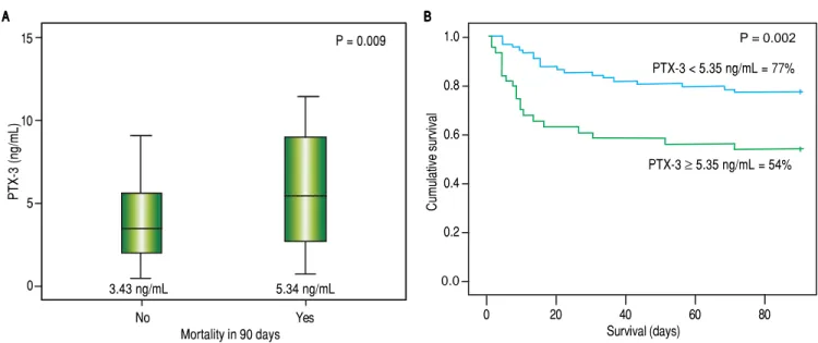
Survival analysis according to serum levels of PTX3
Forty patients died within 90 days (30.8%). It was ob-served that patients who died within 90 days had higher
se-rum levels of PTX3 on admission compared to those who survived (5.3 vs. 3.4 ng/mL; p = 0.009). Figure 5 shows the Kaplan-Meier curves associated with serum PTX3 levels. The probability of survival of patients with serum levels of PTX3 higher than or equal to 5.4 ng/mL was 54.0%, while those with serum levels lower than 5.4 ng/mL showed a 90-day survival rate of higher than 77.0% (p = 0.002).
DISCUSSION
This study has identified unprecedentedly higher PTX3 levels in cirrhotic outpatients compared to healthy con-trols as well as higher levels in cirrhotic patients hospital-ized for disease decompensation compared to cirrhotic outpatients. The higher PTX3 levels in patients with acute decompensation in comparison to cirrhotic outpatients and healthy controls is consistent with the results of stud-ies that showed elevated PTX3 levels in diseases with an
Figure 2. Figure 2.Figure 2.
Figure 2.Figure 2. Pentraxin 3 levels according to the presence or absence of Acute-on-Chronic Liver Failure (ACLF).
PTX-3 (ng/mL)
20
15
10
5
0
None Present
ACLF
Figure 1. Figure 1.Figure 1.
Figure 1.Figure 1. Pentraxin 3 levels according to patient group.
PTX-3 (ng/mL)
Healthy controls Outpatients Inpatients
Figure 3. Figure 3. Figure 3.
Figure 3. Figure 3. Pentraxin 3 levels according to Model for End-Stage Liver Dis-ease (MELD) score.
PTX-3 (ng/mL)
15
10
5
0
< 20 ≥ 20
MELD
Figure 4. Figure 4. Figure 4. Figure 4.
Figure 4. Pentraxin 3 levels according to the number of organ failures.
PTX-3 (ng/mL)
20
15
10
5
0
0 1 ≥ 2
Organ failures (n) 15
10
5
0
P < 0.001 P < 0.001
inflammatory component that affect other organs such as acute myocardial infarction,16 chronic kidney disease,17
acute respiratory distress syndrome (ARDS)18 and severe
infectious diseases affecting patients in intensive care.19
Serum levels are positively correlated with disease severity. This finding is corroborated by the positive correlation between serum PTX3 levels and the scores associated with severity of liver cirrhosis (MELD, Child-Pugh and CLIF-SOFA). In patients in intensive care with systemic inflam-matory response syndrome (SIRS), sepsis, severe sepsis and septic shock, there was also a positive correlation be-tween serum PTX3 levels and the clinical severity scores APACHE II (Acute Physiology and Chronic Health Eval-uation) and SAPS II (Simplified Acute Physiology Score).19 PTX3 expression was evaluated serially in
stud-ies in patients with ARDS18 and in patients with SIRS,
sepsis, severe sepsis and septic shock.19 In both studies it
was observed that PTX3 was the first plasma protein to rise, with a peak at approximately 7.5 h in patients with SIRS or infections in the clinical spectrum of sepsis. Levels declined in patients with ARDS after 24 h of intu-bation.18 CRP, a commonly used inflammation marker,
had a later serum peak at approximately 24 h19 and remained elevated longer.18 Mauri, et al. performed a
pre-liminary retrospective study if patients with ARDS that also showed a positive correlation between PTX3 levels and the number of organ failures,20 consistent with the
results of this study.
In this study a positive correlation was also observed between serum PTX3 levels and 90-day mortality, consist-ent with the results of other groups that evaluated mortali-ty in patients with chronic kidney disease, acute myocardial infarction and ARDS. In these studies, PTX3
proved to be an important and early predictor of mortali-ty.17,18 In patients with SIRS, sepsis, severe sepsis or septic
shock, both PTX3 and IL-6 showed significant positive correlations with mortality.19 IL-6 was not assessed in this
study. However, our group showed that IL-6 levels were positively associated with 90-day mortality (OR 1.002; 95% CI 1.000 - 1.004; p = 0.029).21
Many studies have compared the efficacy of PTX3 to CRP as a predictor of severity and mortality, given the widespread use of CRP for this purpose. The results as mentioned above show positive correlation when serum levels are compared without time reference. However, no correlation was apparent when considering the time to peak of each of these markers. In liver cirrhosis, however, CRP production is affected because it is synthesized al-most entirely in the liver. This may compromise its utility as an inflammation biomarker in these patients.22,23 This
might explain the absence of correlation between CRP and PTX3 in this study. However, Bota, et al. have not found statistically significant differences in CRP levels between intensive care patients with or without liver cirrhosis.24
Lazzarotto, et al. demonstrated that CRP levels are sensi-tive for diagnosis of infection and as predictors of 90-day mortality in patients with cirrhosis.25 Importantly, there
is, to a lesser extent, production of CRP in inflamed tis-sues.26,27 This synthesis may be responsible for incomplete
suppression of CRP levels even in patients with severe he-patic impairment.
There was also a positive correlation between serum levels of PTX3 and creatinine. Due to its high molecular weight (40.6 KD) and multimeric structure, PTX3 levels appear to increase as the glomerular filtration rate (GFR) decreases secondary to reduced clearance.17 The same
Figure 5. Figure 5.Figure 5.
Figure 5.Figure 5. Ninety days survival according to pentraxin 3 levels
No Yes
Mortality in 90 days
0 20 40 60 80
Survival (days) B
BB BB
Cumulative survival
1.0
0.8
0.6
0.4
0.2
0.0
15
10
5
0 A A A A A
PTX-3 (ng/mL)
P = 0.009
3.43 ng/mL 5.34 ng/mL
P = 0.002
PTX-3 < 5.35 ng/mL = 77%
phenomenon occurs with creatinine, it is therefore used as a marker of renal function. Numerous pathological condi-tions that lead to reduced GFR -such as sepsis, which gen-erates numerous shunts in circulation and redistribution of blood volume; major bleeding, by an absolute reduc-tion in blood volume; ascites, by third space volume re-tention and reduction of effective circulating volume-tend to increase serum creatinine. The complications of cirrhosis involve the above hemodynamic dysfunctions. One could imagine that this would be the reason for the positive correlation between serum levels of PTX3 and creatinine. However, more hemodynamic studies are needed to confirm this hypothesis.
Although recently published studies present PTX3 as a marker of severity and/or mortality, little is known about its role in vivo in humans. Garlanda, et al. demonstrated in mice deficient in PTX3 that it has many functions includ-ing regulation of innate immunity against various micro-organisms, discrimination of self- from non-self molecular patterns and tissue repair.27 Other studies
showed that the initial function of PTX3 is protective.28 It
participates in the recognition of harmful stimuli, migra-tion of neutrophils to sites of infecmigra-tion and promomigra-tion of phagocytosis of bacteria by neutrophils,29 enhancement of
nitric oxide production30 and increased expression of
tis-sue factor.31 However, the prolonged persistence of
stim-uli can lead to overexpression of PTX3 and amplification of these inflammatory pathways,28 with resulting damage
to the organism. Thus far, there are no known specific an-tagonists to the action of PTX3. Therefore, there are no data regarding blocking of this pathway and its repercus-sions.
Finally, the relevance of this study relies on the cur-rent difficulty in early determination of the severity of cirrhotic patients. Hemodynamic disturbances hinder the use of SIRS criteria;32 because the hyperdynamic
circulatory state leads to tachycardia, beta-blocker use by many of these patients reduces the heart rate and can mask SIRS, hepatic encephalopathy can lead to tachyp-nea and hypersplenism caused by congestion of the por-tal system can lead to reductions in the number of circulating leukocytes.33 Added to these factors are the
immunological changes associated with advanced liver disease, probably related to deficiencies in the comple-ment system, which impair the elimination of op-sonized bacteria and increased bacterial translocation.34-36 This in turn predisposes patients with
liver cirrhosis to infections. Furthermore, cultures of micro-organisms knowingly take at least 24 to 48 h to present results, which hampers their utility for diag-nosing infectious complications. Therefore, it is im-portant to find early markers of severity and mortality to guide appropriate treatment.
CONCLUSIONS
Circulating levels of PTX3 are increased in patients with liver cirrhosis, particularly those with acute decom-pensation. Serum PTX3 is related to the severity of the disease, the presence of ACLF and 90-day mortality. These results are promising and indicate a potential use for PTX3 as an inflammatory and prognostic biomarker for patients with liver cirrhosis.
AUTHOR CONTRIBUTIONS
Schiavon LL designed the study. Bansho ETO and Silva TE were responsible for data collection. Morato EF, Pinheiro JT, Pereira JG, Wildner LM and Bazzo ML per-formed the pentraxin-3 sample processing. Schiavon LL, Narciso-Schiavon JL and Pereira JG analyzed the data and are responsible for the article as a whole. Narciso-Schia-von JL and Pereira JG wrote the paper and Dantas-Correa EB and Schiavon LL revised the manuscript.
SUPPORTIVE FOUNDATIONS
This study was financed by Conselho Nacional de Desenvolvimento Científico e Técnológico (CNPq).
Institutional review board statement
The study was reviewed and approved by the Federal University of Santa Catarina Institutional Review Board under the number 252709.
INFORMED CONSENT STATEMENT
All study participants or their respective legal guardians provided written informed consent prior to study enroll-ment.
CONFLICTS OF INTEREST
The authors declare no conflicts of interest.
DATA SHARING STATEMENT
No additional data are available.
REFERENCES
1. Scaglione S, Kliethermes S, Cao G, Shoham D, Durazo R, Luke A, Volk ML. The Epidemiology of Cirrhosis in the United States: A Population-based Study. J Clin Gastroenterol
2. Yoon YH, Yi HY. Liver cirrhosis mortality in the United States, 1970-2003: Surveillance Report No. 75. Arlington: National Institute on Alcohol Abuse and Alcoholism 2006. 3. Kim HY, Kim CW, Choi JY, Lee CD, Lee SH, Kim MY, Jang
BK, et al. Complications requiring hospital admission and causes of in-hospital death over time in alcoholic and nonal-coholic cirrhosis patients. Gut Liver 2016; 10: 95-100. [PMID: 26087788 DOI: 10.5009/gnl14363].
4. Silva PE, Fayad L, Lazzarotto C, Ronsoni MF, Bazzo ML, Co-lombo BS, Dantas-Correa EB, et al. Single-centre validation of the EASL-CLIF consortium definition of acute-on-chronic liver failure and CLIF-SOFA for prediction of mortality in cir-rhosis. Liver Int 2015; 35: 1516-23. [PMID: 24840673 DOI: 10.1111/liv.12597].
5. Zhang J, Shan L, Koussih L, Redhu NS, Halayko AJ, Chakir J, Gounni AS. Pentraxin 3 (PTX3) Expression in Allergic Asthmatic Airways: Role in Airway Smooth Muscle Migration and Chemokine Production. Rojas M (Ed.). PLoS ONE 2012; 7: e34965. [PMID: 22529962 DOI: 10.1371/ journal.pone.0034965].
6. Cieslik P, Hrycek A. Long pentraxin 3 (PTX3) in the light of its structure, mechanism of action and clinical implications. Au-toimmunity 2012; 45: 119-28. [PMID: 21988562 DOI: 10.3109/ 08916934.2011.611549].
7. Luchetti MM, Piccinini G, Mantovani A, Peri G, Matteucci C, Pomponio G, Fratini M, et al. Expression and production of the long pentraxin PTX3 in rheumatoid arthritis (RA). Clin Exp Immunol 2000; 119: 196-202. [PMID 10606983 DOI: 10.1046/j.1365-2249.2000.01110.x].
8. Manfredi AA, Rovere-Querini P, Bottazzi B, Garlanda C, Mantovani A. Pentraxins, humoral innate immunity and tissue injury. Curr Opin Immunol 2008; 20: 538-44. [PMID: 18579363 DOI: 10.1016/j.coi.2008.05.004].
9. Ortega-Hernandez OD, Bassi N, Shoenfeld Y, Anaya JM. The Long Pentraxin 3 and Its Role in Autoimmunity. Semin Ar-thritis Rheum 2009; 39: 38-54. [PMID: 18614204 DOI: 10.1016/j.semarthrit.2008.03.006].
10. Rajkovic I, Denes A, Allan SM, Pinteaux E. Emerging roles of the acute phase protein pentraxin-3 during central nervous system disorders. J Neuroimmunol 2016; 292: 27-33. [PMID: 26943955 DOI: 10.1016/j.jneuroim.2015.12.007].
11. Garner JS, Jarvis WR, Emori TG, Horan TC, Hughes JM. CDC definitions for nosocomial infections, 1988. Am J Infect Con-trol 1988; 16: 128-40. [PMID: 2841893 DOI: 10.1016/0196-6553(88)90053-3].
12. Bajaj JS. Review article: the modern management of hepatic encephalopathy. Aliment Pharmacol Ther 2010; 31: 537-47. [PMID: 20002027 DOI: 10.1111/j.1365-2036.2009.04211.x]. 13. Angermayr B, Cejna M, Karnel F, Gschwantler M, Koenig F,
Pidlich J, Mendel H, et al. Child-Pugh versus MELD score in predicting survival in patients undergoing transjugular intra-hepatic portosystemic shunt. Gut 2003; 52: 879-85. [PMID: 12740346 DOI: 10.1136/gut.52.6.879].
14. Kamath PS, Wiesner RH, Malinchoc M, Kremers W, Therneau TM, Kosberg CL, D'Amico G, et al. A model to predict survival in patients with end-stage liver disease. Hepatology 2001; 33: 464-70. [PMID: 11172350 DOI: 10.1053/jhep.2001.22172]. 15. Moreau R, Jalan R, Gines P, Pavesi M, Angeli P, Cordoba J, Durand F, et al; CANONIC Study Investigators of the EASL-CLIF Consortium. Acute-on-chronic liver failure is a distinct syndrome that develops in patients with acute decompensa-tion of cirrhosis. Gastroenterology 2013; 144: 1426-37, 1437 e1421-1429. [PMID: 23474284 DOI: 10.1053/ j.gastro.2013.02.042].
16. Latini R, Maggioni AP, Peri G, Gonzini L, Lucci D, Mocarelli P, Vago L, et al; Lipid Assessment Trial Italian Network (LATIN)
Investigators. Prognostic significance of the long pentraxin PTX3 in acute myocardial infarction. Circulation 2004; 110: 2349-54. [PMID: 15477419 DOI: 10.1161/ 01.CIR.0000145167.30987.2E].
17. Tong M, Carrero JJ, Qureshi AR, Anderstam B, Heimburger O, Bárány P, Axelsson J, et al. Plasma pentraxin 3 in patients with chronic kidney disease: associations with renal func-tion, protein-energy wasting, cardiovascular disease, and mortality. Clin J Am Soc Nephrol 2007; 2: 889-97. [PMID: 17702732 DOI: 10.2215/CJN.00870207].
18. Mauri T, Coppadoro A, Bellani G, Bombino M, Patroniti N, Peri G, Mantovani A, et al. Pentraxin 3 in acute respiratory dis-tress syndrome: an early marker of severity. Crit Care Med
2008; 36: 2302-8. [PMID: 18596636 DOI: 10.1097/ CCM.0b013e3181809aaf].
19. Muller B, Peri G, Doni A, Torri V, Landmann R, Bottazzi B, Mantovani A. Circulating levels of the long pentraxin PTX3 correlate with severity of infection in critically ill patients. Crit Care Med 2001; 29:1404-7. [PMID: 11445697 DOI: 10.1097/ 00003246-200107000-00017].
20. Mauri T, Coppadoro A, Bellani G, Bombino M, Patroniti N, Peri G, Mantovani A, et al. Role of pentraxin 3 (PTX3) and C re-active protein as markers of inflammation in ARDS [abstract].
Intensive Care Med 2005; 31(S1): S216.
21. Fischer J, Silva TE, Silva PES, Colombo BS, Wildner LM, Baz-zo ML, Frode TS, et al. Circulating interleukin 6, 10 and 17 as prognostic markers in patients with liver cirrhosis. In: The 66th Annual Meeting of the American Association for the Study of Liver Diseases: The Liver Meeting 2015, 2015, San Francisco. Hepatology 2015; 62: 1220A.
22. Le Moine O, Deviere J, Devaster JM, Crusiaux A, Durand F, Bernuau J, Goldman M, et al. Interleukin-6: an early marker of bacterial infection in decompensated cirrhosis. J Hepatol
1994; 20: 819-24. [PMID: 7930484 DOI: 10.1016/S0168-8278(05)80155-2].
23. Park WB, Lee KD, Lee CS, Jang HC, Kim HB, Lee HS, Oh MD, et al. Production of C-reactive protein in Escherichia coli-in-fected patients with liver dysfunction due to liver cirrhosis. Diagn Microbiol Infect Dis 2005; 51: 227-30. [PMID: 15808312 DOI: 10.1016/j.diagmicrobio.2004.11.014].
24. Bota DP, Van Nuffelen M, Zakariah AN, Vincent JL. Serum levels of C reactive protein and procalcitonin in critically ill patients with cirrhosis of the liver. J Lab Clin Med 2005; 146: 347-51. [PMID: 16310518 DOI: 10.1016/ j.lab.2005.08.005].
25. Lazzarotto C, Ronsoni MF, Fayad L, Nogueira CL, Bazzo ML, Narciso-Schiavon JL, Schiavon LL, et al. Acute phase pro-teins for the diagnosis of bacterial infection and prediction of mortality in acute complications of cirrhosis. Ann Hepatol
2013; 12: 431-9. [PMID: 23813138].
26. Pepys MB, Hirschfield GM. C-reactive protein: a critical up-date. J Clin Invest 2003; 111: 1805-12. [PMID: 12813013 DOI: 10.1172/JCI200318921].
27. Garlanda C, Bottazzi B, Bastone A, Mantovani A. Pentraxins at the crossroads between innate immunity, inflammation, matrix deposition, and female fertility. Annu Rev Immunol
2005; 23: 337-66. [PMID: 15771574 DOI: 10.1146/ annurev.immunol.23.021704.115756].
28. He X, Han B, Liu M. Long pentraxin 3 in pulmonary infection and acute lung injury. Am J Physiol Lung Cell Mol Physiol
2007; 292: 1039-49. [PMID: 17277044 DOI: 10.1152/aj-plung.00490.2006].
2006; 8: 1321-9. [PMID: 16697676 DOI: 10.1016/ j.micinf.2005.12.017].
30. Dias AA, Goodman AR, Dos Santos JL, Gomes RN, Altmeyer A, Bozza PT, Horta MF, et al. TSG-14 transgenic mice have improved survival to endotoxemia and to CLP-induced sep-sis. J Leukoc Biol 2001; 69: 928-36. [PMID: 11404378]. 31. Napoleone E, di Santo A, Peri G, Mantovani A, de Gaetano G,
Donati MB, Lorenzet R. The long pentraxin PTX3 up-regu-lates tissue factor in activated monocytes: Another link be-tween inflammation and clotting activation. J Leukoc Biol
2004; 76: 203-9. [PMID: 15226367 DOI: 10.1189/jlb.1003528]. 32. Cazzaniga M, Dionigi E, Gobbo G, Fioretti A, Monti V, Salerno F. The systemic inflammatory response syndrome in cirrhot-ic patients: relationship with their in-hospital outcome. J Hepatol 2009; 51: 475-82. [PMID: 19560225 DOI: 10.1016/ j.jhep.2009.04.017].
33. Fernandez J, Gustot T. Management of bacterial infections in cirrhosis. J Hepatol 2012; 56(Suppl. 1): S1-S12. [PMID: 22300459 DOI: 10.1016/S0168-8278(12)60002-6].
34. Garcia-Tsao G, Wiest R. Gut microflora in the pathogenesis of the complications of cirrhosis. Best Pract Res Clin
Gas-troenterol 2004; 18: 353-72. [PMID: 15123075 DOI: 10.1016/ j.bpg.2003.10.005].
35. Wasmuth HE, Kunz D, Yagmur E, Timmer-Stranghoner A, Vi-dacek D, Siewert E, Bach J, et al. Patients with acute on chronic liver failure display "sepsis-like" immune paralysis. J Hepatol 2005; 42: 195-201. [PMID: 15664244 DOI: 10.1016/ j.jhep.2004.10.019].
36. Mookerjee RP, Stadlbauer V, Lidder S, Wright GA, Hodges SJ, Davies NA, Jalan R. Neutrophil dysfunction in alcoholic hepatitis superimposed on cirrhosis is reversible and pre-dicts the outcome. Hepatology 2007; 46: 831-40. [PMID: 17680644 DOI: 10.1002/hep.21737].
Correspondence and reprint request: Janaína L Narciso-Schiavon, M.D., PhD.
Department of Internal Medicine/University Hospital Polydoro Ernani de São Thiago/UFSC.
Rua Professora Maria Flora Pausewang s/n, 3rd floor, Trindade, 88040-900, Florianópolis, SC, Brazil. Tel.: +55-48-37219149. Fax: +55-48-37219014