Olfactory Function is Affected
in Patients with Cirrhosis Depending on the
Severity of Hepatic Encephalopathy
Clemens Heiser,* Bernhard Haller,† Maximilian Sohn,‡ Benedikt Hofauer,* Andreas Knopf,† Tobias Mühling,§
Jessica Freiherr,|| Martin Bender,§ Maximilian Tiller,§ Anna Schmidt,§ Wolfgang Schepp,§ Felix Gundling§
* Department of Otorhinolaryngology, Head and Neck Surgery, Technical University of Munich, Munich, Germany.
† Institute of Medical Statistics and Epidemiology Technical University of Munich, Munich, Germany. ‡ Department of Visceral Surgery, Bogenhausen Academic Teaching Hospital, University of Munich, Germany. § Department of Gastroenterology, Hepatology and Gastrointestinal Oncology,
Bogenhausen Academic Teaching Hospital, Technical University of Munich, Munich, Germany.
|| Institute of diagnostic and interventional neuroradiologiy, RWTH Aachen, Aachen.
September-October, Vol. 17 No. 5, 2018: 822-829
INTRODUCTION
Hepatic encephalopathy (HE) comprises a complex of psycho-motoric symptoms and represents an important complication in cirrhotic patients. While relatively easy and successful therapeutic approaches exist, the valid diag-nosis, especially in early stages, represents a major clinical problem. Therefore, there is a need for new objective
di-agnostic markers to detect HE in early stages.1
Olfactory deficits are frequently described in chronic liver diseases such as cirrhosis, in early studies, however,
results are not conclusive. Interestingly, Bloomfeld, et al.2
demonstrated improvement of chemosensory function af-ter orthotopic liver transplantation (OLT) by deaf-termining taste and smell detection and recognition thresholds be-fore and after OLT in a small cohort of patients with cir-rhosis. Since patients with cirrhosis often suffer from mixed deficiency of proteins, calories and micronutrients due to a progressive loss of fat and muscle mass, malnutri-tion might contribute to olfactory deficits in end-stage
liv-er disease.3 Madden, et al. demonstrated an impaired
gustatory function in cirrhotic patients with significantly
The Official Journal of the Mexican Association of Hepatology, the Latin-American Association for Study of the Liver and
the Canadian Association for the Study of the Liver
Manuscript received: Manuscript received: Manuscript received: Manuscript received:
Manuscript received: March 07, 2017. Manuscript accepted:Manuscript accepted:Manuscript accepted: November 27, 2017.Manuscript accepted:Manuscript accepted:
DOI:10.5604/01.3001.0012.3143 A B S T R A C T A B S T R A C T A B S T R A C T A B S T R A C T A B S T R A C T
Introduction and aim. Introduction and aim.Introduction and aim. Introduction and aim.
Introduction and aim. Olfactory functions are altered to a variable degree by chronic liver disease. Few studies including only small populations of patients emphasized the possibility of hepatic encephalopathy (HE) influencing olfactory nervous tasks. So far, no study has explicitly focused on olfactory function depending on the severity of HE as assessed by objective diagnostic proce-dures. Thus we performed a study using the “Sniffin’ Sticks” test system, critical flicker-fusion frequency (CFF) and clinical West Haven criteria. Material and methods.Material and methods.Material and methods.Material and methods.Material and methods. 54 cirrhotic patients with liver cirrhosis were included. Furthermore, 43 adult volunteers participating as a non-cirrhotic control group. Olfactory testing was performed using the “Sniffin’ Stick” test battery (Burghart Mediz-intechnik, Wedel, Germany) which renders a widely-used tool both in clinical and research settings for the assessment of olfactory threshold, odor identification and discrimination. Several complications of cirrhosis were diagnosed by reference methods. Statistical analysis of cirrhosis-associated complications and their relation to olfactory function was performed. Assessment of HE and classifi-cation of different stages were performed according to clinical criteria (West– Haven criteria) and according to CFF, which was deter-mined using a portable analyzer. Results.Results.Results.Results. Olfactory function was significantly reduced in cirrhotic patients (in 61.1%) compared toResults. controls (p < 0.001). Among cirrhotics patients, the prevalence of olfactory deficits (hyposmia, anosmia) increased with the severity of HE as assessed by CFF and clinical criteria (p = 0.008 and p = 0.097, respectively). No correlation was observed between olfac-tory deficits and severity of liver disease as assessed by Child-Pugh-Score, etiology of cirrhosis and complications of cirrhosis such as ascites and portal venous hypertension. Conclusions. Conclusions. Conclusions. Conclusions. Conclusions. Olfactory testing serves as a screening tool for HE and may faciliate grading of HE-severity.
Key words. Key words.Key words. Key words.
higher thresholds for detection and recognition of several different tastes, together with a higher overall median
gus-tatory score.4 They showed an association of impaired
gus-tatory acuity with hypomagnesemia. Therefore, since malnutrition in cirrhosis is almost universal due to e.g. poor dietary intake or protein losses due to paracentesis and inappropriate long-term protein restrictions, suffi-cient enteral nutrition should be guaranteed if cirrhotic patients report about olfactory deficits. Burch, et al. ana-lyzed the sensory modalities of taste and smell in few pa-tients with cirrhosis by testing different concentrations of sucrose or sodium chloride to evaluate taste acuity show-ing a decreased acuity of taste and smell in in the cirrhotic
subgroup.5 However, they did not find any association of
lack of micronutrients with decreased acuity in percep-tion of the individual test substances. Therefore, the rea-son for olfactory dysfunction in cirrhosis is far from being fully understood.
It is described in the literature that liver dysfunction
af-fects the central nervous system.6 Two newer studies
in-vestigated olfactory dysfunction in liver diseases using the Sniffin’ Sticks. Temmel, et al. showed, that odor identifica-tion, but not odor threshold or odor discrimination is
al-tered in cirrhotic patients.6 While Zucco, et al.
demonstrated that odor threshold and odor identification is compromised in patients suffering from minimal HE, no study so far has focused on olfactory functions in rela-tion to the severity of HE as assessed by objective diagnos-tic procedures.7
Apart from cirrhosis, different neurological diseases such as Parkinson, Alzheimer or Korsakoff are associated with olfactory alterations. In these disorders olfactory test-ing can be a useful screentest-ing tool at an early stage of the disease. HE and several neurological diseases share many similar clinical features, which supports the hypothesis that an impaired olfactory function might be related to the pathophysiology of HE. Surprisingly, there are only few studies investigating olfactory disturbances in cirrhotic patients and simultaneous HE.
The aim of the study was to examine if olfactory defi-cits are related to different grades of severity of HE. Therefore, we tested the olfactory function of cirrhotic patients and non-cirrhotics patients. The severity of the cirrhosis was analysed by measures of several cirrhosis-as-sociated complications including different stages of HE (including sub-clinical stages), ascites and portal hyper-tension in relation to olfactory deficits.
MATERIAL AND METHODS
The study was conducted at the Department of Gastro-enterology, Klinikum Bogenhausen, Munich, Germany. The study protocol was in accordance with the declaration
of Helsinki and approved by the local ethics board (http:// ethikkommission.blaek.de).
Patient Population
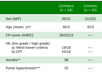
54 patients with liver cirrhosis who were admitted to the Department of Gastroenterology, Hepatology and Gas-trointestinal Oncology, Bogenhausen Hospital, were invit-ed to enter the study between April 2011 and December 2012. Furthermore, 43 adult volunteers participated as a non-cirrhotic control group, who did not suffer from smell disorders in the present and past. The control group was recruited from the Department of Otorhinolaryngol-ogy, Head and Neck Surgery, Technical University of Munich, Munich, Germany and consisted of patients ad-mitted to any kind of ENT surgery except surgery of the nose (mainly ear or neck surgery). The clinical character-istics of all patients (cirrhotic patients and controls) are presented in table 1.
Patients with preexisting smell or taste disorders, rele-vant nasal pathologies such as mucosal inflammation, sig-nificant septal deviation, nasal polyposis and excessive use of nose drops were excluded.
Cirrhosis was established either by histology or by clinical, laboratory, and sonographic or radiographic find-ings demonstrating impaired hepatic function and, mor-phological alterations of the liver typical of cirrhosis or signs of portal hypertension. For assessment of the severi-ty of liver disease, Child-Pugh (CP) classification score was calculated for each individual patient (CP A, -7 points; CP B, 8-10 points; CP C, > 11 points) In the non-cirrhotic control group, cirrhosis was excluded by the same criteria mentioned above.
Table 1. Clinical characteristics of all patients (cirrhotics and controls).
Cirrhotics Controls (n = 54) (n = 43)
Sex (M/F) 33/21 21/222
Age (mean, yr)* 60.8 43.9
CP-score (A/B/C) 26/22/13
—-HE (low grade / high grade)
a) West-haven-criteria 19/18
—-b) CFF 20/16
—-Ascites** 38
—-Portal hypertension*** 25
Assessment of
the severity of hepatic encephalopathy
The classification of HE followed the critical flicker-fusion frequency (CFF) and clinical criteria (West-Haven
criteria).8-9 CFF was measured with a portable analyzer,
and HE was divided into low grade HE (stage 1 according to West-Haven criteria) or high grade HE (> stage 1 ac-cording to West-Haven criteria).
Determination of
Critical Flicker-Fusion (CFF) frequency thresholds
The CFF was measured in a quiet, semidarkened room without distracting noises. A portable, battery-powered analyzer was used (Hepatonorm TM Analyzer, Depart-ment of Gastroenterology, Hepatology, Infectiology, Heinrich Heine University Düsseldorf, Düsseldorf, Ger-many). The analyzer evokes an intrafoveal light stimulus with defined pulses of light at a wavelength of 650 nm, lu-minance of 270 cd/m², and luminous intensity of 5.3 mcd. The frequency of the red light, which is initially generated as a high-frequency pulse (60 Hz) and which gives the pa-tient the impression of a steady light, was reduced gradual-ly until the patient had the impression that the steady light had changed to a flicker. The patient registered this change by pressing a hand-held switch. The process was repeated at least 5 times to ensure that the patient under-stood the procedure. Subsequently, the procedure was re-peated 10 times, and from the resulting data, the mean values for each patient were calculated.
The appropriate cut off to identify abnormal CFF is still not consistently defined. In the pilot study by
Kircheis, et al. the threshold of an abnormal CFF was 39
Hz when discriminating healthy subjects from patients
with cirrhosis and HE.10 Grading of HE according to CFF
was necessary to allow for an objective correlation of
HE-severity with FCCs. Kircheis, et al. found an average CFF
of 36.0 ± 1.4 Hz in subjects with HE I (according to West-Haven-criteria) while the average CFF in subjects with HE II (according to West-Haven-criteria) was 32.1 ± 2.7 Hz and HE III (according to West-Haven-criteria) was < 30 Hz.10,11 In our study, HE was either divided into low
grade HE (average CFF ≤ 39 Hz ≥ 35 Hz) or high grade
HE (average CFF ≤ 35 Hz) while an average CFF > 39 Hz
excluded HE.10,11
Assessment of olfactory function
To evaluate the ability to smell in patients with hepatic
encephalopathy we employed the “Sniffin’ Sticks” test.12
The patients were blindfolded throughout the test. Odor threshold (T), odor discrimination (D) and odor
identifi-cation (I) were tested. Odor thresholds for n-butanol were assessed using a single-staircase, triple-forced choice procedure. Three different pens were presented in a rand-omized order. Two of these pens contained nothing and the third one contained n-butanol at a certain dilution. The patient had to identify the smelling pen. The test started with the lowest concentration of n-butanol and concentration increased until the subject identified the pen containing n-butanol correctly two times. The cor-rect identification triggered a reversal of the staircase, which means that now lower concentrations of n-butanol were presented until the subject did not identify the n-bu-tanol pen. This again triggered a reversal of the staircase. The last four out of seven staircase reversal points were defined as threshold. The subjects’ scores ranged between 0 and 16.
The odor-discrimination test contained triplets of pens. These were presented to the patient in a randomized or-der. Two pens contained the same odorant and the third pen contained a different odorant. The patients had to identify the pen, smelling different compared to the two other pens. As a total of 16 triplets were tested. The sub-jects’ scores ranged from 0 to 16.
Odor identification was assessed by means of 16 differ-ent common odors. From a list of four descriptors an in-dividual odorant had to be identified. The correct answers were added. The scores ranged from 0 to 16.
The sum of these 3 scores were calculated (TDI) to
quantify olfactory function.12,13 The maximally achievable
score was 48 points. This olfactory test is widely and inter-nationally used and has been validated on large samples of healthy subjects and patients.12
Statistical analysis
Statistical analysis was performed using SPSS 21.0 (SPSS Inc., Chicago, IL. USA). Absolute and relative fre-quencies of olfactory deficits were determined for rele-vant patient groups and healthy controls. Continuous data are described by amean and standard deviation or median and 1st and 3rd quartile. Group comparisons for
categori-cal outcomes were performed using χ2 tests. Comparison
of TDI was conducted by a Mann-Whitney U test as a skewed distribution of TDI values was observed. All sta-tistical tests were performed two-sided on a significance level of 0.05.
RESULTS
Cirrhotic group
The clinical characteristics of all patients (cirrhotic and controls) are presented in table 1.
The aetiology of cirrhosis was alcohol abuse in 74.1% (40/54), hepatitis C in 9.3% (5/54), hepatitis B in 3.7% (2/ 54), autoimmune hepatitis in 1.9% (1/54) and others in 11%(6/54). On the basis of CP-classification, 22.2% (12/54) of patients had stage A cirrhosis, 42.6% (23/54) had stage B and 35.2% (19/54) had stage C.
Olfactory function in controls and cirrhotic patients
Olfactory function was reduced in 61.1% of patients compared to controls. When comparing TDI values be-tween both groups by means of a Mann–Whitney U-test, olfactory function was significantly higher in controls
(median 35.5 [1st quartile: 34.3; 3rd quartile: 38.5]) com-pared with cirrhotic patients (29.0 [25.8; 33.5], p < 0.001). Association between cirrhosis-related complications and olfactory function was investigated. All results are summa-rized in table 2.
Association with hepatic encephalopathy
When assessing HE by using CFF, 46.3% of the cirrhot-ic patients (26/54) were classified as low grade and 7 (13%) as high grade HE while HE was ruled out in 38% (21/54) of all patients.
A significant association emerged between olfactory deficits and HE grading as measured by CFF (p < 0.01, Figures 1-2). While normosia was observed for 14 of the
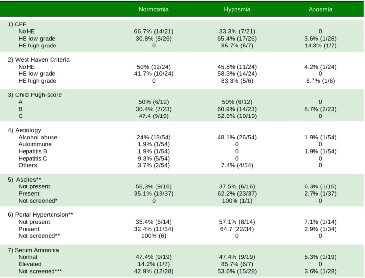
Table 2. Results of olfactory testing using the Sniffin’ Sticks for the detection of complications of cirrhosis.
Normosmia Hyposmia Anosmia
1) CFF
No HE 66.7% (14/21) 33.3% (7/21) 0
HE low grade 30.8% (8/26) 65.4% (17/26) 3.6% (1/26)
HE high grade 0 85.7% (6/7) 14.3% (1/7)
2) West Haven Criteria
No HE 50% (12/24) 45.8% (11/24) 4.2% (1/24)
HE low grade 41.7% (10/24) 58.3% (14/24) 0
HE high grade 0 83.3% (5/6) 6.7% (1/6)
3) Child Pugh-score
A 50% (6/12) 50% (6/12) 0
B 30.4% (7/23) 60.9% (14/23) 8.7% (2/23)
C 47.4 (9/19) 52.6% (10/19) 0
4) Aetiology
Alcohol abuse 24% (13/54) 48.1% (26/54) 1.9% (1/54)
Autoimmune 1.9% (1/54) 0 0
Hepatitis B 1.9% (1/54) 0 1.9% (1/54)
Hepatitis C 9.3% (5/54) 0 0
Others 3.7% (2/54) 7.4% (4/54) 0
5) Ascites**
Not present 56,3% (9/16) 37.5% (6/16) 6.3% (1/16)
Present 35.1% (13/37) 62.2% (23/37) 2.7% (1/37)
Not screened* 0 100% (1/1) 0
6) Portal Hypertension**
Not present 35.4% (5/14) 57.1% (8/14) 7.1% (1/14)
Present 32.4% (11/34) 64.7 (22/34) 2.9% (1/34)
Not screened** 100% (6) 0 0
7) Serum Ammonia
Normal 47.4% (9/19) 47.4% (9/19) 5.3% (1/19)
Elevated 14.2% (1/7) 85.7% (6/7) 0
Not screened*** 42.9% (12/28) 53.6% (15/28) 3.6% (1/28)
R2 = 0.25
21 patients without HE (66.7%), only 29.6% of the patients with low grade HE (8/27) and none of the 6 patients with high grade HE presented with normosia.
When assessing HE by using clinical criteria (West Ha-ven-criteria, WHC), 43.6% (24/54) were classified as low grade-HE, 11.1% (6/54) as high grade-HE, while 43.6% (24/ 54) of all patients were classified as asymptomatic (Figure 2).
In high-grade HE, all patients showed olfactory deficits (Figure 3).
According to those patients, in which WHC and CFF revealed the same grade of HE (70.3%; 38/54), in patients with high-grade HE anosmia was seen in 100% (1/1), and in those with low-grade HE hyposmia appeared in 65% (13/20) and normosia in 35% (7/20). In 28% of our patients (15/54) WHC and CFF did not show the same grade of HE. In those patients, in which CFF showed low-grade HE and WHC did not (46%; 7/15), anosmia occurred in 14% (1/7), hyposmia in 29% (2/7) and normosmia in 57% (4/7). Assessing high-grade HE diagnosed only by CFF (20%; 3/15), all three patients revealed anosmia (100%; 3/ 3). In those patients, in which WHC showed low-grade HE while CFF did not (13%; 2/15), 100% showed nor-mosmia. Moreover, in those patients, in which only WHC revealed high-grade HE (20%; 3/15), hyposmia occured in 100%.
There was an association between olfactory deficits and the severity of HE assessed by West Haven Criteria. Howev-er, this was not statistically significant (p = 0.097, Figure 3).
Plasma ammonia levels (obtained by venous blood sampling) were obtained from 26 patients of our study population. 87.5% (7/8) of patients with elevated plasma levels showed deficits in olfactoric testing (all hypos-mia), while 50.0% (9/18) of patients with normal plasma ammonia level presented with normosmia, 44.4% (8/18) with hyposmia and 1 patient (5.6%) with anosmia (p = 0.119).
Figure 1. Figure 1. Figure 1. Figure 1.
Figure 1. Showing the percentage of patients with anosmia, hyposmia and normosmia classified by the critical flicker frequency (CFF). An associa-tion between olfactory deficits and the severity of hepatic encephalopathy could be detected, which was statistical significance (p = 0.008).
Patients (n)
90
80
70
60
50
40
30
20
10
0
n = 54 p = 0.008
HE high grade HE low grade No HE
Anosmia Hyposmia Normosmia
Figure 2 Figure 2 Figure 2 Figure 2
Figure 2 In addition, we performed an analysis using continuous critical flicker frequency (CFF) as a Boxplot-graph or using continuous CFF and standard deviation (SDI) demonstrating an association between olfactory deficits and the severity of hepatic encephalopathy, which was statistical significance (p = 0.008).
CFC (continuous critical flicker frequeny in hertz)
50
45
40
35
30
25
Anosmia Hyposmia Normosmia
SDI
40
35
30
25
20
15
30 35 40 45
CFF (continuous critical flicker frequeny in hertz) A
A A A
Anosmia Hyposmia Normosmia
Anosmia Hyposmia Normosmia
Association with severity of liver disease
Normosmia and hyposmia occurred each in 50% (6/ 12) of patients with Child Pugh (CP) score A, respec-tively, while anosmia was not observed. In patients suf-fering from Child B cirrhosis anosmia was revealed in 8.7% (2/23), hyposmia in 60.9% (14/23) and normosmia in 30.4% (7/23), respectively. In patients suffering from Child C cirrhosis anosmia was detected in none of the patients, while hyposmia and normosmia were revealed in 52.6% (10/19) and 47.4% (9/19), respectively. (p = 0.41, Figure 4).
When comparing cirrhotic subjects with and without ascites, olfactory deficits occurred in 44.4% (24/54) when ascites was present. Patients with ascites showed anosmia in 2.7% (1/37), hyposmia in 62.2% (23/37) and normosmia in 35.1% (13/37). In patients without ascites anosmia was seen in 63% (1/16), hyposmia in 37.5% (6/16) and normos-mia in 56.3% (9/16). However, these differences were not statistically significant (p = 0.35).
In cirrhotic patients with portal hypertension hypo- or anosmia was seen in 24.1% (13/54). 2.9% (1/34) of patients with portal hypertension showed an anosmia, 64.7% (22/ 34) hyposmia and 32.4% (11/34) a normosmia, while in pa-tients without portal hypertension anosmia occurred in 7.1% (1/14), hyposmia in 57.1% (8/14) and normosmia in 35.7% (5/14). Similar to the cirrhotic patients, there was no significance in correlation between patients with or without portal hypertension (p = 0.1).
In addition, the patient’s ability to identify odors, but not odor thresholds or odor discrimination, was related to
the severity of cirrhosis. Hepatic encephalopathy as as-sessed by psychometric testing correlated inversely with the ability to identify odors, but not with abilities to dis-criminate odors or with odor thresholds.
DISCUSSION
In this study we were able to show, that among cirrhot-ics patients, the prevalence of olfactory deficits (hyposmia, anosmia) increased with the severity of HE as assessed by CFF and clinical criteria. At the same time no correlation was seen between olfactory deficits and severity of liver disease (as assessed by Child-Pugh-Score), etiology of cir-rhosis and complications of circir-rhosis such as ascites and portal venous hypertension. Including all stages, HE is a frequent complication of liver cirrhosis with a high
preva-lence up to 80% of all cirrhotic patients.8 However,
com-pared with e.g. ascites or oesophageal variceal bleeding, HE seems to represent an often overlooked complication of cirrhosis since the possibilities to diagnose this condi-tion in everyday’s practice are limited to only few feasible methods. Several tools have been used to diagnose the se-verity of HE clinically (e.g. West-Haven criteria, Table 1) and technically using objective techniques such as CFF. Olfactory deficits were frequently described in liver cir-rhosis. Some studies demonstrated that an alteration of ol-factory function is not dependent on the severity of
cirrhosis as assessed by CP score.6,7 Since HE and the
neurological diseases share many clinical symptoms, ol-factory deficits in cirrhosis might be related to the cogni-tive dysfunction in HE. However, so far no studies have been published focusing on the olfactory function in
cir-Figure 3. Figure 3.Figure 3.
Figure 3.Figure 3. Showing the percentage of patients with anosmia, hyposmia and normosmia classified by the clinical West Haven-criteria (WHC). There was no statistical significance between these groups (p = 0.097).
Patients (n)
90
80
70
60
50
40
30
20
10
0
n = 54 p = 0.097
HE high grade HE low grade No HE
Figure 4. Figure 4.Figure 4. Figure 4.
Figure 4. Showing the percentage of patients with anosmia, hyposmia and normosmia calssified by Child-Pugh score (CP-score). There was no sta-tistical significance between these groups (p = 0.41).
Patients (n)
90
80
70
60
50
40
30
20
10
0
n = 54 p = 0.41
rhotic patients and its potential alteration by HE of differ-ent grades of severity as assessed by objective procedures such as CFF. The olfactory deficits increases with the se-verity of HE.
In the studies quoted above demonstrating olfactory deficits in patients with liver cirrhosis HE was assessed by using various methodological tests. However, many of those are not appropriate for clinical practice due to e.g. lack of normative data, time needed for administration and limited availability.12 Therefore, the “Sniffin’ Sticks” test
kit has been represents a valid tool for the routine clinical
assessment of olfactory function12-14 and may therefore
serve as the diagnostic gold standard for analyzing olfacto-ry deficits in cirrhotic patients with HE. Temmel et al in-vestigated the relation between olfactory impairment and several laboratory and psychological parameters compati-ble with HE by using the “Sniffin’ Stick test kit”.6 In this
study, the authors demonstrated an inverse correlation of odor identification with, HE. A major limitation of this study, however, was the diagnosis of HE by performing exclusively the single psychometric tests which is not a
valid diagnostic method to rule out or confirm.7 Zucco
performed a pilot study by evaluating olfactory deficits in a
small group of patients with minimal HE.7 In this study,
olfactory function was impaired when minimal HE was present as cirrhotic patients performed significantly worse than a control group for both odor identification and rec-ognition tasks. However, inaccuracies in conducting pa-per-pencil tests and the subjectivity of interpretation require the application of objective diagnostic procedures such as CFF when evaluating a potential correlation of impaired olfactory function and HE. CFF is easy to per-form, reliable and accurate and has the potential to distin-guish reliably between the different severity grades of HE.10,11 To the best of our knowledge, the olfactory
func-tion as assessed by an objective diagnostic procedure such as CFF has not yet been studied to date in cirrhotic pa-tients.
In accordance with previously published data, we found olfactory function to be reduced in the present study in the majority of cirrhotic patients (61.1%) when compared to healthy controls (p < 0.001). Among cirrhot-ics, prevalence of olfactory deficits (hyposmia, anosmia) was increased according to HE-severity as assessed by CFF and clinical criteria (p = 0.008 and p = 0.097, respec-tively). Regarding CFF, the sensitivity was 100% for high grade and 68% for low grade HE. Specificity was 98.7% for high grade and 66.7% for low grade HE. No significant correlation was observed between plasma ammonia values and olfactory deficits. As shown before, we could not ob-serve a correlation between olfactory deficits and severity of cirrhosis (as assessed by Child-Pugh-Score) and under-lying etiology suggesting that impaired olfactory function
is not caused by the liver disease itself but rather by con-current complications. In our study, complications of cir-rhosis such as ascites and portal hypertension were not significantly correlated with olfactory deficits supporting the assumption that an impaired olfactory function in cir-rhosis is related to the pathophysiology of HE.
There are common pathophysiological mechanisms sug-gesting a relationship between HE and olfactory
dysfunc-tion. Butterworth, et al. described neurotoxic substances
(manganese, ammonia) that exert harmful effects on the
brain.15,16 Glutamate and dopamine are involved as
trans-mitters in the olfactory process.17 Manganese and ammonia
can harm these neurotransmitter systems. Behar et al showed that the GABA-ergic systems are altered in HE
pa-tients.18 High concentrations of manganese can accumulate
in the pallidum (globus pallidus) and trigger extrapyramidal
symptoms.19 Glutamate is the neurotransmitter of the
re-ceptor cells in the olfactory system and dopamine is mainly found as neurotransmitter in the periglomerular cells.20,21
In conclusion, our study has demonstrated that olfacto-ry function is significantly altered in cirrhotic patient’s dependent on the severity of HE. Assessing olfactory defi-cits may serve as a screening tool for HE also in sub-clini-cal stages. We observed a significant correlation between loss of olfactory function and increased severity grade of HE. Prospective studies are needed to analyse detection and in-course-control of chemosensibility in sub-clinical and manifest HE in patients suffering from cirrhosis be-fore and after specific medical treatment.
ABBREVIATIONS
• CFF: critical flicker frequency.
• CP: Child PughM.
• ENT: Ear Nose Throat.
• HE: hepatic encephalopathy.
• Hz: Herz.
• OLT: orthotopic liver transplantation.
• SD: standard deviation.
• WHC: West Haven Criteria.
FUNDING SUPPORT
No funding is to be disclosed. For this clinical trial no financial support was received.
CONFLICT OF INTEREST
There is no conflict of interest.
ETHICS STATEMENT
ACKNOWLEDGEMENTS
The authors are indebted to Petra Blankenburg and Doris Decker for their technical assistance.
REFERENCES
1. Dhiman RK, Saraswat VA, Sharma BK, Sarin SK, Chawla YK, Butterworth R, Duseja A, et al.; Indian National Associa-tion for Study of the Liver. Minimal hepatic encephalopathy: Consensus statement of a working party of the Indian Na-tional Association for Study of the Liver. J Gastroenterol Hepatol 2010; 25: 1029-41.
2. Bloomfeld RS, Graham BG, Schiffman SS, Killenberg PG. Al-terations of chemosensory function in end-stage liver dis-ease. Physiol Behav 1999; 66: 203-7.
3. Gundling F, Seidl H, Pehl C, Schmidt T, Schepp W. How close do gastroenterologists follow specific guidelines for nutrition recommendations in liver cirrhosis? A survey of current practice. Eur J Gastroenterol Hepatol 2009; 21: 756-61. 4. Madden AM, Bradbury W, Morgan MY. Taste perception in
cirrhosis: its relationship to circulating micronutrients and food preferences. Hepatol 1997; 26: 40-8.
5. Burch RE, Sackin DA, Ursick JA, Jetton MM, Sullivan JF. De-creased taste and smell acuity in cirrhosis. Arch Intern Med
1978; 138: 743-6.
6. Temmel AF, Pabinger S, Quint C, Munda P, Ferenci P, Hummel T. Dysfunction of the liver affects the sense of smell. Wien Klin Wochenschr 2005; 117: 26-30.
7. Zucco GM, Amodio P, Gatta A. Olfactory deficits in patients affected by minimal hepatic encephalopathy: a pilot study.
Chem Senses 2006; 31: 273-8.
8. Ferenci P, Lockwood A, Mullen K, Tarter R, Weissenborn K, Blei AT. Hepatic encephalopathy – definition, nomenclature, diagnosis, and quantification: final report of the working par-ty at the 11th World Congress of Gastroenterology, Vienna, 1998. Hepatol 2002; 35: 716-21.
9. Gundling F, Schmidtler F, Hapfelmeier A, Schulte B, Schmidt T, Pehl C, Schepp W, Seidl H. Fecal calprotectin is a useful screening parameter for hepatic encephalopathy and spon-taneous bacterial peritonitis in cirrhosis. Liver Int 2011; 31: 1406-15.
10. Kircheis G, Wettstein M, Timmermann L, Schnitzler A, Hauss-inger D. Critical flicker frequency for quantification of low-grade hepatic encephalopathy. Hepatol 2002; 35: 357-66.
11. Kircheis G, Hilger N, Häussinger D. Value of critical flicker frequency and psychometric hepatic encephalopathy score in diagnosis of low-grade hepatic encephalopathy. Gastro-enterol 2014; 146: 961-9.
12. Hummel T, Futschik T, Frasnelli J, Hüttenbrink KB. Effects of olfactory function, age, and gender on trigeminally mediated sensations: a study based on the lateralization of chemosen-sory stimuli. Toxicol Lett 140: 273-80.
13. Wolfensberger M, Schnieper I, Welge-Lüssen A. Sniffin’Sticks: a new olfactory test battery. Acta Otolaryngol
2000; 120: 303-6.
14. Stuck BA, Beule A, Damm M, Gudziol H, Hüttenbrink KB, Landis BN, Renner B, et al.; Committee on Olfaction and Gus-tation of the German Society of Otorhinolaryngology, Head and Neck Surgery. [Position paper Chemosensory testing for expert opinion in smell disorders]. Laryngorhinootol 2014; 93: 327-9.
15. Butterworth RF. The neurobiology of hepatic encephalopa-thy. Semin Liver Dis 1996; 16: 235-44.
16. Butterworth RF, Giguère JF, Michaud J, Lavoie J, Layrar-gues GP. Ammonia: a key factor in the pathogenesis of he-patic encephalopathy. Neurochem Pathol 1987; 6: 1-12. 17. Trombley PQ, Shepherd GM. Synaptic transmission and
mod-ulation in the olfactory bulb. Curr Opin Neurobiol 993; 3: 540-7.
18. Behar KL, Rothman DL, Petersen KF, Hooten M, Delaney R, Petroff OA, Shulman GI, et al. Preliminary evidence of low cortical GABA levels in localized 1H-MR spectra of alcohol-dependent and hepatic encephalopathy patients. Am J Psy-chiatry 1999; 156: 952-4.
19. Krieger D, Krieger S, Jansen O, Gass P, Theilmann L, Lich-tnecker H. Manganese and chronic hepatic encephalopathy.
Lancet 346: 270-4.
20. Berkowicz DA, Trombley PQ. Dopaminergic modulation at the olfactory nerve synapse. Brain Res 2000; 855: 90-9. 21. Doty RL. Olfaction. Annu Rev Psychol 2001; 52: 423-542.
Correspondence and reprint request: P.D. Dr. Med. Felix Gundling,
Department of Gastroenterology, Hepatology and Gastrointesti-nal Oncology, Bogenhausen Academic Teaching Hospital, Technical University of Munich, Englschalkinger Straße 77,
81925 München, Germany.