Otras secciones de este sitio:
☞ ☞ ☞ ☞
☞ Índice de este número ☞
☞ ☞ ☞
☞ Más revistas
Others sections in this web site:
☞ ☞ ☞ ☞
☞ Contents of this number
☞ ☞ ☞ ☞
☞ More journals
Artículo:
Anesthesia for noncardiac surgery in children with congenital heart disease
Derechos reservados, Copyright © 2004: Colegio Mexicano de Anestesiología, AC Revista Mexicana de Anestesiología
Suplemento
Supplement 1 2 0 0 4
Volumen
edigraphic.com
C C
O .
ANTE
S SOC IED
AD MEXICANA DE ANESTE SIOLO
GÍA
Anestesiología
Anestesiología
Revista
Anestesiología
Anestesiología
AUTORES EXTRANJEROS Vol. 27. Supl. 1 2004
pp 63-66
A 4.5 year old, 12 kg female with pulmonary and tricuspid atresia (functionally a single ventricle), transposition of the great arteries, Down syndrome, and radiologic evidence of cervical spine hypermobility needs extensive dental restor-ative surgery under general anesthesia. She has a function-ing right Blalock Taussig shunt, performed at five days of age. Her hematocrit is 55% and the child is visibly cyanotic. She has a history of congestive heart failure for which she takes digoxin.
QUESTIONS FOR CONSIDERATION
1. What are the key issues in this patient’s perioperative management?
2. What are the important pathophysiologic effects of her congenital heart disease? How will they affect your anes-thetic plan?
3. Should you do anything about the hematocrit of 55%? If so, what?
4. Draw a diagram of this child’s heart. How will changes in heart rate, blood pressure, myocardial contractility, or systemic vascular resistance affect this child’s hemody-namic status? Prepare a cardiac grid to demonstrate how to optimize this child’s circulation during the periopera-tive period.
5. How will you anesthetize this child?
GENERAL DISCUSSION
The anesthesiologist providing care to a child with congen-ital heart disease (CHD) must be fully aware of the child’s specific intracardiac and extracardiac defects, the effects of hemodynamic changes on those defects, and the cardiovas-cular effects of the anesthetic agents to be administered. The major pathophysiologic effects of CHD are: shunting, hy-poxemia, heart failure, dysrhythmias, pulmonary hyperten-sion, and outflow track obstruction.
Anesthesia for noncardiac surgery in children with
congenital heart disease
Carol L. Lake, M.D., M.B.A., M.P.H.
Shunting: The excessive pulmonary blood flow associated with shunting may cause congestive heart failure or produce impaired pulmonary function, small airway obstruction, left mainstem bronchial obstruction, increased interstitial/alveo-lar lung water, and vascuinterstitial/alveo-lar obstructive disease. Shunt rever-sal or hypercyanotic episodes may occur in children with int-racardiac shunts. Shunt reversal can occur with any communication between the atriae or ventricles while hyper-cyanotic episodes are most commonly associated with Tetral-ogy of Fallot. Shunt reversal occurs when systemic vascular resistance (SVR) decreases and pulmonary vascular resistance (PVR) increases due to anesthetic effects of increased airway pressures. Hypercyanotic episodes during anesthesia may re-sult from surgical stimulation, dynamic right ventricular out-flow track obstruction, decreased pulmonary blood out-flow as-sociated with hypovolemia, increased airway pressures, or decreased SVR.i. Therapy includes increasing intravascular volume, oxygen, increasing SVR with alpha agonists such as phenylephrine, and decreasing outflow track obstruction with beta blockade (Table I).
Lake CL. Anesthesia for noncardiac surgery in children with congenital heart disease MG
edigraphic.com
flow is redistributed to the brain and heart with decreasedperfusions of splanchnic organs, skin, muscle, and bone. Pulmonary Hypertension: Transient or permanent increas-es in pulmonary vascular rincreas-esistance (PVR) complicate the perioperative care of many infants with CHD such as en-docardial cushion defects, patent ductus arteriosus, ventric-ular septal defects, and aortic outflow anomalies, among oth-ers. An increase in endothelin (endothelial constricting factor) is present in children with pulmonary hypertension second-ary to CHD. The increase in endothelin may result from he-modynamic shear stresses resulting from high pressure in the pulmonary artery. Increased beta receptor density in the lung is also present as is an increase in Factor VIII von Will-ebrand factor.
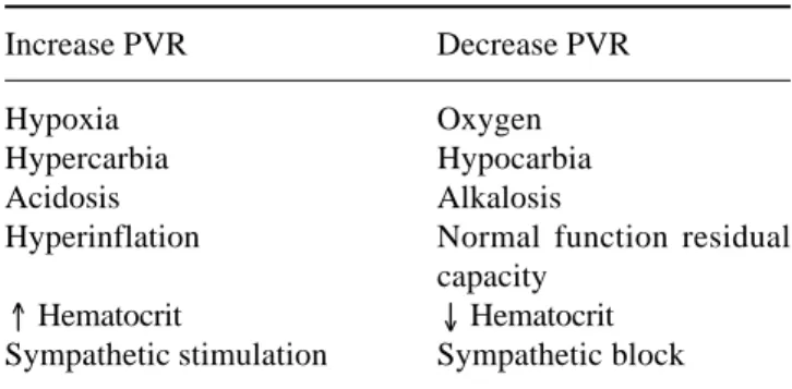
Table I. Factors affecting PVR
Increase PVR Decrease PVR
Hypoxia Oxygen
Hypercarbia Hypocarbia
Acidosis Alkalosis
Hyperinflation Normal function residual
capacity
↑ Hematocrit ↓ Hematocrit
Sympathetic stimulation Sympathetic block
Hyperventilation is the most reliable way to decrease PVR but it is pH, not pCO2 that controls pulmonary vasoconstric-tion. Prostacyclin and nitric oxide will also decrease PVR in children with CHD. Nitrous oxide does not increase PVR in most children with CHD; however, dramatic increases may occur in a few children.
Perioperative management: The approach to any congen-ital heart lesion involves knowledge of three aspects:
• A written description of the cardiac lesions • Diagram of the cardiac lesions
• Hemodynamic grid of changes affecting cardiac perfor-mance
This information can easily be obtained from the echocar-diogram or cardiac catheterization reports. With this infor-mation plus a knowledge of the cardiovascular effects of the proposed anesthetic drugs, a detailed perioperative plan can be made.
The medical history should include questions about cy-anosis, fatigability, frequency of infections, particularly res-piratory and whether the patient can keep up with his/her peers. The need for pharmacologic intervention to maintain cardiovascular performance should be noted. On physical
examination, the stage of growth and development in rela-tion to age should be noted. The absence of peripheral puls-es may indicate prior performance of a subclavian flap an-gioplasty for coarctation, use of the subclavian artery for a Blalock Taussig shunt or prior cannulation with thorombo-sis. Symptoms of heart failure include wheezing, respirato-ry distress, hepatosplenomegaly, regarded growth and de-velopment, diaphoresis, cardiomegaly, and decreased perfusion. The child should also be carefully evaluated for other congenital anomalies such as Down syndrome, Treacher Collins, Pierre Robin, etc.
Morray and coworkers demonstrated increased anesthet-ic risk in children with Down syndrome undergoing cardiac surgery. Children with Down syndrome often have airway problems such as a small mandible, small larynx, large tongue, high arched palate, and may require a smaller tra-cheal tube owing to congenital subglottic stenosis. The need for careful positioning, handling, and stabilization of the neck is essential if spinal cord involvement is associated with the cervical spine hypermobility. Inquiries about gait changes, clumsiness, spasticity should be made and the patient evalu-ated for hyperreflexia and Babinskis that would suggest cord involvement. Other problems relevant to the anesthesiolo-gist in patients with Down syndrome are retardation, in-creased gastroesophageal reflux and dein-creased central/pe-ripheral nervous system activity.
Laboratory evaluations should include a chest X-ray, elec-trocardiogram (ECG) hematocrit, electrolytes, echocardio-gram (if not performed recently), arterial blood gases (if not available from prior catheterization or other evaluation). The chest X-ray should be inspected for heart size, atelectasis, acute respiratory infection, and elevated hemidiaphragms. The ECG should be evaluated for rate, rhythm, ventricular strain (ST-T changes), and hypertrophy. Echocardiography is usually adequate for evaluation of most CHD.
edigraphic.com
:rop odarobale FDP
VC ed AS, cidemihparG
arap
acidémoiB arutaretiL :cihpargideM
sustraídode-m.e.d.i.g.r.a.p.h.i.c cihpargidemedodabor
Preoperative preparation of the pediatric patient concludes with a concise, yet detailed discussion of the cardiac condi-tions and anticipated perioperative plan of action with the child and parents. Adequate perioperative hydration is es-sential in children with cyanotic CHD so preoperative fast-ing should be minimized (clear liquids up to two hours be-fore surgery in infants 0-12 months; clear liquids up to four hours preop in children 12 months or older) or an intrave-nous infusion started. Prophylactic antibiotics should be ad-ministered according to the AHA guidelines. Premedication is usually desirable in children over six months of age with CHD since crying and generalized emotional upset worsen oxygenation and shunting in many lesions. Intramuscular premedications are rarely used and have been largely replaced by oral, nasal, or rectal routes of administration. The table II provides premedication options:
Table II. Premedications
Oral Parenteral
Midazolam 0.5-0.75 mg/kg OR Morphine 0.1-0.2 mg/kg plus
Diazepam 0.15 mg/kg plus Pentobarbital 2 mg/kg plus
Meperidine 1.5 mg/kg Scopolamine 0.01 mg/kg
Nasal Rectal
Midazolam 0.2-0.3 mg/kg Methohexital 25 mg/kg
The arrival of a quiet, sleeping child in the operating suite allows a choice of anesthetic induction techniques, ranging from inhalation of sevoflurane or halothane, through intra-muscular ketamine, to placement of an intravenous catheter and use of narcotics, propofol, or barbiturates. Specific in-duction techniques depend upon the cardiac lesion and the child’s general condition.
The speed of induction is influenced by the presence of an intracardiac or extracardiac shunt. Theoretically, one would expect a faster induction with a volatile agent in pa-tients with a left-to-right shunt due to augmented pulmo-nary blood flow. With a left to right shunt, blood that has already picked up anesthetic recirculates through the lungs picking up even more anesthetic and causing a higher anes-thetic concentration to the leave the heart. However, induc-tion time is essentially unchanged. The distribuinduc-tion and on-set of action of the intravenous agents is slower with a left to right shunt. With a right to left shunt, intravenous induction is rapid and can produce sudden, dramatic effects.
Inhalation induction is prolonged with a right to left shunt due to bypass of the lungs, although the prolongation is only moderate. Theoretically, blood that has left the lungs with a
concentration of anesthetic has that concentration diluted by blood which has bypassed the lungs. Thus, the blood leav-ing the heart has a lower concentration of anesthetic and in-duction is slowed. Such effects are most marked for insolu-ble anesthetic gases such as nitrous oxide and less marked for more soluble gases. The temptation to increase the con-centration too rapidly in children with right to left shunts must be carefully resisted. Another possibility is a change in shunting pattern during use of a volatile inhalation anesthet-ic. Finally, if a very large shunt is present, increasing the FiO2 will not increase arterial pO2.
Monitoring devices for noncardiac surgery in patients with CHD are chosen according to the requirements of the surgi-cal procedure and the severity of the patient’s disease. Usu-ally only the standard American Society of Anesthesiolo-gists’ monitors such as the pulse oximeter, blood pressure cuff, and electrocardiogram are applied prior to anesthetic induction with more invasive monitors such as intra-arterial, central venous, or transesophageal echocardiographic (TEE) transducers applied following induction.
CASE SPECIFIC DISCUSSION
Important anesthetic and perioperative management princi-ples in this child include: 1) Avoidance of air bubbles that might cross through her Blalock shunt into the systemic cir-culation; 2) Maintenance of systemic arterial pressure because
G
17 25/10 58
a=6 v=6
94
82 76
75
59
78
76 63
5
180/0-12 90/70
76
a=12 y= 5 9
100/70 80 F
A
B
C
90/
D
E 0-6
Lake CL. Anesthesia for noncardiac surgery in children with congenital heart disease MG
edigraphic.com
systemic perfusion will control pulmonary blood flow andoxygenation in this patient; 3) Avoidance of drugs or events that might increase PVR and worsen systemic oxygenation; 4) Avoidance of decreased myocardial contractility that might worsen systemic perfusion and heart failure. This child has undergone only a palliative procedure and continues to have both cyanosis and heart failure, major risk factors for adverse events during a noncardiac procedure. The compromise to the arterial circulation of the right arm should be recognized, ne-cessitating measurement of blood pressure and establishment of intravenous access at alternative sites. The function of the patient’s systemic to pulmonary shunts can be monitored with pulse oximetry. Capnography will detect serious alterations in pulmonary blood flow and cardiac output. However, the curvilinear negative correlation between the arterial-end tidal carbon dioxide gradient and arterials oxygen saturation in a child with CHD must be recognized. This gradient increases 2-3 mmHg for each 10% decrease in SpO2. Although TEE would be useful in evaluating this child’s intraoperative cardi-ac function, it is probably not feasible during a dental proce-dure. Nevertheless, a transthoracic echocadiographic evalua-tion could be performed if an adverse event occurs. While there are many satisfactory anesthetic approaches for this child, one possibility is oral midazolam premedication, inhalation induction with sevoflurane, careful nasotracheal intubation
REFERENCES
with in-line neck stabilization, anesthetic maintenance with low dose sevoflurane supplemented with narcotic and neuro-muscular blocker.
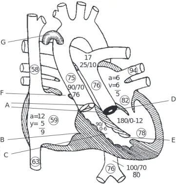
Cardiac diagrams: Cardiologists often display the echocar-diographic or cardiac catheterization findings using a dia-gram of the atria, ventricles, and great vessels that shows the septal defects, valvular stenoses, ventricular outflow obstruc-tion, intracardiac/extracardiac shunts (Figure 1). An exam-ple of such a diagram for this patient is:
Cardiac grids: The physiologic needs of any particular patient with CHD can be analyzed using a cardiac grid, a table in which the hemodynamically desirable changes in heart rate, preload, cardiac output or myocardial contractil-ity, and systemic and pulmonary vascular resistances are plot-ted against the anatomic components of any congenital le-sion. Any example of a cardiac grid for a patient with a hypoplastic right heart, transposition of the great arteries, tricuspid and pulmonic atresia is shown below:
Anatomy Preload PVR SVR HR Contractility
ASD/VSD N ↓ ↑ N N
S→P Shunt → ↓ ↑ ↑ ↑
TGA N ↓ N-↑ N N
1. Moore RA. Preoperative evaluation and preparation of the pediatric pa-tient with cardiac disease. In: Lake CL, Booker P (ed): Pediatric Cardiac Anesthesia. Fourth Edition. Lippincott Williams & Wilkins, 2004 In Press. 2. Frankville DD. Anesthesia for noncardiac surgery in children and adults with congenital heart disease. In: Lake CL, Booker P (ed): Pediatric Cardiac Anesthesia. Fourth Edition. Lippincott Williams & Wilkins, 2004 In Press.
3. Strafford ME, et al. Anesthetic morbidity in congenital heart disease
patients undergoing non-cardiac surgery. Anesthesiology 1991;75: A1056.
4. Morray J, et al. Increased perioperative risk following repair of con-genital heart disease in Down’s syndrome. Anesthesiology 1986;65:221. 5. Nicolson SC, et al. Shortened preanesthetic fasting interval in
pedi-atric cardiac surgical patients. Anesth Analg 1992;74:694. 6. Fletcher R. Gas exchange during anesthesia and controlled ventilation in