P
www.permanyer.com
Rev Invest Clin. 2015;67:5-14 IN-DEPTH REVIEW
Functional Respiratory Assessment
in Interstitial Lung Disease
José Luis Miguel-Reyes
1,2, Laura Gochicoa-Rangel
1, Rogelio Pérez-Padilla
1and
Luis Torre-Bouscoulet
1*
1Department of Respiratory Physiology; 2Asthma Clinic, Instituto Nacional de Enfermedades Respiratorias Ismael
Cosío Villegas, México, D.F., México
Corresponding author:
*Luis Torre-Bouscoulet
Department of Respiratory Physiology
Instituto Nacional de Enfermedades Respiratorias Ismael Cosío Villegas
Tlalpan, 4502, Col. Sección XVI. Del. Tlalpan, C.P. 14080, México, D.F.
E-mail: luistorreb@gmail.com Received for publication: 01-08-2014 Accepted for publication: 11-11-2014
ABSTRACT
Interstitial lung diseases are a heterogeneous group of disorders that affect, to a greater or lesser degree, the alveolus, peripheral airway, and septal interstitium. Functional assessment in patients suspected of having an interstitial lung disease has implications for diagnosis and makes it possible to objectively analyze both response to treatment and prognosis. Recently the clinical value of lung-diffusing capacity and the six-minute walking test has been confirmed, and these are now important additions to the traditional assessment of lung function that is based on spirometry. Here we review the state-of-the-art methods for the assessment of patients with interstitial lung disease. (REV INVEST CLIN. 2015;67:5-14)
Corresponding author: Luis Torre Bouscoulet, luistorreb@gmail.com
Key words: Interstitial lung disease. Idiopathic pulmonary fibrosis. Pulmonary function test. Minimum clinically significant difference. Pulmonary hypertension.
INTRODUCTION
Interstitial lung diseases (ILD) are a heterogeneous group of disorders that affect, to a greater or lesser degree, the alveolus, the peripheral airway, and the septal interstitium. Although ILD share certain physi-ological, clinical, and radiological characteristics, they are distinct entities1,2.The American Thorax Society
and the European Respiratory Society propose a clas-sification into two categories: ILD with known etiol-ogy, and idiopathic ILD. The latter is further classified as: (i) major idiopathic interstitial pneumonias; (ii) rare
idiopathic interstitial pneumonias; and, (iii) unclassifi-able idiopathic interstitial pneumonias1,3,4. Of the
idio-pathic forms, idioidio-pathic pulmonary fibrosis (IPF) is the most common and has the worst prognosis since it is characterized by patches of dense, sub-pleural fibrosis alternating with areas of healthy lung tissue. Other idiopathic ILD are characterized by varying degrees of inflammation and fibroproliferative activity5-7.
As in other respiratory diseases, the functional evalua-tion of patients with suspected ILD has implicaevalua-tions for diagnosis and the assessment of treatment. In addition,
No part of this publication may be reproduced or photocopying without the prior written permission
of the publisher
.
prognosis depends on the patient’s functional condition at the time of diagnosis and the changes that occur in the months following treatment onset8. In recent
years, understanding of the functional changes that occur in patients with ILD9-12 has expanded, so the
objective of the present review is to examine the cur-rent state of knowledge in the field and to analyze the contribution of relevant developments to improve the prognosis. We believe that this information will be of great interest to clinicians who treat patients with ILD. We conducted a thorough review of the publica-tions listed in PubMed, using the following keywords for our search: interstitial lung disease, pulmonary fibrosis, pulmonary function test, spirometry, diffus-ing capacity, 6-minute walkdiffus-ing test, lung volumes, and cardiopulmonary exercise test.
GENERAL CONSIDERATIONS
ON LUNG FUNCTION EVALUATION
Any adequate evaluation of lung function, must include at least an assessment of pulmonary mechanics, gas exchange, and response to exercise (Table 1). The standard test for evaluating respiratory mechanics is spirometry, while lung-diffusing capacity of carbon monoxide (DLCO) is the best test for assessing gas exchange. The cardiopulmonary exercise test (CPET) is the best means available for analyzing integrated responses to exercise, but due to its complexity and cost, it is not very practical. The six-minute walking test (6MWT) is a simple, low-cost alternative.
ABNORMALITIES IN LUNG MECHANICS
In ILD patients, the functional pattern is predominant-ly restrictive, manifested by a progressive reduction of pulmonary volume and capacity. Most patients show a decrease in total lung capacity (TLC), vital capacity (VC), and functional residual capacity (FRC). Some exceptions to this restrictive pattern occur in diseas-es such as lymphangioleiomyomatosis, Langerhans cell histiocytosis and, occasionally, end-stage sarcoidosis. In these diseases, pulmonary volumes may be normal or even increased so these indicators may be useful in differential diagnoses.
Forced spirometry reveals a reduction in forced vital ca-pacity (FVC) with no limitation of the forced expiratory
volume in the first second (FEV1), which produces a normal or elevated FEV1/FVC (Fig. 1). However, spi-rometry does not reliably identify lung restriction because it does not measure static lung volumes10.
For several decades FVC has been used as an outcome when studying patients with IPF. A decline in serial mea-surements of FVC is a widely accepted indicator of dis-ease progression in IPF and an outcome commonly used in therapeutic trials. A decline of at least 10% in FVC indicates disease progression. Other authors suggest that a reduction of 6% in FVC in a period of 24 weeks may be sufficient to affect the prognosis13.
When evaluating respiratory restriction, corporal ple-thys mography is the standard because it allows mea-suring static lung volumes14. This procedure uses
direct measurements of FRC, tidal volume (Vt), vital capacity (VC), and inspiratory capacity (IC), to indi-rectly calculate TLC and residual volume; it is then possible to identify the restrictive ventilatory pattern manifested by TLC, volume of intrathoracic gas, and VC below 80% of that predicted. By using TLC it is possible to assess restriction. Total lung capacity is calculated by adding FRC and IC.
Respiratory restriction may also be determined using imaging methods, the simplest of which is a chest Table 1. Common functional abnormalities in patients with interstitial lung diseases
Abnormalities in lung
mechanics ↓VC↓Compliance (↓FRC and ↓ITGV) Lung restriction (↓TLC) ↑R5Hz
Abnormalities in gas
exchange ↓DLCO↓PaO2, ↓SaO2 ↑A-a O2 gradient Abnormalities during
exercise testing ↓VO↑VE/VCO2 2 ↑VD/Vt ↓MVV ↑VE/MVV ↓VO2/HR
↓distance in 6MWT
VC: vital capacity; FRC: functional residual capacity; ITGV: intrathoracic gas volume; TLC: total lung capacity; R5Hz: resistance measured at 5 Hertz; DLCO: diffusion capacity; PaO2: partial pressure for oxygen; SaO2: arterial saturation of oxygen; A-a O2 gradient: alveolar-arterial oxygen gradient; VO2: oxygen uptake; VE: ventilation; VCO2: carbon dioxide output; VD: dead space; Vt: tidal volume; MVV: maximal voluntary ventilation; HR: heart rate; 6MWT: six minute walking test.
No part of this publication may be reproduced or photocopying without the prior written permission
of the publisher
.
radiograph. In addition, TLC can be quantified using planimetry or by inert gas dilution techniques that may be an alternative when plethysmography is not available.
The pressure-volume curve of patients with ILD shows a reduction of lung compliance; the curve is clearly displaced downwards and to the right compared with the curve of a normal subject (Fig. 2). Likewise, there is an increase in elastic retraction pressure. The re-duction of compliance increases respiratory effort be-cause the system must generate a larger pressure gradient (∆P) to displace the same volume of. Lung compliance decreases as a consequence of loss of lung volume, increase in the amount of interstitial connective tissue, and increase in surface tension due to changes in the surfactant or alveolar size. To assess compliance, an esophageal catheter must be introduced to measure the transpulmonary pressure, which produces discomfort. As pulmonary rigidity
increases, so too does the percentage of fibrosis, leading to a worse prognosis.
The pathological mechanisms involved in the reduction of lung volume seen in most ILD patients include the complete or partial obliteration of some alveolar units, invasion of alveolar spaces by cells and inflammatory exudates, thickening of the alveolar walls due to extracel-lular matrix, and destruction of lung parenchyma (Fig. 3). The increase in elastic retraction pressure reduces the FRC, which, in turn, increases airway resistance.
In the bronchiolocentric forms of ILD, such as hyper-sensitivity pneumonitis, respiratory bronchiolitis as-sociated with ILD, and sarcoidosis, as well as in pa-tients with concomitant chronic obstructive pulmonary disease, an obstructive or mixed (i.e. restrictive plus obstructive) functional pattern may be observed15.
Table 2 summarizes the characteristics of the ob-structive, restrictive, and mixed functional patterns. Volume (l)
1 8
6
4
2
0
Flo
w (l/seg) V
olume (l)
–2
–4
–6
–8
2 3 4
0
1
2
3
0 5 10
Time (sec)
15 20 25
FRC
IC
ITGV
RV Closed
valvule
Spirometry Plethysmography
Predicted REAL %Pred Predicted REAL %Pred
FVC (l) 2.49 1.86 75 TLC (l) 5.09 3.20 62
FEV1 (l) 1.79 1.66 92 TGV (l) 3.22 1.89 58
FEV1/FVC% 72 89 RV (l) 2.14 1.16 54
VC (l) 2.49 2.08 83
Figure 1. Flow volume curve obtained from a forced spirometry. The result suggests restriction and is confirmed by measuring pulmonary volumes by corporal plethysmography. Note that the FEV1/FVC ratio is 89% (normal), with FVC at 75% of the predicted value, and TLC is 62% of predicted. These results confirm restriction. FEV1: forced expiratory volume in the first second; FVC: forced vital capacity; RV: residual volume; ITGV: intrathoracic gas volume; TLC: total lung capacity; VC: vital capacity.
No part of this publication may be reproduced or photocopying without the prior written permission
of the publisher
.
Respiratory patterns and gas exchange
In ILD, the abnormalities in gas exchange generally precede changes in respiration mechanics. The char-acteristic respiratory pattern of ILD patients consists of a decrease in Vt and an increase in respiratory frequency that generates rapid, shallow breathing. This respiratory pattern seems to be mediated by an in-crease in afferent vagal activity, although extra-va-gal influences (i.e. reflexes of the thoracic wall or a behavioral modification dependent on cortical per-ception of the afferent information from the respira-tory system) may also contribute. This rapid, shallow
ventilatory pattern is an adaptive process because it reduces respiratory effort in the presence of reduced lung compliance.
When analyzing gas exchange, the characteristic find-ing in ILD patients is hypoxemia. In places of high alti-tude, such as Mexico City, hypoxemia is considered when the partial oxygen pressure (PaO2)is < 60 mmHg, or the oxygen saturation level (SaO2) is < 90%16. In
the initial stages of ILD, arterial gases at rest tend to be normal. The hypoxemia seen in ILD patients is as-sociated with an increase in the alveolus-arterial oxy-gen gradient (A-a gradient) that worsens with exercise and is often accompanied by a normal or increased ventilatory state (normo- or hypocapnia); the pH val-ues remain within normal ranges. Carbon dioxide re-tention is rare, usually manifesting only in terminal stages of the disease. Occasionally, an adequate oxy-genation at rest may be seen, but hypoxemia be-comes evident once exercise begins since this reduces blood circulation time through the lung capillaries17. It
is important to keep in mind that normal PaO2 orSaO2 levels do not rule out the possibility that significant hypoxemia may occur during exercise or sleep.
The mechanisms involved in the physiopathology of hypoxemia at rest and of the increase of the A-a O2 gradient are almost exclusively due to an imbalance in the ventilation:perfusion ratio (V/Q) accompa-nied, with some areas being poorly ventilated but well perfused and others, more importantly, being
Figure 3. Microphotographs that illustrate: A) A normal lung with an unaltered terminal bronchiole (arrow head), alveolar space (AS)
and the unaltered alveolar septum (arrows); and, B) A section of lung from a patient with interstitial lung disease. Arrows indicate
localized, thickened sub-pleural septa caused by deposits of collagen fibers and scarce lymphoplasmacytic infiltration. Also visible are zones with end-stage lesions (ESL). Hematoxylin-eosin staining; 4× increase from the original (Courtesy: Miguel Gaixola, M.D.).
6.0 TLC
TLC
TLC 4.5
3.0
V
olume (l)
1.5
0
0 10
Emphysema Normal
ILD
Transpulmorary pressure (cmH2O)
20 30 40
Figure 2. Pressure-volume curve from interstitial lung disease patients is characteristically displaced downwards and to the right, compared to normal subjects or to patients with em-physema. This indicates that greater pressure is required to generate the same volume. ILD: interstitial lung disease; TLC: total lung capacity.
A B
No part of this publication may be reproduced or photocopying without the prior written permission
of the publisher
.
well ventilated but poorly perfused, a condition that increases the V/Q ratio. During exercise-induced hy-poxemia a second contributing factor, in addition to the V/Q imbalance, is poor oxygen diffusion through the alveolar capillary units that occurs due to, among other causes, a thickening of the membrane, a reduc-tion in the area available for gas exchanges, and a decrease in contact time between alveolar oxygen and the circulating erythrocytes8-20.
Abnormalities in gas exchanges may also be detected by measuring DLCO; indeed, this seems to be the first functional abnormality that is observed in the course of ILD21,22. The DLCO evaluates the transference of
carbon monoxide (CO) from the alveolar space to the inside of the lung capillary divided by the difference of CO pressures in the two compartments. Factors that determine gas diffusion through a semiperme-able membrane include structural elements such as pulmonary volume, distance to the exchange phase, membrane thickness, exchange surface, effects of the airway closure, and pulmonary capillary blood volume, as well as functional aspects such as absolute V/Q levels, uniformity of the V/Q ratio, alveolar gas com-position, diffusion characteristics of the membrane, hemoglobin bonding properties, and gas pressure in the alveolar capillaries. All of these determinants are included in Fick’s law:
Vgas = A x D x (P1 - P2)/T
This means that the volume of gas (Vgas) which diffuses through a semipermeable membrane is de-termined by the exchange area (A), the diffusion coefficient of the gas involved (D), and the pressure difference on the two sides of the alveolar-capillary
membrane (P1 - P2), while it is inversely proportional to the thickness of the membrane or diffusion dis-tance (T)23-25. This measurement loses accuracy when
assessing patients with extremely reduced pulmonary volumes (VC < 1.0 l) (Fig. 4).
Most ILD patients experience a reduction of DLCO, although this is not specific of a particular type of ILD. Reduced DLCO is due, in part, to the destruction of functionally heterogeneous alveolar capillary units. The lung regions with reduced compliance due to fi-brosis may be poorly ventilated but continue to be well perfused, a condition that produces low V/Q ra-tios. The severity of the reduction in DLCO does not correlate well with the state of the disease because the extensive areas of fibrosis do not always translate into severely reduced DLCO. Similarly, some patients with ILD may have a substantial reduction of lung volumes or severe hypoxemia, even though the DLCO is normal or only slightly reduced. This is specially true in cases of sarcoidosis26. The occurrence of moderate-to-severe
reductions in DLCO in the presence of normal lung volumes should suggest ILD associated with emphy-sema, pulmonary vascular disease, Langerhans cell his-tiocytosis, or lymphangioleiomyomatosis. The DLCO is a useful measure in follow-up to determine prognosis.
Some studies have shown that in the initial stages of sarcoidosis and asbestosis, an increase in the DLCO may be seen26. This increase is made possible by the
membrane diffusion component, which is associated with the initial inflammatory phase. The increase in DLCO has been described as a characteristic finding in diffuse pulmonary hemorrhages due to the presence of erythrocytes in the alveolar spaces. In the early in-flammatory stages of some ILD, an increase in CO Table 2. Physiologic characteristics observed in obstructive, restrictive, and mixed pattern
Functional pattern FEV1/FVC quotient TLC RV DLCO
Obstruction due to COPD ↓ Normal or ↑ Normal or ↑ ↓
Obstruction due to asthma ↓ Normal or ↑ Normal or ↑ Normal or ↑
Lung restriction Normal or ↑ ↓ ↓ ↓
Extrapulmonary restriction Normal or ↑ ↓ Normal or ↓ Normal
Mixed ↓ ↓ Normal ↓
COPD: chronic obstructive pulmonary disease; DLCO: diffusion lung of carbon monoxide; FEV1: forced exhaled volume in 1 second; FVC: forced vital capacity; RV: residual volume; TLC: total lung capacity.
No part of this publication may be reproduced or photocopying without the prior written permission
of the publisher
.
diffusion may be found, possibly associated with an increase in alveolar-capillary permeability. A recently described index of alveolar gas transference called “diffusing capacity of nitric oxide” (DLNO) is being evaluated. Nitric oxide is captured 4.3-times faster than CO using the single-breath method. This obser-vation, although somewhat fortuitous, has stimulated the research into alveolar transference (diffusion) since DLNO is considered to express the true diffusion capacity of the membrane due to its very high affinity for hemoglobin and its independence from pulmonary capillary blood volume (Fig. 4)27.
ALTERATIONS IN PULMONARY
CIRCULATION
Vascular abnormalities are characteristics shared by all ILD. The development of pulmonary hypertension (PH) is a determinant for prognosis, especially in pa-tients with IPF or pulmonary disease associated with scleroderma. The traditional concept that PH in IPF patients is caused by hypoxic pulmonary vasoconstric-tion and loss of pulmonary capillaries has not been fully demonstrated in recent clinical studies. The bio-logical processes that underlie progressive fibrogene-sis may also contribute to vascular remodeling and PH28,29. Recent studies suggest that there is a
relation-ship between fibrosis of the lung parenchyma and the mechanisms that cause structural and functional ab-normalities of the pulmonary blood vessels30. The fact
that there is an anatomical proximity of the blood ves-sels, the interstitial space, and the alveolar epithelium may help to understand the pathogenic mechanisms that lead to the development of PH in ILD patients. The presence of PH in IPF, sarcoidosis, or systemic sclerosis is considered an indicator of poor prognosis in terms of respiratory function, quality of life, and survival.
According to the guidelines for diagnosis and treat-ment of PH, IPF is classified in Group 3 (i.e. PH due to pulmonary disease or hypoxia) to distinguish this ill-ness from other clinical conditions that cause PH. Pul-monary hypertension is defined as ≥ 25 mmHg in the mean pressure of the pulmonary artery (mPAP) mea-sured at rest by right heart catheterization. Although the definition of PH during exercise is less standard-ized, IPF patients may show normal hemodynamics while at rest and develop significant PH only while ex-ercising. Some coexisting disorders, such as obstruc-tive sleep apnea, coronary artery disease with left ventricular dysfunction, and pulmonary embolism, may contribute to the development of PH in IPF patients.
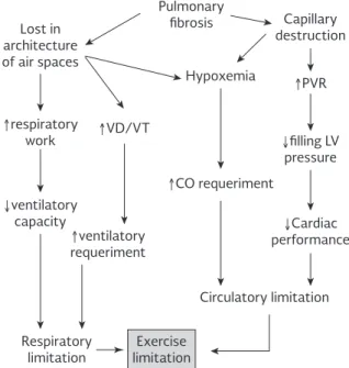
EXERCISE CAPACITY
Exercise deeply affects the respiratory function of ILD patients. Figure 5 shows the factors that contribute to limitations during exercise, a situation that gener-ally produces rapid, shallow breathing with a significant worsening of dyspnea and a significant reduction of PaO2. The ILD patients also show a significant reduction in their exercise capacity. The most commonly used Single breath
0.3% CO 14% He
18% O2
Spirometer FACO
FAHe
Absorption
CO2 + H2O He + COanalysis
(by chemiluminescence)
Dead space wash-out
Volume inspired
Residual volume (RV)
Sample
Effective breath holding time
0 Time (seconds) 10
V
olume
Expiration Inspir
ation
NO 40-8 ppm
NO
Figure 4. Diagram of diffusing capacity of carbon monoxide and diffusing capacity of nitric oxide based on the unique respiration method in which a volume of expiratory reserve maneuver is performed on the basis of stable tidal volume respiration. After that, an inspiratory vital capacity maneu-ver had subjects inhale a mixture of gases (CO 0.3%, He 14%, O2 15% and NO 40 ppm) up to their total lung capac-ity. After an apnea period of 10 seconds, a slow expiratory vital capacity maneuver was performed to take measure-ments of both CO and NO. The analysis of CO was done by mass spectrometry while NO was analyzed by chemilumines-cence. CO: carbon monoxide; FACO: alveolar carbon monox-ide fraction; FAHe: alveolar helium fraction; He: helium; NO: nitric oxide.
No part of this publication may be reproduced or photocopying without the prior written permission
of the publisher
.
exercise tests are the 6MWT and CPET. The most important measurement in the 6MWT is the number of meters that the patient walks, but modifications in oxygen saturation provide additional information to evaluate in more detail the progression of the disease or response to treatment31. The 6MWT may also
pro-vide information that is more accurate than other pul-monary function tests, especially in IPF patients32,33.
Some retrospective cohort studies suggest that a re-duction of 30 meters in the distance walked has im-plications for prognosis32. Another study reports that
after 24 weeks of follow-up, a decrease of 50 meters, or walking fewer than 207 meters, bring a fourfold greater risk of dying within a year, compared with patients who walk more than this distance33. An index
proposed by Lettieria, et al. includes both the distance walked and the degree of desaturation during the 6MWT. The nadir of SpO2 multiplied by the distance walked in meters (the distance-saturation product) has been shown to have high sensitivity and specificity to determine survival at 12 months in IPF patients34.
Studies using the 6MWT have also described a factor called “heart rate recovery”, defined as the reduction in the heart rate during the first minute after finishing the 6MWT; that is, the heart rate is measured at the end of the 6MWT and then compared with the heart rate after one minute. When a subject’s heart rate
Lost in architecture of air spaces
↑respiratory work
↓ventilatory
capacity ↓Cardiac
performance
↓filling LV pressure
↑ventilatory
requeriment
↑VD/VT
↑CO requeriment
Pulmonary fibrosis
Hypoxemia
Capillary destruction
↑PVR
Circulatory limitation
Respiratory
limitation limitationExercise
Figure 5. Factors that limit exercise capacity in ILD patients. CO: cardiac output; PVR: pulmonary vascular resistances; VD: dead space; LV: left ventricle; VT: tidal volume.
does not decrease by at least 14 beats per minute, chronotropic incompetence is suspected due to a disturbance in parasympathetic activity that is asso-ciated with poor prognosis35.
In the CPET, the most common finding in patients with ILD is a reduction in maximum oxygen uptake (VO2max) because the respiratory system is unable to respond to the increased demand for oxygen at tissue level, such that exercise ends early with a low anaerobic threshold. This threshold is defined as the moment in which an-aerobic energy producing mechanisms are activated. Other findings include a progressive increase in dead space during exercise (i.e. an increase in the VD/Vt ratio) and a higher ventilation/VCO2 quotient. The lat-ter reflects the system’s inefficiency in eliminating CO2 at a given level of ventilation; that is, the lesser the inefficiency of the system the greater the ventilation required to eliminate a given quantity of CO2, condi-tions that lead to increases in the ventilation/VCO2 quotient. This quotient increases even more with a co-existing PH because the restriction in the pulmonary vascular bed reduces the availability of CO2.
The ventilation/maximum voluntary ventilation ratio is high in patients with chronic respiratory disorders, including those with ILD. In healthy subjects, maximum ventilation during exercise does not exceed 80% of the amount the subject is capable of breathing (max-imum voluntary ventilation); that is, there is a respi-ratory reserve of approximately 20%. But patients with chronic respiratory disorders have a reduced ventilatory capacity (maximum voluntary ventila-tion), that is, when ventilation increases during exer-cise they quickly reach their maximum respiratory capacity, which means that the ventilation/maximum voluntary ventilation ratio approaches 1, thereby de-pleting the respiratory reserve.
Left ventricular dysfunction is another factor that may reduce an individual’s capacity to exercise. Factors related with it include reduced cardiac output (heart rate × systolic volume). In CPET, this phenomenon may be indirectly assessed by measuring oxygen pulse (VO2/HR), which is equivalent to systolic vol-ume and therefore will be low in patients with ven-tricular dysfunction.
From the clinical perspective, the disproportionate limitation to exercise related with abnormalities in
No part of this publication may be reproduced or photocopying without the prior written permission
of the publisher
.
pulmonary volume and oxygen desaturation during exercise could be an indicator of the development of PH in IPF patients29,36. Since hypoxemia is not always
evident at rest, and it is not always possible to detect severe exercise-induced hypoxemia, it may be impor-tant to consider conducting a CPET that involves se-rial measurement of artese-rial gases, or measuring oxy-gen saturation by pulse oxymetry. Arterial oxyoxy-gen desaturation, or the inability to adequately reduce the volume of dead space by exercising (e.g. a high VD/ Vt ratio), together with an excessive increase in respi-ratory frequency with a lower-than-expected recruit-ment of tidal volume, provide useful information re-garding physiological abnormalities and the extent of disease development. Longitudinal follow-up of pa-tients with IPF has identified a VO2max threshold of 8.3 ml/kg/min, below which the risk of death increas-es significantly (hazard ratio of 3.4)37.
PULMONARY FUNCTION TESTS
IN PULMONARY HYPERTENSION
One method for screening ILD patients for PH relies on a formula that predicts mPAP from standard pulmo-nary function tests. In this equation, pulse oxymetry at room air (SpO2) together with the percentage of FVC and DLCO are found to be predictor parameters for calculating pulmonary artery pressure. Although this formula has a low positive-predictive value, only 51%, it does provide a high negative-predictive value of 96%, thus identifying IPF patients with a low risk for PH. In one study this formula showed to be a useful tool in IPF pa-tients, with a performance similar to that of transesoph-ageal echocardiography38,39. However, the study, which
was conducted in subjects living at sea level, used the pulmonary function test reference equations for sub-jects different from the population living at high alti-tudes. Moreover, it included only young patients who were in the waiting list for lung transplantation and, therefore, represented the most severe cases. In the less severe cases no right cardiac catheterization was performed even though such procedure would have confirmed or ruled out the findings calculated by us-ing the prediction formula.
Pulmonary hypertension is common in the combined syndrome of pulmonary fibrosis with emphysema, which can be considered a distinct entity with its own characteristic functional profile; i.e. lung volume is
preserved, DLCO is severely reduced, and exercise-induced hypoxemia is present. The severe decrease in DLCO probably represents the cumulative or syner-gistic effects of emphysema, fibrosis, and pulmonary vascular disease and is one of the hallmarks of this syndrome. Pulmonary hypertension is a critical deter-minant of poor prognosis40,41.
METHODS FOR FUNCTIONAL
FOLLOW-UP
The clinical course of patients with IPF varies. It may have long periods of stability or periods of acute de-terioration with a gradual but constant decline in pulmonary function42,43. This unpredictable nature
underlines the importance of identifying factors that may help to make more precise prognoses for pa-tients at the time of diagnosis44.
Table 3 shows the functional factors present at diag-nosis and the changes in physiological parameters in the first six months after diagnosis that result in a poor prognosis. One relevant point is that parame-ters such as DLCO and high-resolution computed tomography findings have implications both at the
moment of diagnosis and during follow-up. Clinical efficacy is best evidenced by a beneficial effect on clinically significant outcomes, including perception of symptoms, functions (i.e. the ability to perform daily life activities), or survival. In ILD, and especially in IPF, the main outcomes are mortality due to all causes and non-elective hospitalizations due to all causes. For IPF, there are no validated measures of symptoms or broader constructs such as health status or func-tional status. A surrogate outcome is defined as an indirect measure that may well replace clinically sig-nificant outcomes. Once they are validated, surrogate outcomes may prove to be an appropriate means of prognosis; however, validation requires evidence that the effect of an intervention or a clinically significant outcome is reliably predicted by the effect of an in-tervention in the surrogate outcome. At present, for patients with IPF, there are no validated surrogate outcomes. In subjects with ILD a reduction of FVC of at least 10%, or of DLCO of at least 15%, over a period of 6-12 months is associated with reduced survival rates. While a change in FVC is a good sur-rogate for subsequent mortality, it is imperfect be-cause some patients die without a 10% reduction,
No part of this publication may be reproduced or photocopying without the prior written permission
of the publisher
.
while others may live for long periods of time even after a decline of 10% in FVC. This has led to observa-tions in some recent studies of changes in the per-centage of predicted FVC as an independent predictor of mortality in IPF, together with the concept of the minimum clinically significant difference. This is the smallest difference in a measure that may be seen as important in terms of benefit or risk, and that would lead clinicians to consider a change in treat-ment. Hence, it is an important concept in clinical medicine that will help to interpret the significance of changes observed in one measurement, and could affect the perceived efficacy of an intervention. Minimum clinically significant difference may also have implications for the design of clinical trials in terms of selecting primary and secondary outcomes, as well as in estimating the sample size. In these studies, a minimum clinically significant difference for a predicted FVC between 2 and 6% has been identified.
CONCLUSIONS
The evaluation of respiratory function is of great importance in patients with suspected ILD in relation to differential diagnoses, assessing responses to in-terventions, and establishing prognoses. Among the pulmonary function tests, DLCO is the only one that has implications for prognosis from the moment of diagnosis (baseline measurements). The magnitude of the compromised functionality, especially in terms of hypoxemia and reduced compliance, correlates with the extent of the morphological lesions. It is important to emphasize that alterations in gas exchange tend to precede changes in breathing mechanics; it could in-crease the sensitivity for diagnosing ILD. As these dis-eases progress, the fibrosis extends and, as a result, tissue destruction continues and hypoxemia worsens, especially during exercise. In the final stages, hyper-capnia will develop, which will signal a poor prognosis. For all these reasons, and to determine prognosis, the identification of trends in the different parameters of pulmonary function is a better approach than making baseline measurements.
It is important to stress that the changes observed in ventilatory mechanics and gas exchange are common to most ILD and therefore do not support specific diagnoses. Also, with the possible exception of the k constant of the pressure-volume curve (which shows
only marginal significance) and the increase in A-a gradient, pulmonary function tests do not discrimi-nate between inflammation and fibrosis. An improve-ment in the alveolar-arterial oxygen difference during exercise seems to correlate with minimal fibrosis. Fi-nally, the 6MWT is the most useful, practical exercise test in ILD patients because it is easy to perform, requires no sophisticated equipment, and has signifi-cant prognostic value.
REFERENCES
1. Demedts M, Costabel U. ATS/ERS international multidisciplinary consensus classification of the idiopathic interstitial pneumo-nias. Eur Respir J. 2002;19:794-6.
2. Selman M, Spagnolo P, Richeldi L. Interstitial lung disease in el-derly subjects. Eur Resp Mon. 2009;43:150-62.
3. Travis WD, Costabel U, Hansell DM, et al. An official American Thoracic Society/European Respiratory Society statement: Up-date of the international multidisciplinary classification of the idiopathic interstitial pneumonias. Am J Respir Crit Care Med. 2013;188:733-48.
4. Danoff SK, Terry PB, Horton MR. A clinician’s guide to the diag-nosis and treatment of interstitial lung diseases. South Med J. 2007;100:579-87.
5. ATS/ERS. International multidisciplinary consensus classification of the idiopathic interstitial pneumonias. Am J Respir Crit Care Med. 2002;165:277-304.
6. American Thoracic Society/European Respiratory Society. Up-date of the International Multidisciplinary Classification of the idiopathic interstitial pneumonias. Am J Respir Crit Care Med. 2013;188:733-48.
7. Keir G WA. Assessing pulmonary disease and response to ther-apy: Which test? Semin Respir Crit Care Med. 2010;31:409-18. 8. Vargas-Domínguez C, Gochicoa L, Velázquez M. Pruebas de función
respiratoria, ¿cuál y a quién? Neumol Cir Torax. 2011; 70:99-115. 9. Keir G, Wells AU. Assessing pulmonary disease and response to
therapy: which test? Semin Respir Crit Care Med. 2010;31:409-18. 10. Aaron SD, Dales RE, Cardinal P. How accurate is spirometry at
predicting restrictive pulmonary impairment? Chest. 1999;115: 869-73.
11. du Bois RM, Weycker D, Albera C, et al. Forced vital capacity in patients with idiopathic pulmonary fibrosis: test properties and minimal clinically important difference. Am J Respir Crit Care Med. 2011;184:1382-9.
12. Collard HR, King TE, Bartelson BB, Vourlekis JS, Schwarz MI, Brown KK. Changes in clinical and physiologic variables predict survival in idiopathic pulmonary fibrosis. Am J Respir Crit Care Med. 2003;168:538-42.
13. du Bois R WD, Albera C, et al. Forced vital capacity In patients with idiopathic pulmonary fibrosis and minimal clinically impor-tant difference. Am J Respir Crit Care Med. 2011;184:1382-9. 14. Marciniuk DD, Sridhar G, Clemens RE, Zintel TA, Gallagher CG.
Lung volumes and expiratory flow limitation during exercise in interstitial lung disease. J Appl Physiol. 1994;77:963-73. 15. Selman-Lama M, Perez-Padilla R. Airflow obstruction and airway
lesions in hypersensitivity pneumonitis. Clin Chest Med. 1993; 14:699-714.
16. Perez-Padilla R, Torre-Bouscoulet L, Muino A, et al. Prevalence of oxygen desaturation and use of oxygen at home in adults at sea level and at moderate altitude. Eur Respir J. 2006;27:594-9. 17. Harris-Eze AO, Sridhar G, Clemens RE, Zintel TA, Gallagher CG,
Marciniuk DD. Role of hypoxemia and pulmonary mechanics in exercise limitation in interstitial lung disease. Am J Respir Crit Care Med. 1996;154:994-1001.
18. Rudd RM, Haslam PL, Turner-Warwick M. Cryptogenic fibrosing alveolitis. Relationships of pulmonary physiology and bronchoal-veolar lavage to response to treatment and prognosis. Am Rev Respir Dis. 1981;124:1-8.
19. Agusti AG, Roca J, Gea J, Wagner PD, Xaubet A, Rodriguez-Roisin R. Mechanisms of gas-exchange impairment in idiopathic pulmonary fibrosis. Am Rev Respir Dis. 1991;143:219-25.
No part of this publication may be reproduced or photocopying without the prior written permission
of the publisher
.
20. Hansen JE. Pathophysiology of activity limitation in patients with interstitial lung disease. Chest. 1996;109:1566.
21. Raghu G, Collard HR, Anstrom KJ, et al. Idiopathic pulmonary fibrosis: clinically meaningful primary endpoints in phase 3 clinical trials. Am J Respir Crit Care Med. 2012;185:1044-8.
22. Raghu G, Collard HR, Egan JJ, et al. An official ATS/ERS/JRS/ ALAT statement: idiopathic pulmonary fibrosis: evidence-based guidelines for diagnosis and management. Am J Respir Crit Care Med. 2011;183:788-824.
23. Horstman M, Mertens F, Stam H. Transfer factor for carbon monoxide. Eur Resp Mon. 2005;31:127-45.
24. ATS/ERS TASK FORCE. Standarisation of lung function testing. Eur Respir J. 2005;26:720-35.
25. Krogh M. The diffusion of gases through the lungs of man. J Physiol. 1915;49:271-300.
26. Dujic Z, Tocilj J, Eterovic D. Increase of lung transfer factor in early sarcoidosis. Respir Med. 1995;89:9-14.
27. van der Lee I ZP, Grutters j, Snijder R, van den Bosch J. Diffusing capacity for nitric oxide and carbon monoxide in patients with diffuse parenchimal lung disease and pulmonary arterial hyper-tension. Chest. 2006;129:378-83.
28. Sandoval J, Lupi E, Gaspar J, Seoane M, Casanova JM. [Active and passive factors in the genesis of pulmonary arterial hyper-tension in various cardiopathies and pneumopathies]. Arch Inst Cardiol Mex. 1981;51:67-74.
29. Pitsiou G, Papakosta D, Bouros D. Pulmonary hypertension in idio-pathic pulmonary fibrosis: a review. Respiration. 2011;82: 294-304. 30. Shlobin OA, Nathan SD. Pulmonary hypertension secondary to
interstitial lung disease. Expert Rev Resp. 2011;5:179-89. 31. du Bois RM, Weycker D, Albera C, et al. Six-minute-walk test in
idiopathic pulmonary fibrosis: test validation and minimal clini-cally important difference. Am J Respir Crit Care Med. 2011; 183:1231-7.
32. Flaherty KR, Andrei AC, Murray S, et al. Idiopathic pulmonary fibrosis: prognostic value of changes in physiology and six-min-ute-walk test. Am J Respir Crit Care Med. 2006;174:803-9.
33. Lederer DJ, Arcasoy SM, Wilt JS, D’Ovidio F, Sonett JR, Kawut SM. Six-minute-walk distance predicts waiting list survival in idiopathic pulmonary fibrosis. Am J Respir Crit Care Med. 2006; 174:659-64.
34. Lettieria CJ NS, Browninga R, Barnettb S, Ahmadb S, Shorrc A. The distance-saturation product predicts mortality in idiopathic pulmonary fibrosis. Respir Med. 2006;100:1734-41.
35. Swigris JJ, Olson AL, Shlobin OA, Ahmad S, Brown KK, Nathan SD. Heart rate recovery after six-minute walk test predicts pul-monary hypertension in patients with idiopathic pulpul-monary fi-brosis. Respirology. 2011;16:439-45.
36. Deboeck G, Scoditti C, Huez S, et al. Exercise testing to predict outcome in idiopathic versus associated pulmonary arterial hypertension. Eur Respir J. 2012;40:1410-19.
37. Fell CD, Liu LX, Motika C, et al. The prognostic value of cardio-pulmonary exercise testing in idiopathic cardio-pulmonary fibrosis. Am J Respir Crit Care Med. 2009;179:402-7.
38. Zisman DA, Ross DJ, Belperio JA, et al. Prediction of pulmonary hypertension in idiopathic pulmonary fibrosis. Respir Med. 2007; 101:2153-9.
39. Nathan SD, Shlobin OA, Ahmad S, Urbanek S, Barnett SD. Pul-monary hypertension and pulPul-monary function testing in idio-pathic pulmonary fibrosis. Chest. 2007;131:657-63.
40. Cottin V, Cordier JF. The syndrome of combined pulmonary fi-brosis and emphysema. Chest. 2009;136:1-2.
41. Cottin V, Le Pavec J, Prevot G, et al. Pulmonary hypertension in patients with combined pulmonary fibrosis and emphysema syndrome. Eur Respir J. 2010;35:105-11.
42. Olson AL SJ, Lezotte DC, et al. Mortality from pulmonary fibro-sis increased in the United States from 1992 to 2003. Am J Respir Crit Care Med. 2007;176:277-84.
43. Panos RJ MR, Niccoli SA, et al. Clinical deterioration in patients with idiopathic pulmonary fibrosis: causes and assessment. Am J Med. 1990;88:396-404.
44. Harari S, Caminati A. Update on diffuse parenchymal lung disease. Eur Respir Rev. 2010;19:97-108.
No part of this publication may be reproduced or photocopying without the prior written permission
of the publisher
.


![gases[ – ].Theanthropogenicemissionsofcarbondioxide,CO ,fromfossilfuelcombustionand adsorption;carboncaptureandstorageprocess(CCS);carbondioxide;nanofluids; Cand amassfractionofonly20%ofnanomaterialsincreasedboththesurfaceareaandthemolecularinteractions,so](data:image/gif;base64,R0lGODlhAQABAIAAAP///wAAACH5BAEAAAAALAAAAAABAAEAAAICRAEAOw==)