DEPARTAMENTO DE GENÉTICA
ANÁLISIS DE LA FUNCIÓN MOLECULAR DE LAS
PROTEÍNAS MUSCLEBLIND DE DROSOPHILA
MARTA VICENTE CRESPO
UNIVERSITAT DE VALENCIA
Servei de Publicacions
Novembre de 2007 davant un tribunal format per:
- Dª. Margarita Cervera Jover
- Dª. Mª Isabel Palacios de Castro
- D. Ibo Galindo Orozco
- D. Darren Monckton
- Dª. Nuria Paricio Ortiz
Va ser dirigida per:
D. Rubén Artero Allepúz
©Copyright: Servei de Publicacions
Marta Vicente Crespo
Depòsit legal:
I.S.B.N.:978-84-370-7063-6
Análisis de la función molecular
de las proteínas Muscleblind de
Drosophila
.
Analysis of
Drosophila
Muscleblind molecular function.
MEMORIA PRESENTADA POR Dña. MARTA VICENTE CRESPO PARA OPTAR AL TÍTULO DE DOCTORA EN CIENCIAS BIOLÓGICAS POR LA UNIVERSIDAD DE VALENCIA
Dr. D. RUBÉN D. ARTERO ALLEPUZ, Profesor Contratado Doctor del Departamento de Genética de la Facultad de Ciencias Biológicas de la Universidad de Valencia
CERTIFICA: que Dña. MARTA VICENTE CRESPO, licenciada en Ciencias Biológicas, ha realizado bajo mi dirección el trabajo de investigación recogido en esta memoria bajo el título “Análisis de la función molecular de las proteínas Muscleblind de Drosophila”, para optar al grado de Doctora en Ciencias Biológicas y
AUTORIZA: su presentación en el Departamento de Genética de la facultad de Ciencias Biológicas para tal fin.
Para que conste, y en cumplimiento de la legislación vigente, firma el presente certificado en Burjassot a 12 de julio de 2007.
A mis padres
y a mis abuelos
La única lucha que se pierde es la que se abandona
"Leonardo wrote that a painter should begin every canvas with a wash of black, because all things in nature are dark except where exposed by the light.
[Paul] understands the value of starting with the shadows. The only things people can ever know about you are the ones you let them see."
"Leonardo escribió que un pintor debería comenzar cada lienzo con una capa de negro, porque todo en la naturaleza es oscuro excepto allí donde está expuesto a la luz.
[Paul] entiende el valor de empezar con las sombras. Las únicas cosas que la gente puede saber de ti son aquéllas que tú les dejas ver."
AGRADECIMIENTOS
Por si no vuelvo a escribir un libro, intentaré no dejarme a nadie aquí. Gracias…
A Nuria y Manuel, por acogerme como colaboradora cuando no era más que una pipiola. A Rubén, por presentarme un gran proyecto, porque sin ti no habría tesis que escribir. A Maury y a Chris, por dejarme ir de okupa a sus laboratorios y hacer posible gran parte de esta tesis; a Lei, por acogernos a mí y a mis moscas. A Clare, por enseñarme en tres meses lo que no aprendí en tres años. A Margarita Cervera y Jose, a Carlos García y Ana, y a Joaquim Culí, por su ayuda con la tnt, el tres híbridos y las células S2. A Isabel, por mostrarme un camino hacia delante, mi carril bici sobre el puente.
A mis compañeros de laboratorio. A Lidón, por hacer de nuestro equipo una extraña familia, por llevarme a dar una vuelta por el jardín y ser mi guía en el depósito; a Zaida, Maya y Silvia, por abrir camino en un laboratorio que apenas empezaba; a Vero, por estar ahí desde el principio y aguantar hasta el final; a Use, por ese laboratorio que nos creamos en el espacio que nos dejaron; a Amparo, por ser un encanto de mujer que se merece todo en esta vida; a Fabrice, por recomendarme que fuera a Churchill; a Juanma, por transmitir tanta alegría y sacar partido a mis construcciones, y por hacer posible que mi tesis llegara “a tiempo”; a Yaiza, por hacerme un hueco en su casa; a los que llegaron después, Irma, MªCarmen, Ana los colaboradores…¡buena suerte! Al resto de sufridores habitantes del departamento de genética, sobretodo a los moleculares que por cercanía, por número o por bulliciosos me hicisteis mucha compañía. A los que desde secretaría o cocina nos hacéis la vida más fácil a los demás, porque a todos os debo algo. A la gente del IVIA, por mi primer verano entre pipetas en tan buena compañía. A mis nuevos compañeros, por darme una nueva “segunda casa”, en especial a Kat, por su ayuda con el inglés de la tesis.
A mis compañeros precarios. A todo Joves, por los logros con Consellería y con el CIPF; a mis queridas hormiguitas de la comisión de documentación, por trabajar en la sombra; a la Federación al completo, por seguir en la lucha.
además de primos; y a Maite, Ana y mi tia Rosalía, porque les encantan mis historias.
A Ruth, por sacarme de mí misma, por mostrarme otra cara de la vida, por venir aquella tarde. A Almu, Laura, Nieves, Lidón, Dani, a tanta gente, por salvarme de mí misma, por insistir, por acudir, por todo.
A mis compis de carrera que siguen en mi vida después de tanto tiempo, San, Ni, Ana, Ale, Raquel. Por todos los apuntes compartidos, todos los exámenes sufridos, por Monfragüe, los espaguetis carbonara y por disfrutar de mi pueblo como si fuera vuestro, ¡CODOS EXISTE! A Almu, por tantas horas en la resi y fuera de ella, por no preguntar, por cuidarme como una madre, por la noche antes de Cambridge. A Laura, por volver a mi vida después de tantos años, por abrirme los ojos. A Asun, por ahorrarme un dineral en psicólogos, por adivinarme y hacerme hablar. A Virgilio, por tantas noches de ciber-consejo cuando estaba lejos de casa, por ser mi hotel en Londres.
A mis amigos de siempre. A Andrés, por seguir siendo raro, mantenerse fiel a nuestra cita en nochevieja y poner banda sonora a mi viaje a Italia; a Maxi, por hacerme reír y bailar conmigo; a Raquel, Vero, Marta, Mireia, Lucía, Neus y Ramón, por aquel junior de oro, sin duda, de lo mejor que me ha pasado en la vida; a Jorge, por ser un pedazo de pan; a Toni, por hacerse adicto a nuestras nocheviejas.
A mi familia en Cambridge. A Rafa, por darme un techo cuando no tenía dónde caer en Cambridge y acogerme entre sus amigos sin conocerme. A Silvia, por sacarme de casa cuando no tenía ganas y leerse mi tesis sin tener por qué. A Roxani, por mirarme a los ojos y acompañarme a ver a Peter Pan. A James, por invitarme a una fiesta en su casa apenas unas horas después de conocerme. A la otra Silvia, por ser tal como es y transmitir exactamente eso. A Miriam, por hacer que no tuviera que salir del lab para encontrar una amiga. A mis chicas de Firenze. A Silvia, por encontrarme casa y acompañarme en tierras lejanas. A Valentina, por adoptarme más que hospedarme. A todas las demás, por hacerme un hueco en Careggi. A mis compañeros en Florida. A Yuan, Jihae, Pam y, cómo no, a CP, por acompañarme mientras descubría que en Florida llueve todos los días en verano.
A todos los que me encontré dando vueltas por el mundo, por llenar mi memoria de momentos inolvidables y mis cajones de fotos increíbles.
GRACIAS A TODOS
(y perdón por todos los ratos que os he hechohoy es siempre todavía, toda la vida es ahora,
y ahora,
ahora es el momento de cumplir las promesas que nos hicimos porque ayer no lo hicimos,
porque mañana es tarde, AHORA
Antonio Machado
Según lo exigido en la convocatoria, se hace constar que la
autora de este trabajo “disfrutó” de una beca predoctoral del
programa FPU/MEC, beca que limitó sus derechos sociales y le
negó los derechos laborales de los que disfrutan los
investigadores contratados.
Índice
Índice ...13
Abbreviations and gene symbols ...20
Introduction ...21
1. Regulation and developmental consequences of splicing...22
2. Muscleblind family of proteins: RNA binding proteins with different functions in RNA metabolism. ...24
3. Muscleblind expression is regulated both transcriptional and post-transcriptionaly. ...28
4. Muscleblind proteins in development...29
5. Muscleblind proteins in disease...30
5.1. Myotonic dystrophy...30
5.1.1. Contribution of a mutant RNA gain of function and sequestration of MBNL proteins to DM pathogenesis. ...31
5.1.2. DM: a network of intricate molecular defects. ...33
6. Drosophilamuscleblind...35
Objectives...43
Materials and methods...47
MATERIALS ...48
I. Drosophila melanogaster strains. ...48
II. Escherichia coli strains...48
III. Sacharomyces cerevisiae strains...49
IV. Cell lines...50
V. Drosophila melanogaster cDNAs....50
VI. Vectors. ...51
VII. Constructs...51
VIII. Primers. ...53
I. Drosophila work...56
I.1. Sequence homology searches...56
I.2. Assessment of mexiletine effect...56
I.2.1. Mexiletine administration. ...56
I.2.2. Fly viability. ...57
I.2.3. Lifespan. ...57
I.2.4. Climbing assay...58
I.3. In situ detection of transcripts in Drosophila embryos. 58 I.4. Analysis of alternative splicing defects. ...59
I.4.1. Fly collection. ...59
I.4.2. RNA extraction. ...60
I.4.3. RNA quality on gel. ...60
I.4.4. RT-PCR. ...60
I.5. UV crosslinking and immunoprecipitation (CLIP) of RNA bound to MblC in vivo. ...62
I.5.1. Sample collection and UV crosslink. ...62
I.5.2. Immunoprecipitation and cDNA synthesis...63
II. Yeast methods. ...63
II.1. Yeast three hybrid assay. ...63
II.1.1. Generation of hybrid RNA expressing constructs. 63 II.1.2. RNA structure prediction. ...64
II.1.3. Generation of hybrid protein expressing constructs. ...65
II.1.4. LiAc yeast transformation. ...65
III. Cell culture assays. ...67
III.1. Subcellular localisation assays in mammalian cells. 67 III.1.1. Cloning. ...67
III.1.2. Cell transfections...68
III.1.3. Protein and RNA sub-cellular localisation detection. 68 III.2. α-actinin minigene splicing assay. ...69
III.2.1. Cloning. ...69
III.2.2. Cell culture and transfection of cell lines. ...70
III.2.3. RNA and protein extraction...71
III.2.4. RT-PCR...72
III.3. mouse Tnnt3 minigene splicing assay in human cells. 73 III.4. Cell death assay. ...73
III.4.1. Cell culture and transfection of S2 cells. ...73
III.4.2. Cell viability measurements...75
IV. in vitro techniques...75
IV.1. in vitro UV crosslinking. ...75
IV.1.1. Cloning. ...75
IV.1.2. Protein expression and extraction...76
IV.1.3. Protein purification. ...77
IV.1.4. PCR template for in vitro transcription...78
IV.1.5. in vitro transcription. ...79
IV.1.6. UV crosslinking...80
I. Evaluation of viable muscleblind mutantsand flies
expressing CUG repeat RNA as myotonic dystrophy fly models. ...84
I.1. Adult phenotype of mbl E27/mbl k7103mutants. ...85
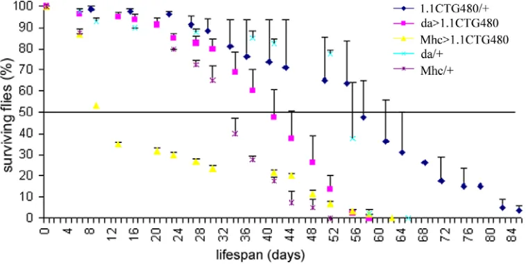
I.2. Lifespan of flies over-expressing CUG repeat RNA. ...87
I.3. Effect of anti-myotonic drug mexiletine in mblE27/mblk7103 mutants. 89 I.3.1. Viability analysis. ...90
I.3.2. Lifespan analysis. ...93
I.3.3. Climbing ability analysis. ...96
I.4. Analysis of alternative splicing in Drosophila DM models. 98 I.4.1. Chloride channel. ...98
I.4.2. α-actinin...101
I.4.3. Troponin T. ...105
II. Analysis of Muscleblind binding to RNA. ...108
II.1.1. Co-localisation of Muscleblind proteins with expanded CUG containing-RNA in mammalian cells....109
II.1.2. Interaction of Muscleblind proteins with CUG repeat containing-RNA and α-actinin transcript in a yeast three hybrid assay. 113 II.1.3. in vitro UV-crosslinking assay for binding to α-actinin RNA. 119 III. Analysis of Muscleblind molecular function...126
III.2. Expression of CUG repeat RNA alters Drosophila α
-actinin minigene splicing in human cells. ...130
III.3. Drosophila Muscleblind isoforms regulate mouse Tnnt3 minigene splicing in human cells. ...132
III.4. Analysis of Mbl protein implication in cell death...134
IV. Molecular basis of isoform specific behaviour. ...136
IV.1. Analysis of sub-cellular localisation of Muscleblind proteins. 137 IV.2. Analysis of the functional relevance of a putative MblC sumoylation site...139
Discussion ...143
1. mblE27/mblk7103mutants, a model to study muscleblind function in late development...146
2. Conservation of Muscleblind molecular function. ...150
3. Functional interaction with Bruno proteins...154
4. Conservation of Muscleblind sequestration by expanded CUG containing RNA in flies. ...156
5. Novel muscleblind targets affected by expression of expanded CUG repeat RNA...158
6. Muscleblind isoforms are functionally distinct. ...161
7. Basis of isoform specific behaviour. ...164
8. Is the binding to physiological targets separable from the binding to pathogenic CUG repeat containing RNA?...167
9. Implication in new processes: apoptosis. ...169
Conclusions ...171
Resumen en castellano...175
INTRODUCCIÓN Y OBJETIVOS...176
1. muscleblind es necesario para el correcto procesado
alternativo de los transcritos de la α-actinina y la TroponinaT de
Drosophila. ...180 2. El efecto tóxico de la expresión de un RNA no codificante portador de repeticiones CUG expandidas está conservado en
Drosophila. ...183 3. Las proteínas Muscleblind modifican el procesado
alternativo de minigenes en cultivo celular y unen por su región N-terminal un fragmento de RNA con secuencias consenso para la unión de MBNL1. ...186 4. La sobreexpresión de las proteínas Muscleblind activa la muerte celular en células de Drosophila. ...188 5. La localización subcelular de las distintas isoformas
Abbreviations and gene symbols
3-AT = 3-aminotriazol A = absorbance α−actn = α-actinin
BDGP = Berkeley Drosophila Genome Project BSA = bovine serum albumin
CUGBP = CUG binding protein
DMPK = Dystrophia Myotonica Protein Kinase
ETR-3 = Embryonically lethal abnormal vision Type RNA binding protein 3 IPTG = Isopropyl β-D-1-thiogalactopyranoside
GFP = Green Fluorescent Protein mbl = muscleblind
Mhc = Myosin Heavy Chain
NSDB = 3-[Benzyldimethyl-ammonio]propanesulfonate] o/n = over night
ORF = open reading frame
PCR = Polymerase Chain Reaction PMSF = phenylmethanesulphonylfluoride PTB = Polypirimidine Tract Binding protein RT = Reverse Transcription
SDS-PAGE = sodium dodecyl sulfate polyacrylamide gel electrophoresis ubi = ubiquitous
UTR = untranslated region w = white
Introduction
Human Muscleblind proteins have emerged as a new family of alternative splicing factors, implicated in many developmental processes and diseases. Drosophila muscleblind shows good signs of being a useful model to study Muscleblind protein function in development and the diseases in which the human homologues are implicated.
1. Regulation and developmental consequences of splicing.
Pre-mRNA splicing involves the removal of introns and ligation of the flanking exons. Through alternative splicing, the exonic sequences of a pre-mRNA are combined generating different transcripts from a single precursor. Alternative splicing can generate more transcripts from a single gene than the number of genes in an entire genome. A dramatic example is Drosophila Down Syndrome Cell Adhesion Molecule (Dscam), which potentially encodes 38016 proteins [1]. 40-60% of human genes are thought to undergo alternative splicing [2].
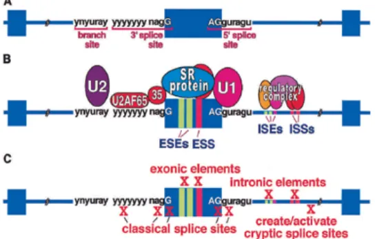
a site will assemble a spliceosome and function in splicing. Additional information and interactions come from many non–splice site regulatory sequences that strongly affect spliceosome assembly (Fig.I.1A,B).
Figure I.1. Classical and auxiliary splicing signalsA) Classical splicing signals found in the major class (>99%) of human introns are required for recognition of all exons. B)
Classical and auxiliary splicing sequence elements and binding factors. Splicing enhancers and silencers in exons (ESEs and ESSs) and introns (ISEs and ISSs) are commonly required for efficient splicing of constitutive and alternative exons. Intronic elements also serve to modulate cell-specific use of alternative exons by binding multicomponent regulatory complexes. C) Cis-acting splicing mutations. Mutations that disrupt cis-acting elements required for pre-mRNA splicing can result in defective splicing that causes disease. (figure from [4]) (n = G, A, U, or C; y = pyrimidine; r = purine).
alternative splicing is spatially and temporally regulated during development, regulating protein expression and generating a complicated network of isoform-dependent protein interactions that establish cell functionality [8-12]. Furthermore, the alternative splicing pattern of a cell can be altered by cell activity [13, 14] and also in response to diverse stimuli [15, 16]. Whereas some splicing decisions are regulated by small variations in general splicing factors, others require specific factors whose expression is highly restricted during development (reviewed in [17-19]). The equilibrium between the levels and activity of general and cell-specific splicing factors decides which isoforms are present in every cell type at each developmental stage. With so much energy dedicated to controlling alternative splicing, it is not surprising that unprogrammed changes in isoform ratio affect cellular functions, frequently leading to disease. Mutations disrupting cis-acting splice sequences (Fig. I.1C), the basal splicing machinery or alternative splicing factors have been linked to cellular transformation, metastasis and various hereditary diseases (reviewed in [4, 20-22]).
2. Muscleblind family of proteins: RNA binding proteins with different functions in RNA metabolism.
Muscleblind proteins are characterized by the presence of Cys3His
5’-UAUU-3’ sub-sites on the ARE region [27]. RNA bases and conserved aromatic residues get intercalated in a stack and both electrostatic and hydrogen-bonding contribute to binding the protein to the RNA.
The characterization of Muscleblind-Like1 (Mbnl1) knockdown (Mbnl1∆E3/∆E3) mice [28] showed that muscleblind function is required for proper alternative splicing regulation. Mbnl1∆E3/∆E3 mice developed myotonia, histological muscle defects, cataracts and impairment of splicing of several muscular transcripts (Table I.1) [28-30]. A set of exons that undergo a synchronized switch between post-natal day 2 and 20 in wild type, were misregulated in Mbnl1∆E3/∆E3 mice [30]. Interestingly, Mbnl1 was translocated from a predominantly cytoplasmic to nuclear distribution during this post-natal interval. In contrast to the Mbnl1 mutant, no myotonia or splicing defects were detected in Mbnl2 knockdown mice and muscle histology was normal.
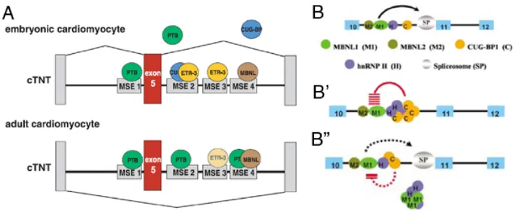
[image:27.612.123.491.377.527.2]
inclusion activators CUG-BP1 and ETR-3 levels, originates a switch in cTNT splicing. E5 is only present in adult cardiomyocytes (figure taken from [31]). B-B”) Model of coordinate regulation of IR splicing in myoblasts. Blue boxes are exons (numbers indicate nomenclature used); lines are introns; circles are proteins. (B) MBNL proteins are required facilitators of IR E11 splicing but a limiting factor might be needed as elevated levels of MBNL proteins do not alter the IR splice equilibrium. (B’) hnRNP H and CUG-BP1 form an RNA-dependent suppressor complex that is required for the maximum repression of IR E11.
(B”) Increased levels of MBNL1 can partially dampen the inhibitory activity of hnRNP H by physical interaction with it.
activators of cTNT E5 inclusion [34, 35], are down-regulated in adult cardiomyocytes. Thus, during the foetal stage, CUG-BP1 and ETR-3 preferentially bind the regulatory regions promoting exon inclusion but, when their levels decrease in adult cardiomyocytes, MBNL1 and PTB repress E5 inclusion. Coordinated physical and functional interactions between hnRNP H, CUG-BP1 and MBNL1 dictate IR splicing in myoblasts [33]. In this case, MBNL1 has been described to activate E11 inclusion by repressing hnRNP H activity (Fig. I.2B). CUG-BP1 and hnRNP H form a repressor complex which is inhibited by binding of MBNL1 to hnRNP H. Hence, the mechanisms by which MBNL1 regulates cTNT and IR splicing seem to be different.
3. Muscleblind expression is regulated both transcriptional and post-transcriptionaly.
Human MBNL1 and MBNL2 expression was detected in brain, kidney, liver, pancreas and muscle tissues [37, 38]. While MBNL2 was found in similar levels in all tissues, MBNL1 was more abundant in skeletal muscle and the heart. MBNL3 transcripts, however, were expressed at much lower levels, with peak expression in the placenta and no expression in skeletal muscle. In contrast to humans, the expression of mouse Mbnl1, Mbnl2 and Mbnl3 genes in adults was more uniform across tissues, although Mbnl1 transcript levels were higher in heart and lower in skeletal muscles [39]. During embryonic development, all three Mbnl genes were prominently expressed in the developing head region and forelimb bud by 9.5 days post conception (dpc). Later in development, the expression patterns of Mbnl1 and Mbnl2 were similar (detected in tongue, mandibular and maxillary regions, lips, thymus, lung and intestines among other tissues) while Mbnl3 expression was stronger in the thymus, lung and intestines. Muscleblind-like2 and 3 (tmbnl2a and tmbnl3) expression patterns have been also characterized in Takifugu rubripes [40]. No differences between juvenile and adult stages were found. Expression of tmbnl2a was ubiquitous but predominant in the heart and brain and mbnl3, in contrast to its vertebrate homolog, was also detected in all analyzed tissues including skeletal muscle. Expression of Drosophila muscleblind mRNA is mainly detected in muscle and the nervous system ([41], see below)
generates transcripts with different motifs in the final protein, which probably confer diverse functional abilities. Nine splicing variants of MBNL1 transcripts, three of MBNL2 and six of MBNL3 have been reported (reviewed in [42]). The C-terminal region was the most variable but some of the isoforms generated also lacked zinc fingers or the linker between them, which are required to interact with RNAs [43]. Alternative splicing also generates four mature transcripts from Drosophila muscleblind, which show different developmental expression patterns and give rise to protein isoforms differing in their C-terminal regions ([44, 45]; see below).
4. Muscleblind proteins in development.
Perhaps murine Mbnl3 is inhibiting the differentiation of hematopoietic stem cells, as it is in the case of human MBNL3 inhibiting myogenesis. Finally, human MBNL3 was upregulated in H295R cells after stimulation with angiotensin. [49].
5. Muscleblind proteins in disease.
Most of the functional data about Muscleblind proteins were generated because of the implication of the MBNL proteins in human diseases. In particular, MBNL proteins are key factors in myotonic dystrophy (DM1 and DM2) [28, 50, 51] and genetic data implicate muscleblind in the Spinocerebellar Ataxia 8 (SCA8) disorder [52]. More recently, MBNL proteins have also been implicated in the pathogenic mechanism of Huntington Disease-Like 2 (HDL2) [53]. Finally, altered levels of MBNL expression have been described in several tumours, squizophrenia, and sporadic idiopathic pulmonary arterial hypertension [36, 54, 55].
5.1. Myotonic dystrophy.
manifestation. The main symptoms of myotonic dystrophies are muscular weakness, myotonia, cataracts, cardiac problems, insulin resistance, male infertility and neurological disorders such as excessive daytime sleepiness. At the molecular level, DM is characterized by a general impairment of alternative splicing regulation, which leads to the presence of foetal transcript isoforms in adult tissues.
5.1.1. Contribution of a mutant RNA gain of function and sequestration of MBNL proteins to DM pathogenesis.
The inability of Dmpk knockout mice to reproduce DM1 symptoms [64, 65], make it unlikely that the disease is a consequence of a lack of DMPK function in the heterozygous condition (haploinsuficiency). Also, CTG repeats have been shown to alter chromatin structure locally, thereby affecting the expression of nearby genes [66]. Extensive similarities between DM1 and DM2, despite the unrelated mutations that cause the diseases, suggested a common mechanism independent of the mutated gene. The observation of nuclear accumulation of mutant RNA into aggregates in the cells of myotonic dystrophy patients led to the RNA gain of function hypothesis: expanded sequences are not translated into a toxic protein but form a secondary structure that stabilizes the mRNA; this mRNA is retained in the nucleus and interferes with the function of diverse nuclear factors, thus leading to disease [67, 68].
Table I.1. Failure of MBNL1-dependent postnatal splicing transitions in DM1 and DM2.
Misregulated alternative splicing in two mouse models of DM1 is concordant with human DM1 and DM2 molecular defects. Mice over-expressing long CUG repeat tract (HSALR) also reproduce these defects. a Indicates the isoform that is preferentially expressed in neonatal muscle at post-natal day 2 (P2) when compared with adult WT muscle. B Denotes exons that show post-natal splicing transition between P2 and P20 in WT hind limb muscle. C Denotes
exons that show miss-regulated alternative splicing in adult (6 month) HSALR transgenic or Mbnl-/- mice when compared with WT mice of appropriate background strain. d Denotes exons that show misregulated alternative splicing in quadriceps muscle from DM1 or DM2 patients compared to healthy individuals (table from [30]).
5.1.2. DM: a network of intricate molecular defects.
changes. One of them seems to involve CUG-BP1. CUG-BP1 is an alternative splicing factor that antagonizes MBNL1 in several RNA targets misregulated in DM [35, 79, 80]. Transgenic mice over-expressing CUG-BP1 also reproduce the typical splicing impairment of DM [81]. Thus, although the mechanism that up-regulates CUG-BP1 activity in DM is not known, it might be contributing to the final systemic effects.
Gene expression is also misregulated in DM. Another factor that is mislocalised in DM1 cells, Sp1, has been implicated in the transcriptional regulation of muscular specific Chloride Channel 1 (CLC1), an RNA that is also mis-spliced in Myotonic Dystrophy. The absence of CLC1 protein is the cause of the myotonia, one of the most severe symptoms of DM patients. Mice over-expressing CUG-BP1 and Mbnl1 knock-down mice also showed strong reduction in CLC1 levels due to an alteration in CLC1 RNA splicing [28, 81] similar to that found in DM patients. This means that at least three factors, MBNL1, CUG-BP1 and Sp1, could be contributing to the absence of CLC1 protein that leads to myotonia in DM. Furthermore, CUG-containing RNA hairpins were described as targets of the RNA interference machinery, which could lead to the misregulation of gene expression by a different mechanism[82].
neither required nor sufficient for foci formation. Moreover, those flies did not show any apparent defect, indicating that RNA foci were not toxic in Drosophila. In contrast, flies expressing 480 CUG repeat RNA showed muscle wasting and degeneration, which were reduced when co-expressing human MBNL1 [84, 85].
It is also very difficult to distinguish the primary effects of expanded RNA nuclear retention and secondary consequences of those primary defects. Fibre type transitions involving changes in gene expression and splicing regulation occur during development and in response to innervation, neuromuscular activity and hormone signalling [86]. In particular, myotonia causes fast muscles to become slower generating characteristic changes in muscular histopathology [87-90]. Therefore, myotonia could be the cause of some of the histological defects and splicing alterations found in DM muscles. Furthermore, it was found that the administration of sera from mothers of children with congenital myotonic dystrophy impairs muscle maturation in rats, suggesting there is a circulatory factor maintaining the immature skeletal muscle found in these patients [91].
6. Drosophilamuscleblind.
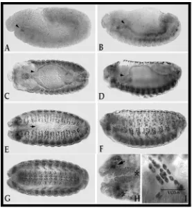
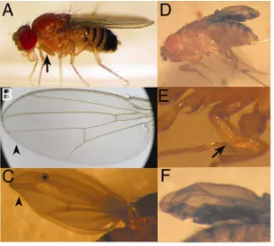
generated during eye development showed that mutant cells fail to differentiate properly. Rhabdomeres were smaller than in wild type tissue and vertical sections showed that they failed to extent into the basal retina [44]. Recently, mutations in the Drosophila muscleblind gene were found to increase fly resistance to starvation [77]. The characterization of mbl null mutant embryos showed disruption of Z bands (Fig. I.3A-B) [41]. Expression of the Z-band specific protein kettin was tested, showing no differences in expression levels between mbl mutants and wild type controls, but the protein failed to assemble properly into the Z bands. mbl mutants also show severe reduction of extracellular tendon matrix at the indirect muscle attachments to the epidermis (Fig. I.3C-F). However, βPS integrin expression and localisation of the tendon matrix component Tiggrin appeared to be normal. These mutants die at a late embryonic stage; in fact, they die as larvae unable to hatch. When the chorion is artificially removed, the larvae show a severe abdominal contraction. With all these results it was concluded that Drosophila muscleblind is implicated in the terminal differentiation of nervous and muscular tissue.
that is typical of genes involved in terminal muscle differentiation as myosin or PS2 integrin [94, 95].
[image:39.612.135.482.121.350.2]Figure I.4. Muscleblind protein is present in somatic, visceral, and pharyngeal musculature. A) Lateral view, stage 11 embryo with Mbl expression in the cephalic mesoderm (arrow) and a barely detectable signal in the remainder of mesoderm. B, C) Mbl expression restricted to visceral (arrow) and somatic mesoderm in a lateral view of a late germ-band retracting embryo (B) and a dorsal view of a late stage 13 embryo (C). D)
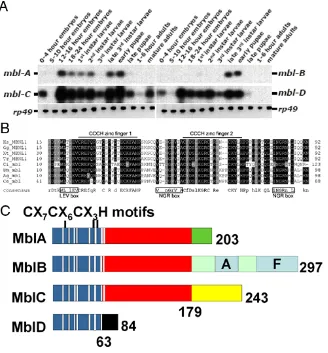
Figure I.5. Alternative splicing generates four mbl transcripts coding four CCCH proteins with specific C-terminal regions. A) The analysis of muscleblind transcript expression by RT-PCR showed isoform-specific developmental regulation (Figure from J. Houseley from the Institute of Biomedical and Life Sciences, University of Glasgow, UK). B)
Amino acid alignment within the first two CCCH zinc fingers from flies (Dm), Anopheles gambiae (Ag), Caenorhabditis elegans (Ce), human (Homo sapiens; Hs), Gallus gallus (Gg),
muscleblind transcripts undergo alternative splicing, giving rise to four transcripts with specific 3’ end sequences [44]. Isoform-specific RT-PCR analysis showed that mbl splicing is regulated during development, as different amounts of the transcript isoforms are detected at different stages of Drosophila life cycle [45] (Fig. I.5A). These transcripts encode four CCCH zinc finger proteins (MblA-D; Fig. I5B-C) that share their amino terminal region [44]. MblA, B and C have two zinc fingers of the CCCH type whereas MblD has just one, as the second is truncated before the last histidine. Several putative sites for post-translational modification were identified recently [42]. Also, two low complexity regions are present in MblB (alanine-rich and phenylalanine-rich) but no functional studies have been performed that demonstrate functionality for any of these domains. Interestingly, rescuing the muscleblind mutant phenotype by expressing UAS-mblA, B and C transgenes showed that the different isoforms rescued to different extent, supporting the idea that Muscleblind isoforms are not functionally redundant [45]. Over-expression of the UAS-mblC transgene in a general embryonic pattern resulted in over 80% rescue of embryonic lethality, whereas mblB and A rescued to a much lower extent.
Objectives
Since their description in 1997 our laboratory has maintained a continued interest in characterizing the molecular functions of Muscleblind proteins in Drosophila and, more recently, their relevance to the mechanism of pathogenesis of myotonic dystrophy. It is within this far-reaching goal that we began this thesis project. We hypothesized that Drosophila could serve as a biomedical model and, in particular, that human and Drosophila Muscleblind proteins performed similar functions in vivo. Therefore, the overall objective of this thesis project was demonstrating the functional conservation between Drosophila and human Muscleblind proteins. We also wondered whether the binding of Muscleblind proteins to physiological targets and to CUG repeat containing RNA had different properties in a way that we could find mutations that inhibited the binding to CUG repeat RNA but left their physiological function uncompromised. This overall aim required addressing the following specific research objectives:
1. Assessment of conservation of the myotonic dystrophy pathogenesis pathway in Drosophila.
2. Assessment of conservation of the alternative splicing regulatory function described for human MBNL proteins in Drosophila Muscleblind.
3. Evaluation of functional diversification of Drosophila Muscleblind protein isoforms as naturally occurring protein variants.
Materials and methods
MATERIALS
I. Drosophila melanogaster strains.
Oregon-R Depto. Genética, Facultad de Biología, U.V.
y1 w1118 Depto. Genética, Facultad de Biología, U.V.
w; mblE27 /CyO [44] mblk7103/CyO [46]
dgo/CyO, ubiquitous (ubi)-GFP available in our laboratory from Muñoz, S. y1w1118; UAS-(CTG)480_1.1 [85]
y1w1118; UAS-(CTG)480_2.1 [85]
w; daughterless-Gal4 Bloomington Stock Center (Indiana) Mhc-Gal4; ry- [98]
mblk7103/CyO, ubiquitous-GFP generated in this work mblE27/CyO, ubiquitous-GFP generated in this work
w,mblE16/CyO, ubiquitous-GFP available in our laboratory from Monferrer, L.
y1w1118; mblE27/CyO, y+; P[w+mC=UAS-mblC:GFP]T15.3 available in our laboratory from Pascual, M.
II. Escherichia coli strains.
relA1. (obtained from Swanson, M., Dept. of Molecular Genetics and Microbiology, University of Florida, USA)
SURE: e14–(McrA–) ∆(mcrCB-hsdSMR-mrr)171 endA1 supE44 thi-1 gyrA96 relA1 lac recB recJ sbcC umuC::Tn5 (Kanr) uvrC [F´ proAB
lacIqZ∆M15 Tn10 (Tetr) Amy Camr] (Stratagene; obtained from Swanson,
M., Dept. of Molecular Genetics and Microbiology, University of Florida, USA)
BL21: F– ompT hsdSB(rB– mB–) gal dcm (obtained from Smith, C., Dept. Biochemistry, University of Cambridge)
BL21 star (DE3): F- ompT hsdSB (rB-mB-) gal dcm rne131 (DE3) (obtained from Smith, C., Dept. Biochemistry, University of Cambridge) Rosetta (DE3): ∆(ara–leu)7697 ∆lacX74 ∆phoA PvuII phoR araD139 ahpC galE galK rpsL (DE3) F'[lac+ lacI q pro] gor522::Tn10 trxB pRARE2 (CamR, KanR, StrR, TetR) (obtained from Smith, C., Dept. Biochemistry, University of Cambridge)
III. Sacharomyces cerevisiae strains.
L40 coat: MATa, ura3-52, leu2-3,112, his3∆200, trp∆1, ade2, LYS::(LexA op)-HIS3, ura3::(LexA op)-LacZ, LexA-MS2 coat (TRP1) [99] (obtained from García, C., Depto. Bioquímica, Facultad biología, U.V.)
IV. Cell lines.
COS: Simian fibroblasts (CV-1 cells) transformed by SV40 that is deficient in the origin of replication region (obtained from Swanson, M., Dept. of Molecular Genetics and Microbiology, University of Florida, USA, and Smith, C., Dept. Biochemistry, University of Cambridge).
HEK293T: Embryonic human kidney containing SV40 large T antigen (obtained from Swanson, M., Dept. of Molecular Genetics and Microbiology, University of Florida, USA, and Smith, C., Dept. Biochemistry, University of Cambridge).
S2: Schneider's Line S2 obtained from dissociated embryos (obtained from Zhou, L. Dept. of Molecular Genetics and Microbiology, University of Florida, USA, and Llorens, J., Depto. Genética, Facultad de Biología, Universidad de Valencia).
V. Drosophila melanogaster cDNAs.
muscleblind (mbl) transcript cDNAs in pBluescript were available in our laboratory [44]. Accession numbers are the following:
• mblA AF001625 • mblB AF001626 • mblC AF001536 • mblD AF001422
bruno cDNAs and putative CLC1 homologues were obtained from the Berkeley Drosophila Genome Project (BDGP):
• CG6942 GH23529 • CG8594 RE63672 • CG5284 LD07266
VI. Vectors.
pEGFP-N3: expresses a protein of interest fused in N-t to GFP in eukaryotic cells (Clontech)
pEGFP-C1: expresses a protein of interest fused in C-t to GFP in eukaryotic cells (Clontech)
pACT2: expresses the protein of interest fused to the Gal4 activation domain in S. cerevisiae [99]
pIIIAMS2.2: expresses RNAs fused to two MS2 binding sequences in S. cerevisiae [99]
pFP105 (modified from pFP98): expresses GST and His6-tagged proteins
in E.coli [44]
pGEX-6P-1: expresses GST-fused proteins in E.coli (Pharmacia) pIEI-4: expression vector for DrosophilaS2 cells (Novagen)
pSG5: vector forminigene expression in eukaryotic cells (Stratagene) pSP72: vector for expanded DMPK 3’UTR expression in eukaryotic cells (Promega)
pGEM4T3: control DNA in cell transfection assays (Amersham)
VII. Constructs.
pIEI-Diap1 obtained from Zhou, L. (Dept. of Molecular Genetics and Microbiology, University of Florida, USA)
MBNL1-GFP obtained from Swanson, M. (Dept. of Molecular Genetics and Microbiology, University of Florida, USA)
pSP72-3’DMPK300 obtained from Swanson, M.
pSG5-tnnt3 obtained from Swanson, M.[29]
pEGFP-N3-MblC∆sumo available in our laboratory from Pascual M.
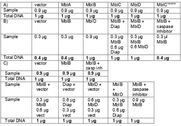
Table M.1. presents a summary of constructs involving Muscleblind isoforms generated in this work
pEGFP-N3 (GFP) pMV1 (myc) pMV2 (myc-GFP) pIEI4 (myc) pFP105
(GST/His6)
pGEX6
(GST)
pACT2
(G4AD)
MblA + + + + + +
MblB + + + + * # +
MblC + + + + + # +
MblD + + + + +
MblC∆SUMO \ + +
ZC +
CCCH +
Table M.1. Epitope tagging of Muscleblind coding sequences generated in this work.
First lane shows vectors used. The epitope to which Mbl is fused to is represented in brackets (GFP = Green Fluorescent Protein; G4AD = Gal4 activation domain). ZC is a fragment containing the common region of MblA, B and C. CCCH is a fragment containing
VIII. Primers.
name sequence use t ºC TNTE2 CGACGATGAAGAGTACAC
TNTE6 CTCTGGATCGCCCTCTCC
Drosophila troponin T
55
TNT4 TTTTCAACCCTAAACCGTAGC
TNT200 ACTCGGTGATGTATTCTTTCAG
Drosophila troponin T
56
6942/E12 GAAGTCCACCACGCTCCAGG
6942/E17 GCTCCCATTGCTTCTGATCCTCCG
Drosophila CG6942
6942/E1 CCGAAGATCAAGTCGCACGCC
6942/E4 GAGCTTTCTGGCTTCATCC
Drosophila CG6942
5284/E7 GGGTGTTATTGGCACATTC
5284/E9 GAAGGTTCTCAACATCGTCC
Drosophila CG5284
60
ACTE5 TTATCCATCGCCATCGTC
ACTE10 TTGAAGTTGGTCTCCAGC
Drosophila α-actinin
65
RP49for ATGACCATCCGCCCAGCATAC
RP49rev ATGTGGCGGGTGCGCTTGTTC
Rp49 65
Xho-Mbl CCGCTCGAGATGGCCAACGTTGTCAA TATG
5’Mbl ORF in pEGFP-N3
See reverse primer
MblA-gc-E CGGAATTCCGAATTGACTTCATTGGA TAC
3’MblA ORF in pEGFP-N3
55
MblB-gc-E CGGAATTCCGGCATGCAACAAAAAAG GC
3’MblB ORF in pEGFP-N3
60
MblC-gc-E CGGAATTCCGTCTTGGCACACCGGGA GGG
3’MblC ORF in pEGFP-N3
65
MblD-gc-E CGGAATTCCGGCAGATTAATTTTTTA CTTAC
3’MblD ORF in pEGFP-N3
60
Eco-Mbl GAATTCCGGCCAACGTTGTCAATATG AACAGCC
5’Mbl ORF in pACT2
See reverse primer
MblA-Xho CCGCTCGAGCAATTGACTTCATTGGA TAC
3’MblA ORF in pACT2
55
MblB-Xho CCGCTCGAGCGCATGCAACAAAAAAG GC
3’MblB ORF in pACT2
60
MblC-Xho CCGCTCGAGCTCTTGGCACACCGGGA GGG
3’MblC ORF in pACT2
65
MblD-Xho CCGCTCGAGCGCAGATTAATTTTTTA CTTAC
3’MblD ORF in pACT2
60
ZC-Xho CCGCTCGAGCCTCTAATCTGTCGGAA CGTGG
3’Common Region in pACT2
CCCH-Xho CCGCTCGAGCCTTGAGGGCCAAATGA TTGCG 3’Zinc Fingers in pACT2 55
Sma-EcoA GGGTATACGCATGCTTCG
Sma-EcoB AATTCGAAGCATGCGTATACCC
3 hybrid adaptor
Xba-SphA CTAGACCATGGATATCCCGGGCATG
Xba-SphB CCCGGGATATCCATGGT
3 hybrid adaptor
ACTE7f CCCACAAGACAATACACC
ACTI7r1 ACATGCATGCCTTAGAACGAGGAAGG C
Actn1 in PIIIA/MS2.2
55
ACTI7f1 GGGCCTTCCTCGTTCTAAG
ACTI7r2 ACATGCATGCGTACCGCTGCGACCTT G
Actn2 in PIIIA/MS2.2
55
Nde-Mbl GGAATCCATATGATGGCCAACGTTGT CAATATG
5'Mbl ORF in pFP105
See reverse primer
MblB-Eco CGGAATTCGCATGCAACAAAAAAGGC 3’MblB ORF in pFP105
65
MblC-Eco CGGAATTCTGGCACACCGGGAGGG 3’MblC ORF in pFP105
65
MblD-Eco CGGAATTCGCAGATTAATTTTTTAC 3’MblD ORF in pFP105
65
ZF-Eco GCATCAGTGAATTCTTGAGGG 3’Zinc Fingers in pFP105
60
T7actn1D CGTAATACGACTCACTATAGGGAACC CACAAGACAATACACC
T7actn1R ATACTTAGAACGAGGAAGGC
Actn1 in vitro transcripti on template
63
T7actn2D CGTAATACGACTCACTATAGGTATGC CTTCCTCGTTCTAAG
T7actn2R CTGCGACCTTGGCAGTG
Actn2 in vitro transcripti on template
57
T7actn5R CTGTGAACGTGTGCGTGTTG Intron 6 in vitro
transcripti on template
65
T7actn6D CGTAATACGACTCACTATAGGGCCCA AAAGGTCTGTTATACG
T7actn6R CTTGAGCACCTTGCAGATCC
Intron 7 in vitro
transcripti on template
65
T7CUGD CGTAATACGACTCACTATAGGTCCTT GTAGCCGGGAATG
T7CUGR AATGGTCTGTGATCCCCC
CUG repeats in vitro transcripti on template
T7CAGD CGTAATACGACTCACTATAGGAATGG TCTGTGATCCCCC
T7CAGR TCCTTGTAGCCGGGAATG
CAG repeats in vitro transcripti on template
myc-dir TCGACGAGCAGAAGCTGATCAGCGAG GAGGACCTGG
myc-rev GATCCCAGGTCCTCCTCGCTGATCAG CTTCTGCTCG
myc tag into pEGFP-N3
5ACTMGE5 GGCGAATTCTTCTGCGCCCTTATCCA TCGC
3ACTMGE9 GGCGAATTCCCAGACGCTCGTACTCC TCCATG
α-actinin minigene
60
1938 GCTGCAATAAACAAGTTCTGCTTT
1956 AGAATTGTAATACGACTCACTATAGG GC
pSG5 56
S-myc-Nf TCGACGAGCAGAAGCTGATCAGCGAG GAGGACCTGGGAGCGGGCCCATAGTA GGC
S-myc-Nr GGCCGCCTACTATGGGCCCGCTCCCA GGTCCTCCTCGCTGATCAGCTTCTGC TCG
myc tag to build pMV
Xho29 GGCCTCGAGATGTTCACCAGCCGCGC TTC
29Eco GGCGAATTCCAGTAGGGCTTCGAGTC CTTGG
Bru1 ORF in pEGFP-N3
55
Xho19 GGCCTCGAGATGATGTTGCAATCCTT GAG
19Bam GGCGGATCCTAAAAATTGCAAGTCGG AAAATGG
Bru2 ORF in pEGFP-N3
60
Xho31 GGCCTCGAGATGGTTCATATTATTGA ATTG
31Eco GGCGAATTCCAATAGGGTCGACTGGC ATC
Bru3 ORF in pEGFP-N3
55
METHODS
I. Drosophila work.
I.1. Sequence homology searches.
Human nucleotide sequences were used to search homologous sequences in the Drosophila genome with the Washington University Blast software package at http://blast.wustl.edu
I.2. Assessment of mexiletine effect.
I.2.1. Mexiletine administration.
The mexiletine dose for humans is 8.6 mg/ kg·per day. Assuming an average larval weight of 1 mg, that supposes a dose of 3.6*10-5 µg/h for
I.2.2. Fly viability.
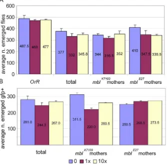
w; mblE27 /CyO and mblk7103/CyO flies were crossed with dgo/CyO, ubi-GFP to obtain mblE27/CyO, ubi-GFP and mblk7103/CyO, ubi-GFP flies. mblE27/CyO, ubi-GFP and mblk7103/CyO, ubi-GFP flies were crossed to obtain the transheterozygous mblE27/ mblk7103, which behaved as a hypomorphic mutation and reduced adult fly viability. Oregon-R (OrR) flies were used as wild type control for the potential toxic effects of mexiletine. 60 virgin females and 36 males were crossed in culture bottles. Emerged adults were counted daily. mblE27/ mblk7103 flies were identified as GFP negative using a GFP fluorescence module mounted on a Leica MZ APO stereo microscope.
Two crosses with 1x and 10x doses of commercial mexiletine were set up with mbl strains in both directions and OrR. Excipient was added at 10x dose as control.
I.2.3. Lifespan.
Viable mblE27/ mblk7103 flies from the viability study were kept in tubes with the same dose of mexiletine and the number of dead flies was counted every day when possible. Fly food was changed twice a week. At least 90 flies, in seven to eight samples, were analysed in this assay from each genotype/mexiletine dose combination.
w; daughterless-Gal4 and Myosin heavy chain-Gal4; ry- males to express the CUG repeat containing RNA in a general or muscle-specific pattern, respectively. Flies carrying the UAS and the Gal4 insertions alone were used as controls. Twenty flies were introduced in each vial and a minimum of two replicas per genotype were made. For those days where dead flies were not counted, the last number registered is taken as an approximation to calculate the percentage of surviving flies that day.
I.2.4. Climbing assay.
mblE27/CyO, ubi-GFP and mblk7103/CyO, ubi-GFP flies were crossed in both directions using 15 virgin females and 8 males in vials containing a 1x or 2x dose of mexiletine or excipient. 30 synchronized flies (collected within 48 h periods) were tested for their climbing ability by counting the number of flies that were above a line at eight cm height after 18 s. Two tubes per cross direction were made at each mexiletine dose. Five replicates per assay were made. The day of the assay flies were changed to a new tube to increase their climbing activity. As the assay tube was larger than the habitual culture tube, flies were allowed ten minutes to get used to it. The experiment was carried out at 25ºC and measurements were around noon.
I.3. In situ detection of transcripts in Drosophila embryos.
M.2. summarizes the labelling reactions performed. Embryos were analyzed with a DM LB2 Leica microscope using bright field microscopy.
Gene cDNA Probe Restriction enzyme
RNA polymerase
CG6942 GH23529 Antisense EcoRI Sp6
CG6942 GH23529 Sense XhoI T7
CG8594 RE63672 Antisense NotI T3
CG8594 RE63672 Sense NruI T7
CG5284 LD07266 Antisense XbaI T7
CG5284 LD07266 Sense KpnI T3
Table M.3. Reactives for the generation of RNA probes for in situ hybridization. The table shows cDNAs used as template, the restriction enzyme that linearized the plasmid and the RNA polymerase DNA dependent that was used to carry out the in vitro transcription.
I.4. Analysis of alternative splicing defects.
I.4.1. Fly collection.
y1w1118;UAS-(CTG)480_2.1 females and w; daughterless-Gal4 males. OrR and y,w flies were collected as controls.
To further characterise tnT alternative splicing, mblE27/CyO, ubi-GFP and mblk7103/CyO, ubi-GFP flies were crossed en masse in culture bottles. 10 to 22 h after egg laying embryos, 1 to 2 day-old pupae and adults of more than six hours expressing CUGs (1.1 strain) with the Myosin heavy chain (Mhc) pattern were also collected. OrR, heterozygous mblE27/ CyO, ubi-GFP and mblk7103/ CyO, ubi-GFP, y1w1118;UAS-(CTG)480_1.1 and Mhc-Gal4; ry- flies were collected at the same stages as controls.
I.4.2. RNA extraction.
Total RNA was extracted with Tri-reagent (Sigma) following the recommendations from manufacturer and treated with DNase I (Invitrogen). RNA concentration was measured by a spectrophotometer (Eppendorf). Only RNA extractions with A260/A280 and A260/A230 ratios over 1.8 were
saved.
I.4.3. RNA quality on gel.
RNA quality was checked loading samples with 50 % formamide and 17.5 % formaldehyde on 1.2 % agarose, 5.5 % formaldehyde gels. 20 mM MOPS, 5 mM NaAc, 1 mM EDTA pH 7 buffer was used to prepare the gel and the samples, and to run the electrophoresis.
Reverse transcription (RT) was performed with 1 µg of total RNA, Superscript II RNase H- and random hexamers following instructions from the provider (Invitrogen). cDNA was used in a standard 20 µl PCR reaction of 25 cycles with Taq DNA polymerase (Need) following the recommendations from the manufacturer to amplify endogenous Drosophila transcripts. Products were separated on 2% agarose gels.
TNT4/ TNT200 primers were used to amplify troponinT (tnT) mRNA from 1 µl of RT reaction. To increase specificity we used a nested PCR approach with the internal TNTE2/TNTE6 primer pair and 1 µl of a 1:100 (or 1:1000 dilution) of the first PCR as template.
Primers 5284/E7 and 5284/E9 were used to amplify alternatively spliced region of CG5284. 2 µl of cDNA were necessary to detect any RT-PCR product. Primer pairs 6942/E12-6942/E17 and 6942/E1-6942/E4 were used to amplify CG6942 mRNA splicing products.
To characterize Drosophila α-actinin (α-actn) splicing pattern, 4 µl of the RT reaction were used as template in a standard 40 µl PCR reaction of 40 cycles with primers ACTE5 and ACTE10. PCR products were purified by NH4Ac precipitation and half of the final volume was digested with SacI to
distinguish α-actn isoforms.
were also amplified with the same primer pairs used to amplify tnT and α -actn cDNA sequences and showed no amplification.
I.5. UV crosslinking and immunoprecipitation (CLIP) of RNA bound
to MblC in vivo.
I.5.1. Sample collection and UV crosslink.
Virgin y1w1118; mblE27/CyO, y+; P{w+mC=UAS-mblC:GFP}T15.3 homozygous female flies were crossed en masse with w; daughterless-Gal4 homozygous males in large laying pots with agar-sucrose petri dishes daubed with fresh yeast paste. Flies were allowed to mate for at least 12 h. Laid eggs, in plates, were incubated 10 additional hours prior to collection. da-Gal4; UAS-mblC:GFP embryos were bleach decorionated and extensively washed with running tap water. Embryos were transferred to a 15 ml conical tube with cold Hank’s Balanced Salt Solution (HBSS; GIBCO). Embryos were gently centrifuged at 800 rcf for 10 min at 4ºC to remove HBSS buffer. Supernatant was decanted and 1 ml of fresh HBSS was used to resuspend embryos prior to crosslink. In order to obtain a cell suspension, in a second experiment embryos were transferred to eppendorf tubes and homogenized with plastic pestles. A third experiment was carried out using a 5 ml homogenizer. GFP signal from individual cells was checked under fluorescence microscope after homogenization. The same washes were done with the cell suspensions. The final ml was placed in 1 cm tissue culture plates on ice/water bath in Stratalinker at same height as sensor. Three 400 mJ/ cm2 irradiations were performed, which were
collected and spinned at 800 rcf for 10 min at 4ºC. HBSS was removed and the samples frozen at -80ºC until used.
I.5.2. Immunoprecipitation and cDNA synthesis.
The following steps of the CLIP protocol were done by Dr. Dan Tutle (Dept. of Molecular Genetics and Microbiology, University of Florida, USA), while I followed his work. The CLIP protocol was performed as previously described [102] with minor modifications. Immunoprecipitation was carried out using the anti-GFP antibody from Roche as it was previously shown in our laboratory that achieved higher efficiency than other anti-GFP antibodies. The immunoprecipitation worked efficiently and an adequate quantity of radiolabelled RNA was obtained, but ligation of linkers required to synthesise the cDNA failed thus precluding from reaching a final result.
II. Yeast methods.
II.1. Yeast three hybrid assay.
II.1.1. Generation of hybrid RNA expressing constructs.
adaptors were included in a 20 µl ligation reaction. E.coli [XLBlue1 or DH5α strain] transformation and plasmid DNA extraction were carried out by standard protocols [103]. After isolating a (CAG)480 pIIIA/MS2 clone, which
expresses CAG-repeat RNA fused to the MS2 RNA, the orientation of the insert was reversed in order to express CUG repeat RNA fused to MS2 RNA. (CAG)480 pIIIA/MS2 DNA and pIIIA/MS2 empty plasmid were
SmaI/SphI-digested and ligated in a standard 20 µl ligation reaction. The resulting constructs showed a distinctive restriction pattern that allowed the quick verification of CTG and CAG repeat DNA-containing plasmids.
The Actn1 fragment was amplified from Drosophila genomic DNA with primers ACTE7f and ACTI7r1 by high fidelity PCR (Pwo DNA polymerase, Roche). PCR product was digested with SphI and ligated to SmaI/SphI digested pIIIA/MS2 plasmid. This ligation does not reconstitute the SmaI site. Actn2 fragment encompassing a cluster of seven MBNL1 consensus binding sequences was also obtained by high fidelity PCR (Pyrobest, Takara) with ACTI7f1 and ACTI7r2 primers. Cohesive ends ligation did not work and phosphorylated PCR product (standard polynucleotide kinase reaction following instructions from provider; Roche) was blunt-end ligated to SmaI digested pIIIA/MS2 plasmid. The two fragments were sequenced to confirm both sequence and orientation.
II.1.2. RNA structure prediction.
II.1.3. Generation of hybrid protein expressing constructs.
High fidelity PCR reactions following the instructions from the manufacturer (Triple Master, Eppendorf) were performed to amplify Muscleblind open reading frames (ORFs). A common 5’end primer (Eco-Mbl) was used in combination with specific 3’ end primers MblA-Xho, MblB-Xho, MblC-MblB-Xho, MblD-MblB-Xho, ZC-Xho and CCCH-Xho to amplify MblA, B, C and D, the common region of MblA, B and C, and a fragment containing the two zinc fingers respectively. PCR products were cloned in frame with the Gal4 activation domain of pACT2. All constructs were sequence confirmed.
II.1.4. LiAc yeast transformation.
• Pick fresh colony (a big one, no older than two days) in 5 ml YAPD 2x and grow o/n
• Next morning, inoculate 100 ml of fresh YAPD 2x with the o/n culture
• Grow until A600= 0.5-0.7
• Spin 5000 rpm, 5 min, room temperature
• Wash with 25 ml of sterile water: 5 min spin 5000 rpm
• Resuspend in 0.5 ml LiAc/TE 1x (10 mM LiAc; 10 mM Tris HCl; 1 mM EDTA; pH 7.5)
• Prepare DNA in eppendorfs; add 10 µl of 10 mg/ ml carrier DNA (salmon sperm) per tube
• Add 50 µl of cell mix per tube
• Add 300 µl of 40% PEG/ 1x TE/ 1x LiAc freshly prepared from 50% PEG, 10x TE, and 10x LiAc solutions
• Heat shock 15 min 42ºC • Spin 1 min 6000 rpm
• Take off supernatant and resuspend in 100 µl of sterile H2O • Plate 50 µl in selective medium (SD + amino acids)
• Incubate at 30ºC until transformants grow (2-5 days)
This protocol, modified from [105], gave better results than others found in TRAFO website (R. Daniel Gietz, University of Manitoba) or Current Protocols in Molecular Biology [106].
Yeasts were co-transformed with pIIIA/MS2 and pACT2 constructs in all combinations. pACT2-IRP and pIIIA/MS2-IRE were used as positive control interaction [99] and as heterologous protein and RNA for negative controls in our experiments. Empty vectors were also used as negative controls.
II.1.5. Confirmation of interaction specificity.
III. Cell culture assays.
III.1. Subcellular localisation assays in mammalian cells.
III.1.1. Cloning.
pSP72 vector carrying human DMPK 3’UTR with an expanded region of 300 CTG repeats was transformed into SURE cells following provider instructions (Stratagene). The lack of recombination in this strain favours the stability of long repeat DNAs. DNA extracted from individual clones was digested with BsaHI to control for CTG repeat length. A fragment around 950 nt encompassing the repeats was obtained from the clone chosen, which corresponded to approximately 160-200 CTG repeats (for convenience we will refer to this clone as CTG197).
ORFs of Drosophila muscleblind splice forms were amplified from full length cDNAs by high fidelity PCR (Pwo DNA polymerase) using isoform-specific primer combinations. A common 5’ primer Xho-Mbl was used in combination with 3´ primers MblA-gc-E, MblB-gc-E, MblC-gc-E, or MblD-gc-E, to amplify the corresponding ORFs. PCR products were purified, digested, and cloned into the XhoI/EcoRI sites of pEGFP-N3 plasmid in frame with the downstream eGFP reporter gene. pEGFP-N3-MblC∆SUMO
was available in our laboratory.
ligated to the annealed oligos. The presence of the epitope was detected by standard colony PCR [103]. This vector (referred to as pMV2) was XhoI/SalI digested and ligated to MblA, B, C, D and C∆SUMO coding regions
(XhoI/SalI digested) obtained from pEGFP-N3 vector.
III.1.2. Cell transfections.
COS and HEK293T cells grown in Dulbecco´s modified Eagle´s medium (DMEM; GIBCO) supplemented with 10% FBS (Foetal Bovine Serum; GIBCO) were seeded in two-well dishes to a density of 80000 cells per well. The transfection mix contained 6 µl of Fugene (Roche), 2 µg of plasmid DNA, and 180 µl of DMEM. After 15 min at room temperature, cells were transfected with 1/6 of the transfection mixture.
In order to analyse the co-localisation of Muscleblind proteins with CUG repeat containing RNA, COS cell transfection mix included 1 µg of CTG197
plasmid and 1 µg of plasmid pEGFP-N3, which expressed Muscleblind protein isoforms fused to GFP. HEK293T cells were transfected with a mix containing 300 ng of GFP-tagged Mbl protein plasmid, 300 ng of CTG197
and 1.4 µg of carrier DNA.
III.1.3. Protein and RNA sub-cellular localisation
detection.
Fluorescent in situ hybridization (FISH) with Cy3 labelled (CAG)10
Cells were analyzed under epifluorescence microscopy using a Zeiss Axioskop2 mot plus microscope.
III.2. α-actinin minigene splicing assay.
III.2.1. Cloning.
Construction of α-actinin minigene for alternative splicing assays was performed as follows: DNA encompassing Drosophila α-actinin cassette exons 6, 7 and 8 was amplified from wild type genomic DNA using the Triple Master kit and oligonucleotide primers 5ACTMGE5 and 3ACTMGE9. The single PCR product was purified with PCR-product cleaning kit (Qiagen) and digested with EcoRI. pSG5 vector (Stratagene) was linearized with EcoRI. Digested vector and PCR product were phenol/chloroform extracted, quantified by agarose gel electrophoresis and mixed in a standard ligation reaction.
myc tagged Muscleblind and MblC∆SUMO proteins were also generated to
perform this assay. The myc epitope was generated by annealing oligonucleotides S-myc-Nf and S-myc-Nr. The hybridization left 3’ and 5' cohesive ends that were ligated to purified SalI/ NotI digested pEGFP-N3/MblC (see section II.3.1.1.) in order to replace the GFP-encoding DNA with the myc epitope. We refer to this derivative as pMV1 vector. pMV1 carrying mblC ORF was digested with EcoRI/XhoI to release the mblC ORF and the myc-encoding vector was purified. The ORFs of the remaining isoforms were obtained from GFP-tagged constructs in pEGFP-N3 (see section II.3.1.1.) by EcoRI/XhoI digestion and ligation to pMV1. GFP tagged MBNL1 was obtained from Prof. Maurice S. Swanson (Dept. of Molecular Genetics and Microbiology, University of Florida, USA).
myc-tagged Muscleblind proteins were BglII and NotI digested from pMV1 vector and cloned into BamHI/NotI digested pIEI4 for protein expression in Drosophila S2 cells. Notice that BamHI site is not recovered after ligation.
III.2.2. Cell culture and transfection of cell lines.
HEK293T cells were grown to 40-60% confluency in DMEM. HEK293T cells were co-transfected with 2 µg of Drosophila α-actinin minigene and 100 ng of pEGFP-N3 expressing Mbl isoforms, or pMV1 vector, using 2 µl of Lipofectamine (Invitrogen) per well and 200 µl Optimem media (Life Technologies). Four hours after transfection the media was changed to antibiotic-free DMEM media supplemented with 10% FBS.
of Drosophila α-actinin minigene, 0.5 µg of CUG197 and 0.5 µg of
pEGFP-N3 (control) or GFP-tagged Muscleblind protein constructs. Empty pGEM42 vector was used to reach a final 1.5 µg of transfected DNA whenever necessary. 1.5 µl of Lipofectamine (Invitrogen) were used per well.
Drosophila S2 cells were grown in Schneider’s medium supplemented with 10% FBS and 0.1% Streptomycin and Penicillin. For transfection, cells were seeded with a density of 1.8* 106 cells/ well in serum-free medium. 24
h after seeding, cells were transfected with 2 µg of Drosophila α-actinin minigene and 100 ng of Mbl isoforms in pMV1 vector using 8 µl of Cellfectine reagent (Invitrogen).
For Bruno protein activities assessment, COS cells grown in DMEM with 10 % FBS were transfected with 0.5 µg of α-actinin minigene and 0.25 µg of GFP-tagged proteins.
III.2.3. RNA and protein extraction.
Total protein from HEK cells co-expressing GFP-tagged Muscleblind proteins and CUG repeat RNA was extracted with 150 µl of hot 2xSDS loading buffer with freshly added 10% β-mercaptoethanol. Two cycles of hot (>95ºC) and dry ice incubation were made to lysate cells. 20 µl of protein extract were loaded on a 15 % SDS-PAGE gel and scanned for densitometry. Equal amounts of protein were then loaded on a 15 % SDS-PAGE gel and treated as described in the previous paragraph.
III.2.4. RT-PCR.
5 µg of total RNA were used in an RT reaction performed as described in section I.4.4. To detect transcripts arising from the Drosophila α-actinin minigene we used 4 µl of cDNA (7 µl when working with S2 cells) in combination with primers 1938 and 1956 in a 40-cycle PCR reaction (for primer sequences and annealing temperatures see table M.2). PCR products were purified by NH4Ac precipitation and half of the final volume
was digested with SacI to differentiate α-actinin isoforms A and B, which otherwise have the same size. The remaining PCR products and entire digestions were loaded in a 2% agarose gel. 1DAdvanced Phoretix software from Nonlinear dynamics was used to quantify the bands.
Alternatively, PCR products were radiolabelled by including 32.5 µCi of (α32P)-dCTP (PerkinElmer Life Sciences) in the 27-cycle PCR.
visualized by autoradiography using Biomax MS film (Eastman Kodak). Bands were quantified with a Molecular Dynamics Phosphorimager.
III.3. mouse Tnnt3 minigene splicing assay in human cells.
HEK293T cells grown and seed as above (section II.3.2.2.), were transfected with 0.5 µg of mouse Tnnt3 minigene and 0.5 µg of GFP-tagged Muscleblind and Bruno proteins, or empty pEGFP-C1 vector, using 1 µl of Lipofectamine in presence of Optimem Media. Proteins were extracted with 2xSDS loading buffer (see section II.3.2.3) and total RNA was extracted with Tri reagent (see section II.1.4.2). As Tnnt3 minigene is also in pSG5 vector, detection of minigene products was performed with non-radioactive PCR as described in section II.3.2.4.
III.4. Cell death assay.
III.4.1. Cell culture and transfection of S2 cells.
Drosophila S2 cells were grown in Schneider’s medium with 10% FBS in 75 cm2 flasks. 24 h prior to transfection, cells were seeded to 4.5 * 105
cells/ well in 24-well plates.
Prior to transfection, two 1.5 ml sterile tubes were UV irradiated per sample; the tube containing the expression vectors will be called A and the one containing the transfection reagent, B.