In situ
Raman characterization of minerals and degradation processes in a variety of
cultural and geological heritage sites
F. Gázquez
a,⁎
, F. Rull
b, A. Sanz-Arranz
b, J. Medina
b, J.M. Calaforra
c, C. de las Heras
d, J.A. Lasheras
d aDepartment of Earth Sciences, University of Cambridge, Downing Street, Cambridge, Cambridgeshire CB2 3EQ, United Kingdom
bDepartment of Condensed Matter Physics, Crystallography and Mineralogy, University of Valladolid, Paseo de Belén, 7, 47011 Valladolid, Spain c
Water Resources and Environmental Geology Research Group, University of Almería, Crta.Sacramento s/n, 04120, La Cañada de San Urbano, Almería, Spain
d
Museo de Altamira, Ministerio de Educación, Cultura y Deportes, 39330 Santillana del Mar, Cantabria, Spain
a b s t r a c t
a r t i c l e i n f o
Article history:
Received 20 September 2015 Received in revised form 8 January 2016 Accepted 12 April 2016
Available online xxxx
We test the capabilities ofin situRaman spectroscopy for non-destructive analysis of degradation processes in in-valuable masterpieces, as well as for the characterization of minerals and prehistoric rock-art in caves. To this end, we have studied the mechanism of decay suffered by the 15th-century limestone sculptures that decorate the retro-choir of Burgos Cathedral (N Spain).In situRaman probe detected hydrated sulfate and nitrate minerals on the sculptures, which are responsible for the decay of the original limestone. In addition,in situRaman anal-yses were performed on unique speleothems in El Soplao Cave (Cantabria, N Spain) and in the Gruta de las Maravillas (Aracena, SW Spain). Unusual cave minerals were detected in El Soplao Cave, such as hydromagnesite (Mg5(CO3)4(OH)2·4H2O), as well as ferromanganese oxides in the black biogenic speleothems recently discov-ered in this cavern. In the Gruta de las Maravillas, gypsum (CaSO4·2H2O) was identified for thefirst time, as part of the oldest cave materials, so providing additional evidence of hypogenic mechanisms that occurred in this cave during earlier stages of its formation. Finally, we present preliminary analyses of several cave paintings in the renowned“Polychrome Hall”of Altamira Cave (Cantabria, N. Spain). Hematite (Fe2O3) is the most abun-dant mineral phase, which provides the characteristic ochre-reddish color to the Altamira bison and deer paint-ings. Thus, portable Raman spectroscopy is demonstrated to be an analytical technique compatible with preserving our cultural and natural heritage, since the analysis does not require physical contact between the Raman head and the analyzed items.
© 2016 Elsevier B.V. All rights reserved.
Keywords:
In situRaman spectroscopy Cultural heritage Speleothems Rock-art pigments Burgos Cathedral Altamira Cave El Soplao Cave Gruta de las Maravillas
1. Introduction
In most cases, mineralogical investigations of cultural and geological items require an initial phase of mineral sampling, which may some-times pose a threat to the preservation status of a protected heritage site. For instance, detailed mineralogical characterization of building materials has enabled the detection of the source of stone pathologies in a variety of historical buildings and masterpieces [1–6, among others]; however, the need for material sampling (in the order of grams) for subsequent destructive analysis in the laboratory can dam-age the masterpieces or other elements of interest.
Another example of potentially destructive sampling is given by mineralogical studies in protected caves, which are usually
sites of significant aesthetic, geological and touristic relevance[7]. Oversampling and bad practice on the part of researchers during field work might produce severe damage in the caverns[8,9], in particular, to speleothems [10]. Remarkably, some caves host both relevant geological and archeological features that lead cave managers and authorities to maximize preservation measures. The restrictive policies for material sampling and the impossibility of remaining for long periods in these subterranean environments, due to visitor perturbations on microclimate[11], are in direct contrast with the need to characterize the composition and origin of the elements that are subject to protection.
In this context, portable, and non-invasive analytical techniques pro-vide new opportunities for studying the mineralogical composition of delicate and precious materials, without the need for sample prepara-tion. In the present study, we test the capabilities of a miniaturized Raman spectrometer forin situnon-destructive analysis of minerals in Spectrochimica Acta Part A: Molecular and Biomolecular Spectroscopy xxx (2016) xxx–xxx
⁎ Corresponding author.
http://dx.doi.org/10.1016/j.saa.2016.04.035
1386-1425/© 2016 Elsevier B.V. All rights reserved.
Contents lists available atScienceDirect
Spectrochimica Acta Part A: Molecular and Biomolecular
Spectroscopy
degraded limestone sculptures in Burgos Cathedral (N Spain) and in three Spanish caves that accommodate valuable speleothems and prehistoric rock-art. Raman analyses were utilized in each case to shed light on particular mineralogical issues and unanswered questions.
2. Study sites
2.1. The retro-choir of Burgos Cathedral
In situRaman spectroscopy was used to perform a preliminary screening of the type of materials responsible for the decay of the 15th-century sculptures that decorate the retro-choir of Burgos Cathe-dral (N Spain). The retro-choir comprisesfive retables carved in lime-stone by the French sculptor and architect, Felipe Vigarny. Among them, the ones representing theCrucifixion of Christand theDescent from the Crossare affected intensively by degradation mechanisms, in-cluding limestone disaggregation,flaking, peeling, and stone cracking (Fig. 1).
Burgos Cathedral was declared a World Heritage Site by UNESCO in 1984 (http://whc.unesco.org/en/list/316) and represents the top tourist attraction of the city of Burgos and of the Castile and Leon region, wel-coming up to 350,000 visitors each year. In this case, preliminaryin situRaman analyses allowed for the selection of representative areas
for subsequent sampling and further detailed laboratory analysis using other mineralogical techniques[6].
2.2. El Soplao Cave
El Soplao Cave was opened as a show cave in 2005 and receives up to 200,000 visitors each year. Spectacular helictites and huge speleothems are the most relevant aesthetic features of this mining show-cave[12]. Other unusual speleothems have also been described, including amberine stalactites[13],flowstones containing layers of cemented de-trital materials[14], black ferromanganese crusts and stromatolites[15, 16], frostwork-type speleothems and moonmilk deposits[17], some of which were studied in ourin situsurvey.
2.3. Gruta de las Maravillas
The Gruta de las Maravillas (Cave of Wonders), located in the village of Aracena (Huelva, SW Spain) has been open to the public since 1914, and currently welcomes about 150,000 visitors every year. This cavern boasts a wide variety of speleothem types, including anthodites, sub-aqueous and subaerialflowstones and coralloids, cave raft cones and huge columns. In this study, we analyzed the mineralogical characteris-tics of rare bluish helictites in the Palmatoria sector of the cave. The presence of subaqueous speleothemic calcite crusts and erosive forms, such as cupolas and scallops (usually related to phreatic mechanisms)
has been interpreted as evidence of subaqueous conditions that would have occurred in the earlier stages of formation of the cave[18]. Al-though it has been suggested that thermal-hypogenic mechanisms op-erated during these initial speleogenetic phases[18], no mineralogical or geochemical evidence has yet been found. To shed light on the cave origin, we usedin situRaman to analyze mineral crusts deposited on the marble bedrock that are vestiges of these earlier stages.
2.4. Altamira Cave
In situ Raman analysis of prehistoric rock-art (some 36,160– 15,329 cal BP) in the renowned“Polychromes Hall”of Altamira Cave (rated as the“Sistine Chapel”of the Paleolithic), were also performed, with the purpose of determining the mineralogical nature of the pig-ments used by the Paleolithic artists. This cave was declared World Her-itage Site by the UNESCO in 1985 (http://whc.unesco.org/en/list/310) and was recently reopened to the public under an extremely restrictive visitor regime.
3. Methodology and sampling strategies
3.1. In-situ Raman spectroscopy
In situRaman spectroscopy analyses were done at the sites described above during several surveys between 2012 and 2014. The Raman anal-yses were carried out using a BWTEK Raman device, composed of a BWTEK BRM-OEM-785 diode laser (785 nm), a BWTEK BAC100-785E Raman head, and a BWTEK Prime T BTC661E-785CUST spectrometer with a Hamamatsu CCD (S10141-1107S, 2048 pixels). The equipment covers a spectral range in Raman displacement of 150–3000 cm−1, with a spectral resolution of 5 cm−1measured as FWHM. Spectra were acquired using commercial software provided by BWTEK. The maximum laser power was 270 mW (100%) but was adjusted to lower values using the BWTEK software. The laser was initially set at 10% (~ 30 mW) of its nominal power and was gradually increased up to 40–60% (~100–160 mW) in most analyses. Under these conditions, no thermal degradation of the materials was observed, which is nor-mally evidenced by a drastic increase of the Raman spectrum baseline. The mean integration time ranged from 1 to 10 s and 20–50 accumula-tions were performed for each analysis. The Raman head was coupled with a photographic tripod adapted for analyses of vertical surfaces. In most cases, the analyses were done without the need for the Raman head to touch the materials to be analyzed.
Spectra treatment, including manual baseline correction, was per-formed using OPUS software. Mineralogical identification used a set of algorithms and a Raman spectra database that are being developed in
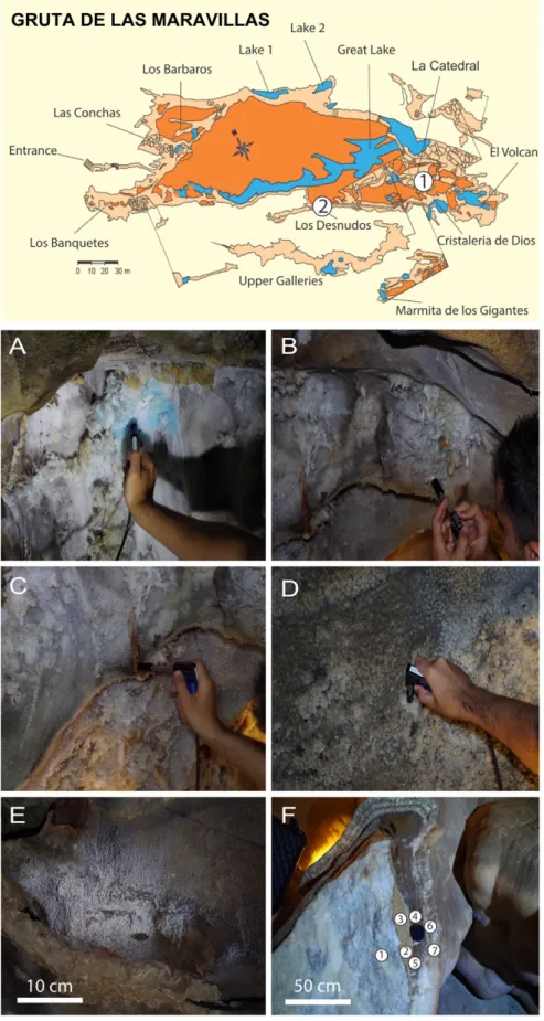
Fig. 2.Topography of El Soplao Cave and location of the study site. A. In situ Raman analysis of a stalactites; B. Moonmilk deposits; C. Frostwork speleothem; D. Alteration materials on the bedrock; E. Ferromanganese stromatolites.
Table 1
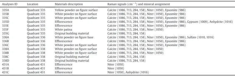
Results ofin situRaman spectroscopy analyses performed in the retro-choir of Burgos Cathedral.
Analyses ID Location Materials description Raman signals (cm−1) and mineral assignment
335A Quadrant 335 Yellow powder onfigure surface Calcite (1086, 713, 284, 158), Niter (1050), Epsomite (986) 335B Quadrant 335 White powder onfigure surface Calcite (1086, 713, 284, 158), Niter (1050), Epsomite (986) 335C Quadrant 335 White powder onfigure surface Calcite (1086, 713, 284, 158), Niter (1050), Epsomite (986)
335D Quadrant 335 Efflorescence Calcite (1086, 713, 284, 158), Niter (1050), Epsomite (986), Gypsum (1009), Anhydrite (1016) 335E Quadrant 335 Efflorescence Calcite (1086, 713, 284, 158), Niter (1050), Epsomite (986)
335F Quadrant 335 White patina Calcite (1086, 713, 284, 158), Niter (1050) 335G Quadrant 335 Original building material Calcite (1086, 713, 284, 158)
336A Quadrant 336 White powder onfigure base Calcite (1086, 713, 284, 158), Niter (1050), Epsomite (986), Sulfate (1010, 1016) 336B Quadrant 336 Efflorescence Calcite (1086, 713, 284, 158), Niter (1050), Epsomite (986)
336C Quadrant 336 White powder onfigure surface Calcite (1086, 713, 284, 158), Niter (1050), Epsomite (986) 338A Quadrant 338 White powder onfigure surface Calcite (1086, 713, 284, 158), Niter (1050)
338B Quadrant 338 White powder onfigure surface Calcite (1086, 713, 284, 158), Niter (1050) 338C Quadrant 338 Original building material Calcite (1086, 713, 284, 158)
338D Quadrant 338 Original building material Calcite (1086, 713, 284, 158) 431A Quadrant 431 Efflorescence Niter (1050)
431B Quadrant 431 Efflorescence Niter (1050)
the framework of the 2018 European Space Agency ExoMars mission [19]. Raman spectra were compared with the ExoMars Raman standard database, which currently comprises over 300 minerals[20], and the RRUFF mineralogical database (www.rruff.info [21]).
3.2. The retro-choir of Burgos Cathedral
A square sampling grid was established over the surface of each of thefive retro-choir panels (each 9 m high and 5 m wide), giving 45 quadrants per panel and a total of over 250, covering a surface of 220 m2. The analyses in this study were conducted on the most deteri-orated panels (theCrucifixion of Christand theDescent from the Cross), especially in the lower and central parts of the panels (quadrants 335, 336, 338 in theCrucifixion of Christand quadrant 441 in theDescent from the Cross). The location of each analysis is given inFig. 1and the mineralogical characterization inTable 1. Seventeenin situanalyses were performed on different materials, including the apparently unal-tered original limestone, as well as efflorescences andflaky patinas.
3.3. El Soplao Cave
Raman analyses in El Soplao Cave were focused on four study areas (Fig. 2): (1) Gorda Gallery (SOP-GOR; 11 analyses) where several
analyses were carried out on the carbonate bedrock of the cave and its overlying alteration materials. In addition, soft globular efflorescences (moonmilk), millimetre-sized acicular aggregates (frostwork) and sugary-texture patina and ochre-color alteration crust on the bedrock, were analyzed (Table 2andFig. 2). (2) Opera Hall (SOP-OPE; 2 analy-ses): moonmilk aggregates and white helictites on the cave ceiling. (3) The Italianos Gallery (SOP-ITA; 6 analyses): frostwork and moonmilk, helictites, stalactites and the carbonate bedrock were stud-ied. (4) False Floor Hall (SOP-FAL; 5 analyses): carbonate helictites and frostwork, as well as ferromanganese stromatolites were analyzed.
3.4. Gruta de las Maravillas
The Raman study in Gruta de las Maravillas focused on two sites (Table 3andFig. 3): (1) Palmatoria Sector (ARA-PAL; 7 analyses), where we analyzed bluish aragonite helictites, moonmilk aggregates and frostwork. (2) Hall of the Nudes (ARA-DES; 7 analyses), in which the marble bedrock, dark alteration crust, brownish laminated carbon-ate layer and whitish carboncarbon-ateflowstones were analyzed (Fig. 3). The analyses were performed on a wall where these materials were exposed as a result of work to adapt the cave for tourism in the early 20th century.
Table 3
Results ofin situRaman spectroscopy in the Gruta de las Maravillas.
Analyses ID Location Materials description Raman signals (cm−1
) and mineral assignment
ARA-PAL-01 Palmatoria sector Blue helictites Aragonite (1086, 706, 207) + Cuprite (217) ARA-PAL-02 Palmatoria sector Blue helictites Aragonite (1086, 706, 207) + Cuprite (217) ARA-PAL-03 Palmatoria sector White helictites Aragonite (1086, 706, 207)
ARA-PAL-04 Palmatoria sector White helictites Calcite (1086, 712, 282)
ARA-PAL-05 Palmatoria sector White helictites Calcite (1086, 712, 282) + Aragonite (1086, 706, 207) ARA-PAL-06 Palmatoria sector Brownish boxwork Calcite (1086, 712, 282) + Quartz (462)
ARA-PAL-07 Palmatoria sector Moonmilk Aragonite (1086, 706, 207) ARA-DES-01 Desnudos sector Marble hostrock Calcite (1086, 712, 282)
ARA-DES-02 Desnudos sector Alteration crust Calcite (1086, 712, 282) + Gypsum (1137, 1007, 493, 411) ARA-DES-03 Desnudos sector Alteration crust Calcite (1086, 712, 282) + Gypsum (1007, 493, 411) ARA-DES-04 Desnudos sector Laminated darkflowstone Calcite (1086, 712, 282)
ARA-DES-05 Desnudos sector Laminated darkflowstone Calcite (1086, 712, 282) ARA-DES-06 Desnudos sector Whiteflowstone Calcite (1086, 712, 282) ARA-DES-07 Desnudos sector Whiteflowstone Calcite (1086, 712, 282)
Table 2
Results ofin situRaman spectroscopy in El Soplao Cave.
Analyses ID Location Materials description Raman signals (cm−1) and mineral assignment
SPL-GOR-01 Gorda gallery Moonmilk Hydromagnesite (1117, 724, 753, 244, 266) SPL-GOR-02 Gorda gallery Frostwork Aragonite (1086, 706, 207)
SPL-GOR-03 Gorda gallery Saccharide textured crystals Aragonite (1086, 706, 207) + Calcite (1086, 712, 282) SPL-GOR-04 Gorda gallery Helictite Aragonite (1086, 706, 207)
SPL-GOR-05 Gorda gallery Moonmilk Aragonite (1086, 706, 207)
SPL-GOR-06 Gorda gallery Moonmilk Aragonite (1086, 706, 207) + Hydromagnesite (1117, 724, 753) SPL-GOR-07 Gorda gallery Moonmilk Aragonite (1086, 706, 207) + Hydromagnesite (1117, 724, 753) SPL-GOR-08 Gorda gallery Moonmilk Aragonite (1086, 706, 207) + Huntite (1122, 315, 272) SPL-GOR-09 Gorda gallery Frostwork Aragonite (1086, 706, 207)
SPL-GOR-10 Gorda gallery Alteration materials on bedrock Dolomite (1097, 719, 285)
SPL-GOR-11 Gorda gallery Bedrock Dolomite (1097, 719, 285)
SPL-OPE-01 Opera hall Helictite Calcite (1086, 712, 282)
SPL-OPE-02 Opera hall Helictite Calcite (1086, 712, 282) + Aragonite (1086, 706, 207) SPL-ITA-01 Italianos gallery Helictite Aragonite (1086, 706, 207)
SPL-ITA-02 Italianos gallery Frostwork Aragonite (1086, 706, 207) SPL-ITA-03 Italianos gallery Frostwork Aragonite (1086, 706, 207) SPL-ITA-04 Italianos gallery Alteration materials on the bedrock Calcite (1086, 712, 282) + Oxides
SPL-ITA-05 Italianos gallery Stalactite Aragonite (1086, 706, 207) + Calcite (1086, 712, 282) SPL-ITA-06 Italianos gallery Stalactite Aragonite (1086, 706, 207)
SPL-FAL-01 Falsefloor hall Frostwork Aragonite (1086, 706, 207) SPL-FAL-02 Falsefloor hall White stalactite Aragonite (1086, 706, 207) SPL-FAL-03 Falsefloor hall Ferromanganese stromatolites Birnessite (576, 680)
3.5. Altamira Cave
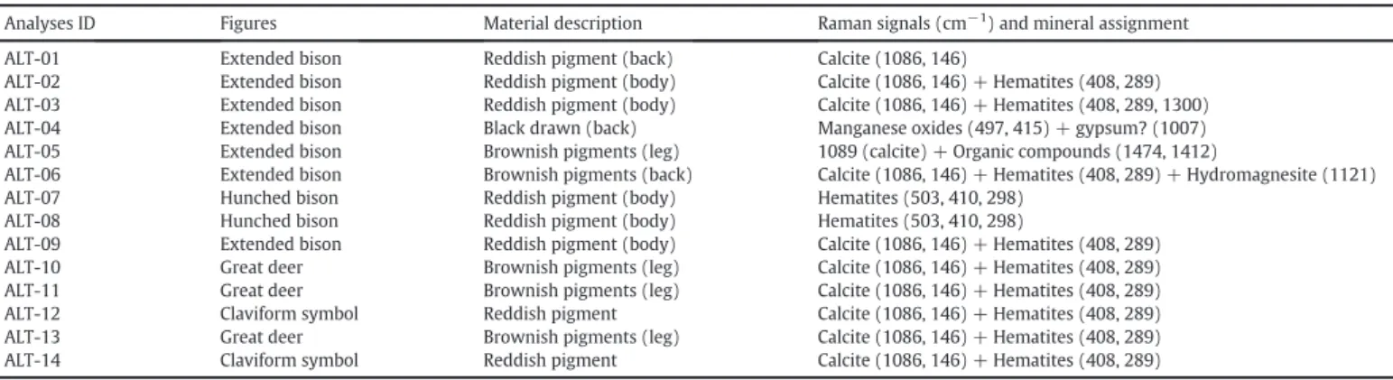
The Raman analyses in Altamira Cave addressed the mineralogical characterization of the pigments that compose certain rock-art features on the ceiling of the Polychrome Hall. This preliminary sampling was devoted to the analyses of deep reddish pigments and blackish traces of an extended bison (7 analyses) and a hunched bison (2 analyses). The brownish pigments of the great deer (3 analyses) and the reddish claviform symbols that appear on the legs of the great deer and the
central part of the chamber (2 analyses) were also studied (Table 4 andFig 4).
4. Results and discussion
4.1. The retro-choir of Burgos Cathedral
In situRaman spectroscopy detected 5 different minerals on the sur-faces of the 15th-century sculptures that decorate the retro-choir of the
Fig. 4.A. Position forin situRaman analysis in the ceiling of the Polychromes Hall of Altamira Cave. B, C. Details of analysis of the cave paintings.
Table 4
Results ofin situRaman spectroscopy of the rock-art paintings of the Polychrome Hall of Altamira Cave.
Analyses ID Figures Material description Raman signals (cm−1) and mineral assignment
ALT-01 Extended bison Reddish pigment (back) Calcite (1086, 146)
ALT-02 Extended bison Reddish pigment (body) Calcite (1086, 146) + Hematites (408, 289) ALT-03 Extended bison Reddish pigment (body) Calcite (1086, 146) + Hematites (408, 289, 1300) ALT-04 Extended bison Black drawn (back) Manganese oxides (497, 415) + gypsum? (1007) ALT-05 Extended bison Brownish pigments (leg) 1089 (calcite) + Organic compounds (1474, 1412)
ALT-06 Extended bison Brownish pigments (back) Calcite (1086, 146) + Hematites (408, 289) + Hydromagnesite (1121) ALT-07 Hunched bison Reddish pigment (body) Hematites (503, 410, 298)
ALT-08 Hunched bison Reddish pigment (body) Hematites (503, 410, 298)
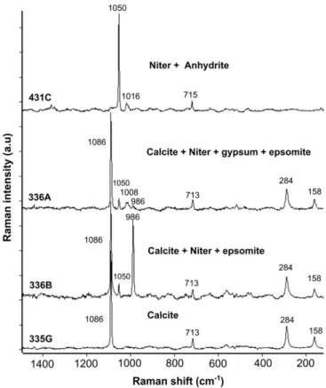
Burgos Cathedral (Fig. 5andTable 1). Calcite (CaCO3) was identified as the primary building material, from which thefigure were carved, whereas niter (KNO3), epsomite (MgSO4·7H2O) and small amounts of gypsum (CaSO4·2H2O) and anhydrite (CaSO4) are present in the effl o-rescences and alteration patina (Fig. 1). These results are in close agree-ment with the results from the detailed mineralogical characterization of alteration materials in this masterpiece, carried out using diverse lab-oratory techniques (Raman and FT-Raman, X-ray diffraction and infra-red spectroscopy)[6]. Though this thorough investigationflagged the presence of 20 different degradation compounds that are responsible for the decay of the original stone[6], Mg-sulphate (mainly epsomite) and K-nitrate (along with Na-nitrate) were found to be the most abun-dant detrimental materials and the main cause of stone disaggregation in the retro-choir. This decay process is attributed to the hydration-dehydration and dissolution-precipitation cycles of certain minerals, such as those in the epsomite-hexahydrite (MgSO4·7H2O/ MgSO4·6H2O) system. The derived stress in the capillary network has been identified as one of the main cause of ageing of building materials [22,23]. In the case of Burgos Cathedral, the damage observed is con-nected to the capillary rise of salts-bearing water from the subsoil[6].
Remarkably, thein situRaman survey in the retro-choir took approx-imately 6 h, whereas over 300 work hours were required for the labora-tory analyses by different techniques of nearly 150 samples[6]. Taking into account that both methodologies produced similar results in terms of identifying the main problem affecting the limestone sculp-tures and mineralogical identification of the most abundant degradation compounds, thein situRaman technique is demonstrated to be a fast and efficient tool for studying stone pathology without the need for ma-terial sampling or sample preparation, as also suggested in recent inves-tigations[3,4,24, among others].
4.2. El Soplao Cave
Thein situRaman analyses in El Soplao Cave detectedfive different carbonate minerals, as well as metallic oxy-hydroxides (Fig. 6and Table 2). Aragonite (CaCO3) is the most common mineral forming part of the speleothems in this cavern, as revealed by the analysis of anthodites, stalactites and frostwork speleothems at the three study sites (Fig. 6). These results agree with the mineralogy offlowstones and frostwork speleothems reported in previous studies of this cave [12]. Aragonite is a rather common mineral in dolostone caves such as El Soplao, where the dolomitic nature of the bedrock has been corrobo-rated by Raman spectroscopy. Dolomite (CaMg(CO3)2) is the main con-stituent of the cave walls which, in places, display a brownish crust also composed of microcrystalline dolomite and calcite (Fig. 6). According to previous investigations [25,26], the high Mg2 +/Ca2 + ratio in the dripwater favors the precipitation of aragonite and inhibits the nucle-ation of calcite. This could explain the overwhelming presence of arago-nite in El Soplao Cave.
On the other hand, hydromagnesite (Mg5(CO3)4(OH)2·4H2O) has been detected as cottonball-like globular aggregates (moonmilk) on the cave walls (Fig. 6), frequently placed at the apex of aragonite acicules. In places, hydromagnesite is accompanied by aragonite and huntite (Mg3Ca(CO3)4). Precipitation of Mg-carbonates in caves has usually been attributed to the evaporation of Mg-rich solutions. In some cases, the presence of these minerals in caves is related to micro-bial activity, though this has not been confirmed for the moonmilk de-posits of El Soplao Cave[17].
The dark laminated deposits of the False Floor Hall are composed of Fe-Mn oxides that in previous studies, were identified by XRD as goe-thite (α-FeO(OH)) and birnessite ([AyMnO2-y·z(H2O)] (where A repre-sents an interlaminar cation). Indeed, the Raman signals at around 500 and 700 cm−1(Fig. 6) can be assigned to birnessite, which is a rather common mineral in subterranean environments[27,28]. The precipita-tion of this metal oxide in the El Soplao Cave is related to the mobiliza-tion of manganese from the host rock probably under phreatic
anaerobic conditions and subsequent precipitation on the walls and floors of the cave as oxides when the water table fell and conditions were again oxygenic[15]. The oxidation of manganese was probably mediated by microorganisms (as suggested by the presence of fossil bacteria inside ferromanganese speleothems discovered in this cave in previous studies[16]).
4.3. Gruta de las Maravillas
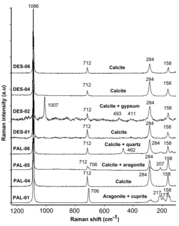
Aragonite and calcite were the most abundant minerals found byin situRaman spectroscopy in the Gruta de las Maravillas. Both minerals form part of helictites, frostwork andflowstones in this cavity, with no clear spatial distribution or differentiation in terms of speleothem mor-phologies. Remarkably, the bluish aragonite anthodite of the Palmatoria Sector displays a weak Raman signal at 217 cm−1that has been assigned to the minor presence of cuprite (Cu2O) (Fig. 7). This signal did not appear in spectra from other whitish speleothems at the same site. Thisfinding is in accordance with earlier trace elements analyses of these speleothems in the Gruta de las Maravillas reported by Del Val et al. (1998) who found up to 183 ppm of copper to be present in these aragonite speleothems[29]. Despite being relatively rare, bluish aragonite speleothems have been described in several caves of Sardinia, Italy[30,31]and France[32]. Cabrol (1978) claimed that copper concen-trations above 50 to 100 ppm in aragonite might give rise to their bluish color.
At the same location in the cave, we analyzed boxwork-type features comprising calcite and quartz (SiO2) (Fig. 3). The term“boxwork”refers to mineral veins in the bedrock which, due to the greater resistance of these infillings (usually calcite, quartz or metallic oxides), protrude from the cave wall after dissolution and/or corrosion of the surrounding host rock[33,34]. In the Gruta de las Maravillas, these veins represent a primitive subaqueous stage in which calcite and quartz precipitated in the cracks of the marble host rock. More recently, subsequent to the
speleogenetic mechanisms that produced the cave galleries and the fall of the water table to below the cave level, subaerial dissolution-corrosion processes preferentially affected the carbonate host rock, which has a microcrystalline structure. In contrast, the veins of calcite and quartz were more resistant to corrosion and were less affected by erosion[34]. In this way, the calcite blades project into the cave in the form of a boxwork.
At the second study site in Gruta de las Maravillas, we analyzed the mineralogical sequence in the Hall of the Nudes, from the Precambrian marble bedrock to the more recent speleothemic calcite crusts (Fig. 3). All the phases showed a predominantly calcite composition, including the marble stone of the bedrock and theflowstones that later grew over the cave walls underwater, due to slow CO2-degassing and conse-quent supersaturation in calcite.
Remarkably, gypsum (CaSO4·2H2O) has been detected, along with calcite, at the contact between the bedrock and thefirst of the laminated flowstone layers. This brownish lamina has a sugary texture and is a vestige of a primitive phase in the cave formation. The presence of gyp-sum at this stage unequivocally indicates that SAS (sulfuric acid speleogenesis) mechanisms were involved during the cavity formation, very probably in a thermal-water environment. Oxidation of pyrite (FeS2), widely hosted in the Precambrian marble of the Aracena Massif
[35], produced a lowering in the water pH, which favored the corrosion
of the marble and simultaneous precipitation of gypsum, following the reaction:
4FeS2+ 15O2+ 18H2O + 8CaCO3→8CaSO4· 2H2O + 4FeOOH + 8CO2. SAS has been proposed as being responsible for the genesis of dozens of caves worldwide[36–40], which in most cases also host gypsum de-posits. Although iron oxyhydroxides are not detected by Raman in the points studied in the Gruta de las Maravillas, ocher-colored limonite-type substances (FeO(OH)·nH2O) have been observed in many parts, infilling cracks in the bedrock; along with corrosion forms typical of hy-pogene speleogenesis. This suggests that the mechanism of pyrite oxi-dation and coupled gypsum/Fe-oxides precipitation described above took place during an earlier stage of development of this cave.
4.4. Altamira Cave
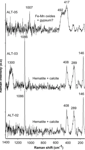
In situanalyses of the outstanding rock-art in the Polychromes Hall of Altamira Cave were performed for thefirst time using Raman spec-troscopy. Hematite was the most common mineral utilized by the Pa-leolithic artists for the representations of bison, deer and claviform symbols on the chamber's ceiling (Fig. 8). The majority of the spectra flagged the presence of calcite, which could be attributed either to cal-cite Raman signals from the substrate limestone bedrock or to second-ary calcite coatings more recently deposited on the ocher pigments, which has been used to date these paintings in recent studies[41].
In one of the spectra, we found a weak signal at 1121 cm−1. This has been attributed to the presence of hydromagnesite on the paintings (Table 4). This mineral was also detected in previous studies on moonmilk deposits in this cave, and pointed out as being a microbial mediated product, potentially detrimental to the conservation of the paintings[42]. It is worth noting that in some spectra we found several
Fig. 7.Results ofin situRaman spectroscopy in the Gruta de las Maravillas. Helictites and carbonate concretions were analyzed in the Palmatoria Sector, showing the Raman spectra of calcite and aragonite (PAL-01, PAL-04, PAL-05, PAL-06). Note the presence of cuprite in the bluish aragonite helictites. The Raman signals observed in the spectra obtained from different materials in the Hall of the Nudes have been mainly assigned to calcite. Remarkably, gypsum has been detected at the contact between the marble bedrock and thefirst subaqueous speleothems.
sharp, weak signals at 1474 and 1412 cm−1associated with calcite, which can be attributed to the presence of Mg-oxalate or glushinskite (Mg(C2O4)·2(H2O)) and NH4+-oxalate or oxammite ((NH4)2 (C2O4)·(H2O)), which are typical metabolic intermediates and bio-markers of bacterial activity[43].
The black traces analyzed in this study showed the Raman spectra of metallic oxides, probably poorly crystalline manganese oxy-hydroxides (Fig. 8), also utilized in other Paleolithic art-rocks (i.e.in Lascaux and Ekain Caves[44]) as pigments for blackish features.
The great abundance of hematite in thesefigures matches the results from previous laboratory analyses of these paintings performed by Mar-tín (1977)[45]. This author found mainly hematite in the ocher-colored drawings and MnO/Mn2O3in the blackish lines, as well as a lesser pres-ence of carbonaceous compounds attributed to the use of charcoal as pigment. Likewise, this mineralogy roughly agrees with the results from the analysis of pigments and pictorial tools of the Altamira site– stored in the collection of the Altamira Museum–that were initially an-alyzed using XRD by Cabrera (1980)[46], and more recently using Raman spectroscopy, XRD and FTIR by Rull et al. (2014)[47].
5. Conclusions
In situRaman spectroscopy has enabled non-destructive mineralog-ical characterization of materials in deteriorated areas of the Burgos Cathedral's retro-choir and in three Spanish caves of geological and pre-historic/historical relevance. Thein situsurvey in Burgos Cathedral de-tected a variety of hydrated sulfate and nitrate minerals on the limestonefigures, which are responsible for the decay of the limestone
sculptures. The mineralogical analysis byin situRaman spectroscopy in El Soplao Cave and the Gruta de las Maravillas found a variety of differ-ent minerals, including anhydrous and hydrated carbonates, as well as metallic oxides. In the case of the Gruta de las Maravillas, gypsum was detected for thefirst time. Its presence at the contact between the mar-ble bedrock and thefirst speleothemic materials suggest that the initial cave stages were linked with SAS (sulphuric acid speleogenesis), which had been previously proposed for the origin of this cavity, but not until now demonstrated by empirical data. Finally, we found that hematite (Fe2O3) is the most abundant mineral phase in the rock-art of the Poly-chromes Hall of Altamira Cave.
Our results from a variety of examples demonstrate that Raman is a non-destructive technique suitable for the analysis of minerals in the field, without the need for material sampling or sample preparation. Thus, it is compatible with the conservation of sites that are protected due to their cultural and/or geological value. In summary, Raman spec-troscopy is confirmed as an alternative methodology to the traditional sampling and gathering of material for the study of minerals and degra-dation mechanisms affecting our cultural heritage, as well as for inves-tigations of geological processes in caves.
Acknowledgments
Financial support for this study was provided by the Banco Santan-der Foundation, through project VA373A12-1, granted by the Regional Government of Castille and Leon, the funds of the Water Resources and Environmental Geology Research Group (University of Almería) and the Project “RLS Exomars Science” (AYA2011-30,291-C02-02; funded by the Ministry of Science and Innovation, Spain and EU FEDER funds). The authors are grateful to the incumbents of Burgos Ca-thedral for providing access to the temple during sampling, and to the managers of El Soplao S.L. for providing access to El Soplao Cave and allowing us to use their facilities. Wenceslao Martinez-Rosales is ac-knowledged for his help during sampling in the Gruta de las Maravillas. Our special thanks to Carlos Sanz for his kind permission to use his photograph.
References
[1] O. Buj, J. Gisbert, B. Franco, N. Mateo, B. Bauluz, J. Geol, Soc. London 331 (2010) 195–202.
[2] A. Duran, M.D. Robador, J.L. Perez-Rodriguez, Int. J. Archit Herit. 6 (3) (2011) 342–358.
[3] J. Dewanckele, M.A. Boone, T. De Kock, W. De Boever, L. Brabant, M.N. Boone, G. Fronteau, J. Dils, L. Van Hoorebeke, P. Jacobs, V. Cnudde, Sci. Total Environ. 447 (2013) 403–414.
[4] O. Gómez-Laserna, M.A. Olazabal, H. Morillas, N. Prieto-Taboada, I. Martinez-Arkarazo, G. Arana, J.M. Madariaga, J. Raman Spectrosc. 44 (2013) 1277–1284.
[5] M. Veneranda, M. Irazola, M. Diez, A. Iturregui, J. Aramendia, K. Castro, J.M. Madariaga, J. Raman, Spectroscopy 45 (2014) 1110–1118.
[6] F. Gázquez, F. Rull, J. Medina, A. Sanz-Arraz, C. Sanz, Environ. Sci. Pollut. Res. 22 (2015) 15677–15689.
[7] A. Cigna, E. Burri, Development, management and economy of show caves, Int. J. Speleol. 29 (2000) 1–27.
[8] S.A. Truebe, J.E. Cole, M. Lee, H.R. Barnett, Proceedings, National Cave and Karst Man-agement Symposium, Midway, Utah, 2011 149–153.
[9] S.A. Truebe, 20th National Cave and Karst Management Symposium, 2013 47–50.
[10] G.S. Springer, NSS News, June 2012 12–14.
[11] A. Fernández-Cortés, J.M. Calaforra, F. Sánchez-Martos, J. Gisbert, Int. J. Climatol. 26 (2006) 691–706.
[12] F. Gázquez, A. Delgado-Huertas, P. Forti, H. Stöll, J.M. Calaforra, J.J. Durán, F. Carrasco, in: Cuevas: Patrimonio (Ed.), Asociación de Cuevas turísticas, Naturaleza, Cultura y turismo, Madrid 2010, pp. 293–304.
[13] F. Gázquez, J.M. Calaforra, F. Rull, P. Forti, A. García-Casco, Int. J. Speleol. 41 (1) (2012) 113–123.
[14] F. Gázquez, J.M. Calaforra, P. Forti, H. Stoll, B. Ghaleb, A. Delgado-Huertas, Earth Surf. Process. Landf. 39 (10) (2014) 1345–1353.
[15] F. Gázquez, J.M. Calaforra, P. Forti, Int. J. Speleol. 40 (2) (2011) 163–169.
[16] C. Rossi, R.P. Lozano, N. Isanta, J. Hellstrom, Geology 38 (2010) 1119–1122.
[17] F. Gázquez, J.M. Calaforra, L. Sanna, Las cuevas turísticas como activos económicos: conservación e innovación, in: y J.J. Durán, P.A. Robledo (Eds.),Asociación Española de Cuevas Turísticas 2012, pp. 47–60.
[18] W. Martinez-Rosales, M. Lopez-Chicano, J.M. Calaforra, S.E. Laurizten, F. Saez, Rodriguez, El Karst de Andalucía, Geoespeleología, Bioespeleología y Presencia
Humana, Calaforra, J.M and Berraocal J.A. Consejería de Medio Ambiente de la Junta de Andalucía, Sevilla 2008, pp. 209–215.
[19] I. Hermosilla, G. Lopez-Reyes, A. Catalá, A. Sanz, D.R. Llanos, F. Rull, EPSC Abstracts, 72012 EPSC2012-567-1.
[20] A. Sansano, R. Navarro, A.J.A. Manrique, J. Medina, I. Hermosilla, F. Rull, 45th Lunar and Planetary Science Conference, 2014 #2803.
[21] R.T. Downs, Program and Abstracts of the 19th General Meeting of the International Mineralogical Association in Kobe, Japan, 2006 O03–13.
[22]E. Ruiz-Agudo, F. Mess, P. Jacobs, C. Rodriguez-Navarro, Environ. Geol. 52 (2007) 269–281.
[23] M. Steiger, K. Linnow, Cryst. Growth Des. 8 (2008) 336–343.
[24] P. Colomban, J. Raman Spectrosc. 43 (2012) 1529–1535.
[25] J.L. Bischoff, W.S. Fyfe, Am. J. Sci. 266 (1968) 65–79.
[26] E.A. Burton, L.M., Walter, Geology 15 (1987) 111–114.
[27]A.Z. Miller, A. Dionísio, M.A. Sequeira-Braga, M. Hernández-Mariné, M.J. Afonso, V.S.F. Muralha, L.K. Herrera, J. Raabe, A. Fernández-Cortés, S. Cuezva, B. Hermosin, S. Sanchez-Moral, H. Chaminé, C. Saiz-Jimenez, Chem. Geol. 222-223 (2012) 181–191.
[28] F. Gázquez, J.M. Calaforra, P. Forti, J. De Waele, L. Sanna, F. Rull, A. Sanz, Geomorphol-ogy 198 (2013) 133–146.
[29]J. Del Vall, J.J. Duran, F. Ramirez, in: y J. J. Durán, J. López Martínez (Eds.), Karst en Andalucía, Instituto Tecnológico Geominero de España 1998, pp. 183–187.
[30] R. Cervellati, P. Forti, R. Zavatti, Speleologia Emiliana, 2 3 (17) (1971) 43–60.
[31] G.A. Caddeo, J. De Waele, F. Frau, L.B. Railsback, Int. J. Speleol. 40 (2) (2011) 181–190.
[32] P. Cabrol, Mémoires Recherches Géologiques Hydrogéologiques, 12, Université Montpellier, 1978 275 p.
[33]C. Hill, P. Forti, Cave Minerals of the World, National Speleological Society, Hunts-ville, AL, 1997 433 pp.
[34] F. Gázquez, J.M. Calaforra, F. Rull, Geomorphology 177–178 (2012) 158–166.
[35]R. Piña, R. Lunar, L. Ortega, F. Gervilla, Revista Sociedad Española de Mineralogía, 112009 153–154.
[36] S. Galdenzi, T. Maruoka, J. Caves Karst, Stuifmail 65 (2003) 111–125.
[37] A.S. Engel, L.A. Stern, P.C. Bennet, Geology 32 (2004) 369–372.
[38] J.M. Calaforra, J. DeWaele, Geomorphology 134 (2011) 43–48.
[39] P. Forti, Z, Geomorphology 54 (2010) (2010) 115–135.
[40]Ph. Audra, F. Gázquez, F. Rull, J.-Y. Bigot, H. Camus, Geomorphology 249 (2015) 25–34.
[41] A.W.G. Pike, D.L. Hoffmann, M. García-Idez, P.B. Pettitt, Alcolea, R.D. Balbín, C. Gónzalez-Saiz, C. De las Heras, J.A. Lasheras, R. Montes, J. Zilhao, Science 336 (6087) (2012) 1409–1413.
[42] J.C. Cañaveras, S. Sanchez-Moral, E. Sanz-Rubio, J. Bedoya, V. Soler, I. Groth, Schumann P., L. Laiz, I. Gonzalez, C. Saiz-Jimenez, Geomicrobiol J. 16 (1999) 9–25.
[43] R.L. Frost, Anal. Chim. Acta 517 (1–2) (2014) 207–214.
[44] E. Chalmil, M. Menu, C. Vignaud, Meas. Sci. Technol. 14 (9) (2003) 1590.
[45] J. Martin, Informe sobre los estudios realizados en la cueva de Altamira, Instituo de Catalisis y Petroleoquimica del CSIC, Madrid, 1977.
[46] J.M. Cabrera, Altamira Symposium, Ministerio de Cultura, Madrid, 1980 621–641.
[47]F. Rull, F. Gázquez, J. Medina, A. Sanz, C. De las Heras, A. Prada, J.A. Lasheras, J.M. Calaforra, Revista Sociedad Española de Mineralogía, 192014 (in press).