Prenatal prevalence of skeletal dysplasias
and a proposal ultrasonographic diagnosis approach
Mario E. Guzmán-Huerta,* Alfredo S. Morales,* Andrés Benavides-Serralde,*
Lisbeth Camargo-Marín,* Berenice Velázquez-Torres,* Juan M. Gallardo-Gaona,*
Sandra Acevedo-Gallegos,* Alejandro Martínez-Juárez,** José A. Ramírez-Calvo*
* Departamento de Medicina Materno Fetal, Unidad de Investigación en Medicina Fetal (UNIMEF), ** Departamento de Genética, Instituto Nacional de Perinatología, Isidro Espinosa de los Reyes.
ARTÍCULO ORIGINAL
ABSTRACT
Objective. To determine the prevalence of fetal bone dyspla-sias diagnosed at the Department of Maternal Fetal Medicine (UNIMEF) of the Instituto Nacional de Perinatología (INPer); and to describe the most frequent skeletal dysplasias and to propose a diagnostic flow chart. Materials and methods. This is a case series study including skeletal dysplasias cases from January 1995 until December 2009 at the UNIMEF. Sta-tistical analysis was performed using SPSS 12 staSta-tistical soft-ware. Results. A total of 81,892 births were registered at the institution during the study period. The prevalence of bone dysplasia was 8.1 per 10,000 births. We used a diagnostic flow chart that was developed at our institution to diagnose skele-tal dysplasias. Micromelia (n = 40, 59.7%) and both rhizome-lia and mesomerhizome-lia (n = 17, 25.3%) were highly prevalent. We found other structural anomalies in 40 cases (61.1%), which were associated with different skeletal dysplasias; these other anomalies were mainly congenital heart diseases (12 cases) with a predominance of ventricular septal defects. There was polyhydramnios in 43.2% of cases. The mean of the gestatio-nal age at diagnosis was 24.5 weeks (SD 5.66). The karyotype was obtained in 11.9% (8/67) of cases. A total of 7 stillbirths and 11 neonatal deaths were registered, of which only 10 ca-ses received a necropsy. Births occurred in the third trimester for 88% of cases, of which 85% were born via Cesarean sec-tion, whereas in the second trimester, the vaginal approach was chosen in 100% of cases. Conclusions. The prenatal diagnosis of bone dysplasias is challenging due to the late de-velopment of the diagnostic features. Nevertheless, using ul-trasonography in a systematic approach, in conjunction with a multidisciplinary approach, is a key factor in the diagnosis of this disease during the fetal period.
Prevalencia prenatal de displasia esquelética y una propuesta de diagnóstico ultrasonográfico
RESUMEN
INTRODUCTION
Bone dysplasias are a wide and heterogeneous group of genetic disorders that are characterized by bone anomalies in morphology, growth or integrity with different inheritance patterns, presentation, natural history and prognosis.1-3 The prevalence of
skeletal dysplasias at birth has been estimated at 2.4/10,000 births.4,5
The classification of bone dysplasias, as defined in the 2007 publication “Nosology and Classification of Genetic Skeletal Disorders”,3 includes 372 different
alterations divided into 37 groups based on molecu-lar, biochemical and radiologic criteria. Of these conditions, 215 conditions are associated with one or more of 140 different genes.
The fetal skeleton can be reliably evaluated by two-dimensional (2D) ultrasound starting at 14 weeks. Ultrasonographic femoral and humeral evalua-tions are critical during the second half of preg-nancy.
Any fetus with tubular bones whose length is in-ferior to the 5th percentile or below 2 standard de-viations for the corresponding gestational age during the second trimester must be evaluated by experts in the field in order to obtain a more precise diagnosis.5
In addition to the bone evaluation, other para-meters must be considered, such as facial profile (frontal prominence, depressed nasal bridge, microg-nathia), presence or flattening of vertebral bodies and hand and feet morphology (polydactyly, adac-tylia, and finger malformations). It is also very important to screen for signs of lethality, such as micromelia, abdominal circumference/femoral length ratio < 0.16, thoracic circumference below the 5th percentile, thoracic/abdominal circumference < 0.6 and cardiac circumference/thoracic circumfe-rence > 0.6.6
When abnormalities in other organs are also de-tected, an increase in morbidity and mortality can be expected. Of note, the precise diagnosis in bone dysplasias reaches nearly 40%.7,8 Therefore, it is of
utmost importance that the prenatal ultrasound
diagnosis is made by an expert in morphological ul-trasound.
MATERIAL AND METHODS
This was a case series study. We reviewed charts of patients seen at the Department of Maternal Fe-tal Medicine of the National Institute of PerinaFe-tal Medicine, according to institutional admission crite-ria. Patients with ultrasonography level results and data suggestive of structural bone alterations were included.
The prenatal diagnosis, which confirmed or ex-cluded structural bone alterations, inex-cluded an eva-luation of the different components of the fetal skeleton, including abnormalities in growth, shape, size, texture (bone mineralization and remodeling) and the number and presence of associated anoma-lies.
All diagnostic tests were performed by maternal-fetal medicine specialists utilizing any of the follo-wing equipment: an ultramark 9 HDI with a 3.5 MHz abdominal transducer, a Philips ATL HDI5000 with a 5.2 multifrequency abdominal transducer or a General Electric Voluson 730 Expert. The bone structural evaluation in all suspicious cases requi-red an additional evaluation, which took 30 to 40 min.
Given the presumptive diagnosis of fetal skeletal dysplasia, the cases were discussed in a multidiscipli-nary section by the Maternal-Fetal, Genetics, Psy-chology and Social Work Departments. Parents were counseled on diagnostic methods (invasive and non-invasive), treatment options and the follow-up required for each case.
During the ultrasonographic follow-up, we eva-luated growth curves, lethality criteria, associated anomalies, delivery method and any additional stu-dies performed on the neonate or stillborn infant.
RESULTS
A total of 81,892 births occurred between Janua-ry 1995 and December 2009, with 67 cases of fetal
dado que la integración diagnóstica definitiva en la mayoría de los casos se completa al nacimiento; sin embargo, la eva-luación ultrasonográfica sistemática en conjunto con el abordaje multidisciplinario son determinantes en el diag-nóstico de esta patología en su etapa fetal.
Palabras clave. Ultrasonido. Displasias esqueléticas. Pre-valencia. Diagnóstico prenatal.
a aaa aaa aaa aaa aaa aaa aaa aaa aaa aaa aaa a a a a a a a a a a a a a aaaaaaaaaaaaaaaaaaaaaaaaaaaaaaaaaaaaaaaaaaaaaaaaaaaaaaaaaaaaaaaaaaaaaaaa aaaaaaaaaaaaaaaaaaaaaaaaaaaaaaaaaaaaaaaaaaaaaaaaaaaaaaaaaaaaaaaaaaaaaaaa aaaaaaaaaaaaaaaaaaaaaaaaaaaaaaaaaaaaaaaaaaaaaaaaaaaaaaaaaaaaaaaaaaaaaaaa aaaaaaaaaaaaaaaaaaaaaaaaaaaaaaaaaaaaaaaaaaaaaaaaaaaaaaaaaaaaaaaaaaaaaaaa a aaa aa
aaaaaaaaa aaaaaaaaa aaaaaaaaa aaaaaaaa a a a a aaaaaaaaaaaaaa aaaaaaaaaaaaaa aaaaaaaaaaaaaa aaaaaaaaaaaaaa a a a a aaaaaaaaaaaaaa aaaaaaaaaaaaaa aaaaaaaaaaaaaa aaaaaaaaaaaaaa aaaaaaaaaaaaaaaaaaaaaaaaaaaaaaaaaaaaaaaaaaaa aaaaaaaaaaaaaaaaaaaaaaaaaaaaaaaaaaaaaaaaaaaa aaaaaaaaaaaaaaaaaaaaaaaaaaaaaaaaaaaaaaaaaaaa aaaaaaaaaaaaaaaaaaaaaaaaaaaaaaaaaaaaaaaaaaaa aaaaaaaaaaaaaaaaaaaaaaaaaaaaaaaaaaaaaaaaaaaa aaaaaaaaaaaaaaaaaaaaaaaaaaaaaaaaaaaaaaaaaaaa aaaaaaaaaaaaaaaaaaaaaaaaaaaaaaaaaaaaaaaaaaaa aaaaaaaaaaaaaaaaaaaaaaaaaaaaaaaaaaaaaaaaaaaa
a a a a a a a a a a a a a a a a a a a a a a a a a a a a a a a a a a a aaaaaaaaaaaaaaaaaaaaaaaaaaaaaaaaaaaaaaaaaaaaaaaaaaaaaaaaaaaaaaaaaaaaa aaaaaaaaaaaaaaaaaaaaaaaaaaaaaaaaaaaaaaaaaaaaaaaaaaaaaaaaaaaaaaaaaaaaa aaaaaaaaaaaaaaaaaaaaaaaaaaaaaaaaaaaaaaaaaaaaaaaaaaaaaaaaaaaaaaaaaaaaa aaaaaaaaaaaaaaaaaaaaaaaaaaaaaaaaaaaaaaaaaaaaaaaaaaaaaaaaaaaaaaaaaaaaa
1
Figure 1. Prenatal diagnosis and frequency of bone dysplasia.
bone dysplasias, resulting in a prevalence of 8.1 per 10,000 newborns per year (Table 1).
The most common skeletal dysplasias in our stu-dy were: osteogenesis imperfecta (MIM 166210) (OI) followed by achondroplasia (MIM 100800) and tha-natophoric dysplasia (MIM 187600). Femoral hypo-plasia, xiphomelic dyshypo-plasia, Apert syndrome (MIM 101200) and pelvic hemiatrophy were each diagno-sed once, and skeletal dysplasias without a specific diagnosis occurred in 21.8% (15/67) of patients (Figure 1 and Table 2).
The main non-lethal bone dysplasia was achon-droplasia (MIM 100800) (13.4%), whereas the most common lethal one was OI type II (20.8%).
The gestational age at the time of diagnosis va-ried between 13.2 and 38.4 weeks of gestation (me-dian 24.5 ± 5.66 weeks). Diagnoses were made in the second trimester in 46.33% of cases and in the third trimester in 49.2% of cases.
The sonographic markers of skeletal dysplasias identified were: a femur with pronounced curvature (n = 40, 59.7%), a decrease in bone density and changes in the cardiothoracic index (n = 21 each, 31.3%), a shortening of tubular bones (n = 20,
29.8%) and a frontal prominence (n = 14, 20.8%) (Figures 2 and 3).
The most representative anomalies were craniosy-nostosis and frontal prominences in 27 cases (40.2%). Spinal alterations were present in 4 cases (5.9%). Extremities alterations with micromelia oc-curred in 40 cases (59.7%), and either rhizomelia or mesomelia occurred in 17 cases (25.3%). Finger normalities were present in 5 cases. In 40 cases, ab-normalities other than bone dysplasia were found, with cardiopathy (specifically interventricular and interauricular septum defects) being the most com-mon (30%), such was an expected finding in this kind of pathology. Abnormal amniotic fluid was found in 29 cases (61.1%), polyhydramnios was pre-sent in 43.2% of cases, oligohydramnios was prepre-sent in 11 cases and there was 1case of anhydramnios (Table 1).
Nine cases of achondroplasia type skeletal dyspla-sias were detected. In three of these patients, there was a positive family history. In the first case, both parents were affected (heterozygous for gen FGFR3 mutation), and the infant was diagnosed at 16 weeks of gestational age with severe short extremities, was born at 36 weeks and experienced early neonatal death. In the second case, the parents had a live daughter with achondroplasia (both parents affec-ted), in whom the diagnosis was made at 26.4 weeks of gestation; the daughter was born alive at 30.6 weeks but suffered early neonatal death. In the third case, the parents (mother affected) had a female affected newborn who died at 6 months of age due to pneumonia, and the diagnosis was made at 22.1 weeks of gestation.
Regarding OI, only one case had a direct family history in which the maternal grandmother, mother
1
Table 1.Table 1. Affected structures in fetuses with bone dysplasia diagnosis. 1 .
Bone abnormality Number (%)
Cranial 1 (1.5)
Cranial and extremities 26 (38.8)
Spine 2 (3)
Spine and extremities 2 (3)
Extremities 36 (53.7)
Total 67 (100)
Ostogenesis imperfecta
Thanatophoric dysplasia
Achondroplasia
Camptomelic dysplasia
Short rib polydactyly syndrome
Metaphyseal chondrodysplasia Unspecified dysplasia 20.80% 13% 13% 4.40% 4.40% 2.90% 21.80%
Table 2.Table 2. Ultrasonographic diagnosis of bone dysplasia in conjunction with clinical (genetic) and necroscopic confirmation.
Ultrasound diagnosis (MIM, n) Prenatal, n (%) Necropsy Clinical evaluated Integral diagnosis
Yes No Yes No Yes No
Osteogenesis imperfecta (166210) 14 (20.8) 0 14 14 0 11 3
Achondroplasia (100800) 9 (13.4) 0 9 9 0 9 0
Thanatophoric dysplasia (187600) 9 (13.4) 4 5 9 0 7 2
Campomelic dysplasia (114290) 3 (4.5) 0 3 3 0 2 1
Short rib polydactyly syndrome (263510) 3 (4.5) 2 1 2 1 2 1
Metaphyseal chondrodysplasia (156500) 2 (3) 0 2 2 0 1 1
Achondrogenesis (200600) 2 (3) 2 0 2 0 1 1
Hypochondroplasia (146000) 2 (3) 0 2 2 0 1 1
Pfeiffer syndrome (101600) 2 (3) 0 2 2 0 2 0
Spondylocostal dysostosis (277300) 2 (3) 0 2 2 0 1 1
Femoral hypoplasia 1 (1.5) 0 1 0 1 0 1
Xyphomelic dysplasia 1 (1.5) 0 1 0 1 0 1
Apert syndrome (101200) 1 (1.5) 0 1 0 1 0 1
Pelvic limb hemihypertrophy 1 (1.5) 0 1 0 1 0 1
Unspecified skeletal dysplasia 15 (22.4) 2 13 5 10 5 10
Total 67 (100) 10 57 52 15 42 25
g e
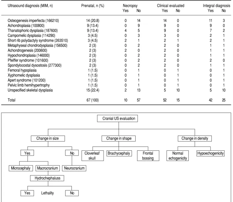
Figure 2. The diagnostic approach for evaluation cranial bone dysplasias.
and brother were diagnosed with OI (type unk-nown); in this case, the fetus was diagnosed with OI type IV. Of the nine cases of thanatophoric dyspla-sia, only one case (11.1%) had a family history of it (skeletal dysplasia different to thanatophoric in a se-cond degree relative). When analyzing the total ca-ses of skeletal dysplasia (67), only 5.8% of them had a family history of skeletal dysplasia or any other birth defect.
Karyotyping was performed only in those fetu-ses with suspected chromosomal alteration, it was performed in 11.9% (7/67) of cases. The results of the karyotype of the seven cases with fetal
skele-tal dysplasia were 46, XX (4/7, 57%), 46, XY (3/7, 43%).
Regarding perinatal outcomes, a total of 7 still-birth sand 11 early neonatal deaths accounted for 26.8% of the infants. Neonatal deaths were found in 9.0% (1/11) of cases diagnosed with achondrogene-sis, 36.3% (4/11) of cases diagnosed with thanato-phoric dysplasia and 54.5% (6/11) (Figure 4) of the unspecified skeletal dysplasia cases.
Anatomopathological studies were performed in 45.4% (5/11) of early neonatal deaths, reaching a comprehensive classification diagnosis in only 70% of them. The Genetics Department reviewed 77% of
Cranial US evaluation
Change in size Change in shape Change in density
Yes No Cloverleaf Brachycephaly Frontal Normal Hypoechogenicity
skull bossing echogenicity
Microcephaly Macrocranium Neurocranium
Hydrochephaluss
cases (52/67) and integrated (clinical, radiologic and pathologic when possible) was performed in 94% of cases (Table 2). For the 6% of cases in which the analysis was not complete, these analyses were not performed due to incompletely described necropsies, lack of assistance of genetics department during the postnatal consultation (deliver occurs in weekend or during night) and incomplete clinical records.
In this study we found a concordance in the pre-natal and integrated postpre-natal diagnosis of 68% (46/ 67), in 62% (42/67) with the complete postnatal inte-grated protocol of diagnosis (genetics evaluation, ne-cropsy when possible, neonatologist evaluation) and 6% (4/67) without the complete protocol, in the re-maining 32% (21/67) the diagnosis of skeletal dys-plasia was confirmed but no the specific diagnosis suspected in the prenatal evaluation.
The mean of gestational age at birth was 36.3 ± 6.33 weeks. The pregnancies resolved in the third
trimester in 88% (59/67) of women; of these preg-nancies, 85% (51/59) of infants were delivered through Cesarean section. Ten of 67cases (14%) were resolved in the second trimester, with vaginal birth in 100% of them. The main indication for birth in the second trimester was fetal demise.
DISCUSSION
Currently, over 200 skeletal dysplasias have been classified according to phenotype, and a total of 400 cases of skeletal dysplasia have been characterized through their genotype and/or proteins and are re-flected in the Nosology and Classification of Genetic Skeletal Disorders (2007). Skeletal dysplasias are the second most common fetal structural disease in prenatal diagnostic centers. The diagnosis of skele-tal dysplasia requires additional studies to clarify its origin; guide parents regarding its monitoring, treatment, prognosis and postnatal care; and imple-ment a multidisciplinary manageimple-ment by a group of experts, including a fetal physician, a geneticist, an obstetrician, an anesthesiologist, a neonatologist and an orthopedist, to offer patients a comprehensi-ve approach to improcomprehensi-ve conditions in pre-and post-natal stages.
Genetic counseling is a communication process that relies on clinical history, family history, labo-ratory and imaging studies and specialized genetic studies (cytogenetic, enzymatic and molecular) that aims to define the etiology of the disorder, establish prognosis and determine the recurrence risk. For fa-milies who previously had a fetus with a confirmed skeletal dysplasia and are at risk of recurrence, the molecular analysis of DNA from chorionic villus sampling (CVS) between 11 and 13 weeks9 or 3
gg 3
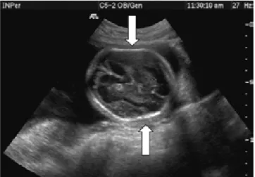
Figure 3.Figure 3. Cranial depression.
. g e
5 g
Figure 5. Flowcharts of the diagnostic approach for evaluating dysplasias. A A. Facial bone dysplasias. B. Thoracic bone dysplasias. C. Extremity bone dysplasias. DD. Bone dysplasias in the hands and feet. E.E. Spinal bone dysplasias.
A A
B B
C
D
centesis between16 to 20 weeks of gestation is an available option to directly look for mutations.
In Mexico, Ávila, et al., at the La Raza National Medical Center, has retrospectively reviewed repor-ted birth defects from 1984 to 2003, totaling 14,986 newborns with a total of 3,682 malformations (2.46%). The average number of births per year was 7,494, with 184 infants presenting defects each year; congenital abnormalities that affected the musculos-keletal system (19.3% of infants) were second in pre-valence only to cardiovascular system defects 26% of the infants.10 The overall frequency of skeletal
dysplasia in perinatal deaths is 9.1/1,000 births. Despite the increased of validated methods for diagnosing skeletal dysplasia including histological, radiological, ultrasound and molecular techniques, around 7% of cases do not corroborate the diagno-sis.11
In our review, the prevalence of skeletal dysplasia is high (8.1 per 10,000 newborns per year vs. 2.4 per 10,000 births reported in international literature),4,5
which is primarily due the fact that the INPerIER is a national referral hospital for tertiary care, and many high-risk pregnancies in which the fetus has skeletal dysplasia are resolved here. Thus, these re-sults do not reflect the current status of these disor-ders in our country.
In this review, the four most frequently diagnosed dysplasias were OI (20.8%), followed by achondro-plasia (13.4%), thanatophoric dysachondro-plasia (13.4%), and short rib polydactyl syndrome (4.4%). This fin-ding coincides with that shown in previous series conducted by Goncalves and Jeanty in 1993 and Schramm in 2009.12,13 In the 67 cases studied, there
were 20 different types of dysplasia. Maternal age was not a risk factor for the development of fetal skeletal dysplasia, as previously shown. Most wo-men were between 25 and 29 years of age. These women’s infants were most commonly diagnosed du-ring the third trimester, which was a result of late referral by the primary care level, both for confirma-tion or exclusion of a diagnosis and resoluconfirma-tion. Ul-trasonographic markers of skeletal dysplasia that were more frequently found in the study were fe-murs with pronounced curvature (n = 40, 59.7%), followed by a decrease in bone density and abnormal cardiothoracic ratio (n = 21 each, 31.3%), the pre-valence of these markers for diagnosis of skeletal displasia is similar to that reported by Romero in 1990.14 In 29.8% (20/67) of infants, there was
shor-tening of tubular bones, and 14 (20.8%) patients presented with frontal bossing. By analyzing the type of ultrasonographic markers, it is clear that a
thorough review that is sensitized to these changes will be enough for suspected or confirmed cases and will classify the fetal skeletal alteration (Figure 3). We found a total mortality rate of 26.08% (11/67), of which there were a total of 7 deaths and 11 early neonatal deaths. Thanatophoric dysplasia was res-ponsible for 54.5% of deaths in this study (6/11). Within the data reported in this review, only 5.8% of cases had a family history of skeletal dysplasia or other structural defects, which leads to two con-clusions: de novo mutations account for the presen-ce of disease without a corresponding family history (excluding the cases of skeletal dysplasia with auto-somal recessive inheritance), and these defects must be analyzed in the whole population and not only patients who have a family history of the disease.
As showing the results, two complementary eva-luations for confirming the diagnoses were often performed incompletely: the karyotype was determi-ned in only 11% of cases, there were no molecular genetic analysis in any of the cases by the lack of availability at the Institute and pathological analy-sis after fetal or neonatal death was performed in less than half of the cases due the lack of acceptance of the parents. Many of the records had incomplete postnatal outcomes. Similarly, postnatal support was infrequently given to patients. Thus, it neces-sary to work cooperatively, especially between the medical and paramedical groups, which will subse-quently impact the couple and their family and pro-vide continuity to the monitoring of all cases of fetal skeletal dysplasia. We must therefore adhere to diagnosis and management flow charts that are de-signed to standardize the diagnosis, monitoring, as-sessment and resolution of cases of fetal skeletal dysplasia. The proposed schemes used in the Fetal Medicine Department of our institution are one op-tion to resolve the issues that we have raised and standardize screening and confirmation/exclusion criteria of the disease (Figure 5).
REFERENCES
1. Dora B, Favre R, Virile B, Langer B, Dreyfus M, Stoll C. Pre-natal sonographic diagnosis of skeletal dysplasias. A report of 47 cases. Annals de Genetique 2000; 43: 163-9.
2. Superti-Furga A, Bonafé L, Rimoin DL. Molecular-pathogene-tic classification of geneMolecular-pathogene-tic disorders of the skeleton. Am J Med Genet 2001; 106: 282-93.
3. Superti-Furga A, Unger S. Nosology and classification of ge-netic skeletal disorders: 2006 revision. Am J Med Genet A
2007; 143: 1-18.
4. Hurst JA, Firth HV, Smithson S. Skeletal dysplasias. Seminars in Fetal & Neonatal Medicine 2005; 10: e233-e241.
6. Campbell J, Henderson A, Campbell S. The fetal femur/foot length ratio: a new parameter to assess dysplastic limb reduc-tion. Obstet Gynecol 1988; 72: 181-4.
7. Donnenfeld AE, Mennuti MT. Second trimester diagnosis of fetal skeletal dysplasias. Obstet Gynecol Surv 1987; 42: 199-217.
8. Krakow D, Williams J, Poehl M, Rimoin DL, Platt LD. Use of threedimensional ultrasound imaging in the diagnosis of pre-natal-onset skeletal dysplasias. Ultrasound Obstet Gynecol
2003; 21: 467-72.
9. Golden CM, Ryan LM, Holmes LB. Chorionic villus sampling: a distinctive teratogenic effect on fingers? Birth Defects Res A Clin Mol Teratol 2003; 67(8): 557-62.
10. Avila J, Lievano SA, Santo I, Ahumada E. Incidence of birth defects for 20 years in a highly specialized unit. Bol Med Hosp Infant Mex 2006; 63(1): 23.
11. Sharony R, Browse BS, Lachman RS. Prenatal Diagnosis of the Skeletal dysplasias. Am J Obstet Gynecol 1993; 169: 668-75. 12. Goncalves L, Jeanty P. Fetal biometry of skeletal dysplasia: a
multicentric study. J Ultrasound Medicine 1994; 13: 977-85.
13. Schramm T. Prenatal sonographic diagnosis of skeletal dyspla-sia. Ultrasound Obstet Gynecol 2009; 34: 160-70.
14. Romero R, Athanassiadis AP. Fetal skeletal anomalies. Radiol Clinic of North America 1990; 28: 75-99.
Correspondence and reprint request:
Dr. Mario E. Guzmán-Huerta
Departamento de Medicina Materno-Fetal, 6o. piso Instituto Nacional de Perinatología
Isidro Espinosa de los Reyes Montes Urales, Núm. 800 Col. Lomas Virreyes 11000, México, D.F.
Tel.: (52 55) 5520-9900, Ext. 112 E-mail: mguzmanhuerta@yahoo.com.mx