Otras secciones de este sitio:
☞ ☞ ☞ ☞
☞ Índice de este número ☞
☞ ☞ ☞
☞ Más revistas ☞
☞ ☞ ☞
☞ Búsqueda
Others sections in this web site:
☞ ☞ ☞ ☞
☞ Contents of this number ☞
☞ ☞ ☞
☞ More journals ☞
☞ ☞ ☞ ☞ Search
Artículo:
Microwave irradiation for shortening the processing time of samples of flagellated bacteria for scanning electron microscopy
Derechos reservados, Copyright © 2003: Asociación Mexicana de Microbiología, AC
Revista Latinoamericana de Microbiología Número
Number3 - 4
Julio-Diciembre
July-December 2 0 0 4
Volumen
Volume 4 6
edigraphic.com
Rev
ista
Lat
inoamer
Microwave irradiation for shortening the processing
time of samples of flagellated bacteria for scanning
electron microscopy
Francisco Hernández-Chavarría*
* Facultad de Microbiología and Centro de Investigación en Estructuras Microscópi-cas, Universidad de Costa Rica, San José, Costa Rica.
Received March 22, 2004; received in revised form June 29, 2004; accepted Septem-ber 9, 2004.
ABSTRACT. Microwave irradiation (MWI) has been applied to the development of rapid methods to process biological samples for scanning electron microscopy (SEM). In this paper we propose two simple and quick techniques for processing bacteria (Proteus
mira-bilis and Vibrio mimicus) for SEM using MWI. In the simplest
meth-odology, the bacteria were placed on a cover-glass, air-dried, and submitted to conductivity stain. The reagent used for the conductiv-ity stain was the mordant of a light microscopy staining method (10 ml of 5% carbolic acid solution, 2 g of tannic acid, and 10 ml of sat-urated aluminum sulfate 12-H2O). In the second method the samples were double fixed (glutaraldehyde and then osmium), submitted to conductivity stain, dehydrated through a series of ethanol solutions of increasing concentration, treated with hexamethyldisilazine (HMDS), and dried at 35°C for 5 minutes. In both methods the steps from fixation to treatment with HMDS were done under MWI for 2 minutes in an ice-water bath, in order to dissipate the heat generated by the MWI. Although both techniques preserve bacterial morphol-ogy adequately, the latter, technique showed the best preservation, including the appearance of flagella, and that process was complet-ed in less than 2 hours at temperatures of MWI between 4 to 5 °C.
Key words: Microwave irradiation, Proteus mirabilis, Vibrio
mim-icus, swarming, flagella, conductive staining, hexamethyldisilazine.
RESUMEN. La irradiación con microondas (IMO) se ha aplicado en el desarrollo de métodos rápidos para procesar muestras biológicas para microscopía electrónica de barrido. En este artículo, proponemos dos técnicas rápidas y simples para procesar bacterias (Proteus mirabilis y
Vibrio mimicus) para MEB usando IMO. En la metodología más
sim-ple, las bacterias se colocaron sobre un cubreobjetos, se secaron al aire y se sometieron a tinción conductiva. El reactivo empleado para la tin-ción conductiva fue el mordiente de un método de tintin-ción para mi-croscopía óptica (10 ml de una solución de ácido carbólico al 5%, 2 g de ácido tánico y 10 ml de una solución saturada de sulfato de aluminio en agua). En el segundo método, las muestras se fijaron dos veces (con glutaraldehído y luego con osmio), se sometieron a tinción conductiva, se deshidrataron por tratamiento en una serie de soluciones de etanol de concentración creciente, se trataron con hexametildisilazina (HMDS) y finalmente se secaron a 35°C por 5 minutos. En ambos métodos, los pa-sos desde fijación hasta tratamiento con HMDS se realizaron con IMO por 2 minutos en un baño de hielo, para disipar el calor generado por la IMO. Aunque ambas técnicas preservaron adecuadamente la mor-fología bacteriana, la última técnica llevó a una mejor conservación de la morfología bacteriana, incluyendo la apariencia de los flagelos. Una ventaja adicional es que el proceso puede completarse en menos de dos horas a temperaturas de IMO entre 4 y 5 °C. (Traducido por la Revista).
Palabras clave: Irradiación con microondas, Proteus mirabilis,
Vibrio mimicus, movimiento en enjambre, flagelos, tinción
conduc-tiva, hexametildisilazina.
INTRODUCTION
Processing of biological samples for electron microsco-py is shortened by using microwave irradiation (MWI), that reduces the time required for fixation, washing and
de-hydration,2,6,7 Fixation, and each step of the washing or
de-hydration process can be completed in only two minutes
using MWI in a domestic microwave oven.3 MWI
increas-es molecular rotation rincreas-esulting in more frequent molecular collisions, thus facilitating chemical reactions and shorten-ing processshorten-ing times. Nevertheless, MWI heats samples, but it is possible to dissipate such heat by immersing
sam-ples in an ice-water bath during irradiation.4
Processing methods to observe bacteria under scanning electron microscopy (SEM) are easier than those for eu-karyotic cells, due to the presence of the bacterial cell wall. Gram positive and Gram negative bacteria have a cell wall composed of peptidoglycan, also called murein or murein-sacculus; it forms a sack that covers the surface of the cell and protects it against osmotic damage. Bacteria can be rapidly processed with MWI, as was described for
Entero-coccus1, and it is also possible to dry the samples using
hexamethyldisilazine (HMDS) instead of a critical-point dryer. Nevertheless, Gram negative bacteria present a chal-lenge, because their cell walls are structurally more com-plex. Bacteria with flagella pose additional difficulties, be-cause these threadlike organelles are very slender (ca. 10 nm in diameter) and are disrupted easily when the bacteria are suspended to make a smear.
Two examples of flagellated Gram negative rods are Proteus mirabilis and Vibrio mimicus; the former is a straight rod that exhibits swarming phenomenon, during
Hernández-Chavarría Microwave irradiation for processing flagellated bacteria
Rev Latinoam Microbiol 2004; 46 (3-4): 81-84
82
edigraphic.com
sustraídode-m.e.d.i.g.r.a.p.h.i.c cihpargidemedodabor
which short bacilli of about 3µm with less than ten flagella are transformed into giant rods of about 80 µm long with
more than a thousand flagella.11 V. mimicus is a curved rod
with a sheathed polar flagellum. We describe in this paper two simple techniques to process flagellated bacteria using these two species.
MATERIAL AND METHODS
Bacterial samples:
Both strains used were cultured as follows: P. mirabilis, a small drop (20 µl) taken from a 24 hour culture in trypti-case broth was inoculated in the center of a blood agar plate, making a small dot without spreading, and incubated at 35°C for 24 hours to promote swarming. In the case of V. mimicus the sample was streaked out over the surface of tripticase agar plates to obtain isolated colonies after an in-cubation period of 24 hours at 35°C.
The more important aspect to avoid detachment of flagella is a careful manipulation of the culture to make a suspension directly on the support used for processing the sample. The sample was obtained by touching the end of the swarming growth film or the colony with a bacteriological needle. The tip of the needle with adhered bacteria, was slowly immersed for 30 seconds in a small drop of distilled water (20 µl), placed on a plastic cover-slip, keeping the needle tip from touching
the plastic, following the recommendation of Kodaka et al.5 in
their description of the staining of flagella for light microsco-py. In this procedure the bacteria attached to the tip of the nee-dle swim away in a drop of water, which was then air dried (first method) or in a drop of poly-L-lysine left for 10 minutes at room temperature to promote the adhesion of bacteria to the plastic support (second method). The support used to process
the bacterial suspension for SEM were small squares (5 mm2)
of plastic cover-slip, previously ion sputtered with 20 nm of gold, in order to obtain a conductive substrate for the bacteria, and avoid the charged up phenomenon, caused by insulated surface that imparts a negative charge not dissipated, resulting
in bright flashes on the screen of the SEM.10 For the second
method, the bacterial suspension was made in a drop of poly-L-lysine, and was fixed, dehydrated and finally dried with hexametil disilazane (HMDS).
Fixation and washing with microwave irradiation
All steps of the processing, fixation, washing, and dehy-dration were conducted in a microwave oven for periods of two minutes each; the plastic square coverslips with the samples were placed in a plastic Petri dish that was floated in an ice-water bath in order to dissipate the heat generated
during MWI.4 This procedure of MWI was followed for the
fixation, washing, conductive staining and dehydration in an ascending gradient of ethanol (30% to 100%).
A domestic microwave oven was used (Daytron 2450 MHz, 700 W) at a power level of 125.6 W calculated
ac-cording to the formula P=mγ∆T/t.6 Processing methods
were as follows:
First method (Air drying and conductive staining):
The bacterial suspension on the plastic cover-slip was kept at room temperature until the drop was dried (15 to 30 min-utes) and were rinsed for 30 seconds in tap water. Then, a
drop (20µl) of 4% OsO4 was placed over the dry film and
microwaved for 2 minutes in order to fix the cells, and im-mediately the plastic supports were rinsed for 30 seconds in tap water. A drop (20µl) of Kodaka´s mordant (10 ml of 5% carbolic acid solution, 2 g of tannic acid, and 10 ml of
saturated aluminum sulfate 12-H2O) was added, washed
with tap water, and again treated with osmium, then rinsed again in tap water, dried at 35°C for 15 minutes and ana-lyzed in a SEM without the ion sputter-coating of gold or covered with only 5 to 10 nm of gold. The sequential treat-ment with osmium, Kodaka´s mordant and osmium again
(Os-Kodaka-Os) was carried out in a MWI for 2 minutes in
an ice-water bath, and was done in order to get a
conduc-tive stain as described previously for tissue samples.9
Second method (Conductive stain and drying with HMDS): The plastic cover-slip squares used to place the
bac-teria were previously treated with a drop of poly-L-lysine as
was described by Mazia et al;8 but, instead of the period of 2
hour at room temperature or overnight at 4°C to make the poly cationic membrane, the support was covered with the poly L lysine in a microwave oven for 5 minutes and then washed with distilled water. The procedure is as follows:
1. A drop of water was placed on each plastic cover-glass
square and the bacteria were deposited as described above and incubated 10 minutes at room temperature into a moist chamber to avoid drying.
2. The water was removed touching the surface of the
drop with a filter paper.
3. A drop (20µl) of 2.5% glutaraldehyde was placed on
the preparation to fix de bacteria (MWI 2 min).
4. The fixer was drained and washed three times with
dis-tilled water (DW) for 2 min under MWI.
5. Post fixation with a drop (20µl) of 4% OsO4 (MWI 2 min).
6. The plastic cover slips were washed with DW (MWI 2
min, for three times).
7. Conductive staining (Os-Kodaka-Os): A drop (20µl) of
Kodaka´s mordant was added (MWI 2 min), washed (DW under MWI 2 min, for three times), a drop of 4%
OsO4 (MWI 2 min).
8. Washed (MWI 2 min) and dehydrated through an
edigraphic.com
:rop odarobale FDP
VC ed AS, cidemihparG
arap
acidémoiB arutaretiL : cihpargideM sustraídode-m.e.d.i.g.r.a.p.h.i.c
9. The dehydrated preparation was treated with a drop
(20µl) of HMDS, followed by 2 min of MWI, drained, and dried at 35°C for 5 minutes.
10. Finally the specimen was sputter-coated with gold (10 nm) in an ion sputter (Eiko IB-3), or were observed without gold.
All the samples were analyzed using a SEM (Hitachi S-570) at 15 KV, with a tilt angle of 45°, and working distance of 12 mm.
RESULTS
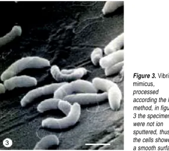
There were differences in the preservation of bacterial flagella according to the processing method used. For ex-ample, the morphology of bacteria in samples processed for the simplest method, that include air-drying and con-ductive stain, the flagella were visible but sometimes ap-peared disrupted. Thus, the hyperflagellated giant bacilli of P. mirabilis appeared covered with fragments of flagella (Fig. 1), which at high magnification appeared as an amor-phous material that covered the cells (inset of fig. 1). The best images were obtained in the samples dehydrated and dried with HMDS (Second method), in which the flagella are well preserved and appeared as a conspicuous tangled net (Fig. 2). The specimens of V. mimicus showed one clearly defined flagellum, that was evident in specimens without ion sputtering (Fig. 3), with images exhibiting a smoother surface than specimens covered with gold in the ion sputter (Fig. 4).
DISCUSSION
The morphological analysis of Gram-negative bacteria using SEM requires addressing problems associated with
the conservation of the structure of the outer membrane; also, if the agents exhibit bacterial appendages, such as fla-gella, they pose additional challenges. Bacterial flagella are slender structures less than 20 nm wide, only about 4 times the resolution of the majority of standard SEM, and a tenth of the resolution limit of the light microscope. These are delicate structures very sensitive to electron bombardment, heat exposure, and ion damage generated in the ion-sput-ter. For observation under light microscope, the flagella are stained using methods that promote deposition of some materials, thus increasing their thickness. One of those
techniques uses Ryion´s stain.5 Under light microscopy,
the flagella are visible with this stain because the complex of mordant and stain increase their diameter. This mordant containing tannic acid and aluminum sulphate, the first component is used in most of the conductive staining
pro-cedures,10 and the second has an element with an atomic
number of 13, that can generate secondary electrons. For this reason, the mordant can be used as a conductive stain in SEM, that allows the observation of tissue samples
with-out a layer of gold.9 The mordant treatment on flagella,
in-creases their outer diameters and also enhances osmium impregnation as well as preventing flagellar damage during ion-sputtering, thus enhancing the images obtained by SEM described in this paper.
Although bacterial cells are easily processed due to the protection of the cell-wall, the analysis of their flagella re-quires special drying methods and HMDS is a good option. Furthermore, the use of MWI shortens the processing time, since each step of the treatment, from fixation to HMDS, including the washing was completed in periods of 2 min-utes at temperatures between 4 to 5 °C, by dissipating the
heat in an ice bath.4 The whole process of the second
meth-od described in this report, including fixation, dehydration,
1
Figure 1.
Swarmer cells of Proteus mirabilis. Fig. 1: Conductive staining and air drying (Bar = 2 µm), the swarmer cells appeared covered with fragments of flagella, that are pasted to the cells as shown in the inset (Bar = 1 µm).
Figure 2.
Hernández-Chavarría Microwave irradiation for processing flagellated bacteria
Rev Latinoam Microbiol 2004; 46 (3-4): 81-84
84
edigraphic.com
and drying with HMDS, took less than two hours, required only drops (20 µl) of each reagent, and exhibited good preservation of bacterial flagella.
Another important aspect in preserving flagellae is the method we used for obtaining the sample, which consisted in touching the surface of a colony with the tip of a bacterial needle; a routine practice used by bacteri-ologists when they stain flagella for light microscopy. Other methods, such as removing the bacterial colony to suspend the cells in water disrupt and detach flagella. When the colony is touched by the bacteriological nee-dle, the bacteria adhere to the tip of the needle and are easily released and become motile when suspended in a drop of water. Under these conditions, the flagella were not disrupted during the preparation of the bacterial sus-pension. In the first method, the bacterial cells were air dried and then treated with a conductive staining
proce-dure using the mordant of Ryion´s stain,5 which permits
observation of flagella; however, they could appear dis-rupted. In contrast, in the second method, the bacteria was double fixed, first with glutaraldehyde and then with osmium, treated with the conductive staining, de-hydrated and dried in HMDS. This more sophisticated procedure better preserves the structure of flagella, as is shown in this paper.
ACKNOWLEDGEMENTS
The author is grateful to Dr. Jorge D. García, Dr. George M. Smith, and the anonymous referee for their suggestions and critical review of the manuscript. This study was sup-ported by the Vice-presidency of Research of the University of Costa Rica (Project N° 430-99-215).
REFERENCES
1. Fox, N. E. & Demaree Jr., R. S. 1999. Quick bacterial microwave technique for scanning electron microscopy. Microsc. Res. Tech. 46:338-339.
2. Gilberson, R. T. & Demaree Jr., R. S. 1995. Microwave fixation: understanding the variables to achieve rapid reproducible results. Microsc. Res. Tech. 32:246-254.
3. Hernández, F. & Guillén, R. 2000. Microwave processing for scan-ning electron microscopy. Eur. J. Morphol. 38:109-111.
4. Hernández, F. & Vargas-Montero, M. 2001. Rapid contrasting of ultrathin sections using microwave irradiation with heat dissipa-tion. J. Microsc. 203:227-230.
5. Kodaka, H., Armfield, A. Y., Lombard, G. L. & Dowell, V. R. 1982. Practical procedure for demonstrating bacterial flagella. J. Clin. Microbiol. 16:948-952.
6. Kok, L. P. & Boon, M. E. 1992. Microwave cookbook for micros-copists. Art and science of visualization. 3rd ed. Coulomb Press Leyden, Leyden. p: 311-314.
7. Login, G. R. & Dvorak, A. M. 1994. The microwave tool book. Beth Israel Hospital Boston. USA. p: 91-114.
8. Mazia, D., Schatten, G. & Sale, W. 1975. Adhesion of cells to sur-faces coated with polylisine: Applications to electron microscopy. J. Cell Biol. 66:198-200.
9. Manzo, L. & Hernández-Chavarría, F. 1994. A new conductivity staining method for tissue analysis under scanning electron micro-scope. Rev. Biol. Trop. 42:183-186.
10. Postek, M. T., Howard, K. S., Johnson, A. H. & McMichael, K. 1980. Scanning electron microscopy. A student book. Ladd Re-search Industries, Inc. p: 166-168.
11. Velas, R. 1992. The swarming phenomenon of Proteus
mirabi-lis. Intercellular communication and multicellular interactions
may provide clues to this 100 years-old mystery. ASM News 58:15-22.
Correspondence to:
Francisco Hernández-Chavarría. Facultad de Microbiología and Centro de Investigación en Estructuras Microscópicas, Universidad de Costa Rica, San José, Costa Rica. E-mail: hchavarr@cariari.ucr.ac.cr
Figure 3. Vibrio
mimicus, processed according the last method, in figure 3 the specimens were not ion sputtered, thus, the cells showed a smooth surface. 3
Figure 4. Are the
same process, but, ion sputtered (5-10 nm of gold). In both figures each cell shows a polar flagellum. (Bar = 1 µm).