R E S E A R C H A R T I C L E
Open Access
Antiviral mode of action of bovine dialyzable
leukocyte extract against human
immunodeficiency virus type 1 infection
Humberto H Lara
*, Liliana Ixtepan-Turrent, Elsa N Garza-Treviño, Jose I Badillo-Almaraz and
Cristina Rodriguez-Padilla
Abstract
Background:Bovine dialyzable leukocyte extract (bDLE) is derived from immune leukocytes obtained from bovine spleen. DLE has demonstrated to reduce transcription of Human Immunodeficiency Virus Type 1 (HIV-1) and inactivate the nuclear factor kappa-light-chain-enhancer of activated B cells (NF-B) signaling pathway. Therefore, we decided to clarify the mode of antiviral action of bDLE on the inhibition of HIV-1 infection through a panel of antiviral assays.
Results:The cytotoxicity, HIV-1 inhibition activity, residual infectivity of bDLE in HIV-1, time of addition experiments, fusion inhibition of bDLE for fusogenic cells and the duration of cell protection even after the removal of bDLE were all assessed in order to discover more about the mode of the antiviral action.
HIV-1 infectivity was inhibited by bDLE at doses that were not cytotoxic for HeLa-CD4-LTR-b-gal cells. Pretreatment of HIV-1 with bDLE did not decrease the infectivity of these viral particles. Cell-based fusion assays helped to determine if bDLE could inhibit fusion of Env cells against CD4 cells by membrane fusion and this cell-based fusion was inhibited only when CD4 cells were treated with bDLE. Infection was inhibited in 80% compared with the positive (without EDL) at all viral life cycle stages in the time of addition experiments when bDLE was added at different time points. Finally, a cell-protection assay against HIV-1 infection by bDLE was performed after treating host cells with bDLE for 30 minutes and then removing them from treatment. From 0 to 7 hours after the bDLE was completely removed from the extracellular compartment, HIV-1 was then added to the host cells. The bDLE was found to protect the cells from HIV-1 infection, an effect that was retained for several hours.
Conclusions:bDLE acted as an antiviral compound and prevented host cell infection by HIV-1 at all viral life cycle stages. These cell protection effects lingered for hours after the bDLE was removed. Interestingly, bDLE inhibited fusion of fusogenic cells by acting only on CD4 cells. bDLE had no virucidal effect, but could retain its antiviral effect on target cells after it was removed from the extracellular compartment, protecting the cells from infection for hours.
bDLE, which has no reported side effects or toxicity in clinical trials, should therefore be further studied to determine its potential use as a therapeutic agent in HIV-1 infection therapy, in combination with known antiretrovirals.
Background
The pandemic of Human Immunodeficiency Virus Type 1 (HIV-1) infection, the cause of Acquired Immunodefi-ciency Syndrome (AIDS), is a grave public health issue and ranks among the greatest infectious disease scourges in history [1]. There were more than 33.3 million people worldwide with HIV-1 infection or AIDS, according to the
latest estimates by the Joint United Nations Program on HIV/AIDS (UNAIDS) [2].
The use of highly active antiretroviral therapies has dramatically reduced morbidity and mortality among patients infected with HIV-1 [3,4]. However, the success of antiretroviral treatment is frequently restricted by the emergence of HIV-1 drug resistance [5]. Therefore, the search for new drugs to inhibit viral replication [6] or to restore the immune system in HIV-1 patients continues. Newly discovered naturally derived or chemically synthe-sized substances are continuously being evaluated as * Correspondence: [email protected]
Laboratorio de Inmunología y Virología, Departamento de Microbiología e Inmunología, Universidad Autonoma de Nuevo Leon, Nuevo Leon, Mexico
therapeutic drug candidates with antiviral activity. These potential drugs are eagerly awaited and may prove benefi-cial for the growing number of HIV-infected individuals who have developed resistance to the currently available antiretrovirals [7].
Dialyzable Leukocyte Extract (DLE) is derived from immune leukocytes and contains low molecular weight proteins (< 10, 000 Da) [8]. DLE possess three chromato-graphic fractions (Fa, Fb and Fc), Fraction Fb inhibits viral production more than 80%. Therefore, fractions Fa and Fc did not show inhibitory effect for any viral dose used [9].
This preparation is a modulator of the immune response that is able to transmit the ability to express delayed-type hypersensitivity (DTH) and cell mediated immunity (CMI) from sensitized donors to immune deficient recipients [10]. DLE also mediates effects on immune system func-tions, further influencing its response. These effects include cytokine modulation [11,12], the activation of monocyte and macrophage chemotaxis [13] and natural killer activity enhancement [14]. The therapeutic and pro-phylactic applications have been the most important and interesting aspects of DLE [15], principally because there has been no reported side effects or toxicity in humans [16].
DLE has demonstrated to be effective in those diseases in which Cell-Mediated Immunity (CMI) plays a relevant role in protection against and control of the disease, such as viral infections ((herpes zoster [17], hepatitis B [18], intracellular bacterial diseases like tuberculosis [19] and leprosy [20], parasite infections, such as leishmaniasis [21] or cryptosporidiosis [22], and fungal infections (mucous-cutaneous candidiasis [23]), as well as in primary immuno-deficiencies (Wiskott Aldrich syndrome [24], Behçet’s syn-drome [25]), bronchial asthma [26], otitis media [27], uveitis [28]) and some types of cancer [29,30].
Previously we reported that bovine DLE (bDLE) was useful as an adjuvant in breast cancer patients under-going chemotherapy, demonstrating protective effects against myelosuppression secondary to antitumoral drugs by improving cellular and humoral immunity, as well as in regulating the production of different cytokines involved in cellular proliferation [16,29,30]. Furthermore, in vitroassays demonstrated that bDLE affected the regu-lation of the expression of p53, bab-1, c-myc, bax, bcl-2 and bad mRNA [31,32]. Nowadays, the majority of the studies on DLE are limited to diseases that occur with chronic inflammation [33], like HIV-1 infection.
A main feature of HIV infection is the expression of several proinflammatory cytokines expressed as soluble factors or membrane-bound molecules that regulate both HIV replication and T cell apoptosis. Proinflammatory cytokines have key roles in the HIV lifecycle, especially at the level of transcription, by enhancing the ability of HIV
to establish latent reservoirs on HIV infected patients. In addition, several HIV proteins, such as Nef, Tat, and Vpr hijack proinflammatory cytokine signaling, further under-lining the potential importance of inflammation in HIV pathogenesis. Moreover, anin vivochronic inflammatory state has been correlated to increased levels of viremia and accelerated disease progression [34]. DLE has been used to treat HIV-1 infected patients, either asympto-matic or at the AIDS phase, resulting in a partial immune reconstitution [35,36], a lower incidence of opportunistic infections [37], and clinically relevant improvement [16,38].
Viruses have evolved to modulate the NF-kB pathway to enhance viral replication, improve host cell survival, and evade the immune response [39]. With HIV, viral and cellular membrane fusion activates NF-B, a process that requires CD4+
T cells. HIV-1 contains regulatory regions in its long terminal repeat (LTR) implicated in the control of viral gene expression that contain three Sp1 core promoter binding sites and two NF-B core enhancer motifs [40] that are recognized by endogenous host cell transcription factors. These are important regu-latory elements in the LTR that control expression of the promoter along with Tat, a viral transactivator protein necessary for HIV-1 replication [41].
Previous studies have reported DLEin vitroreduced HIV-1 transcription [42] by regulating activation of NF-B and Sp1 transcription factors [42-44]. Other studies reported DLE induced the production of leukocytes and reduced TNF-a[44] and TFG-b1 [45] secretion, which are cytokines that play a pivotal role in HIV-1 pathogenesis by up-regulating the transcription of HIV-1 and increasing the expression of HIV co-receptor CXCR4, respectively. Additionally, envelope glycoprotein gp120 can signal B by engaging the CD receptor in a pathway that involves p56 and activates NF-B and HIV-1 LTR transcription [46]. HIV-1 gene expression and transcription is an essen-tial step in the viral life cycle and is considered to be a possible target for the inhibition of HIV-1 replication [43]. The exact mechanism of action of bDLE is still unclear, however we focused on the bDLE mode of action against HIV-1 infection.
Results
Cytotoxic effect
The half cytotoxic concentration (CC50) of bDLE when exposed to HeLa-CD4-LTR-b-gal cells was 3.41 ± 0.1 IU (P < 0.0001) (Figure 1A).
Range of antiviral activity
bDLE was tested against an HIV-1IIIBisolate using indi-cator cells in which infection was quantified by a lucifer-ase-based assay. The concentration of bDLE at which HIV-1IIIB infectivity was inhibited by 50% (IC50) was
Laraet al.BMC Research Notes2011,4:474 http://www.biomedcentral.com/1756-0500/4/474
found to be 1.53 ± 0.1 IU (P < 0.0001) (Figure 1B). In addition, bDLE inhibited HIV-1IIIBinfectivity at doses that were not cytotoxic for HeLa-CD4-LTR-b-gal cells. The therapeutic index (TI = CC50/IC50) for bDLE in these cells was then calculated to be 2.23. The therapeu-tic index reflects a compound’s overall efficacy by relating cytotoxicity (CC50) with effectiveness, measured as the ability to inhibit infection (IC50), under the same assay conditions.
Virucidal activity
To determine if the bDLE might have effects on the virus itself, HIV-1IIIBisolates were treated with different concentrations of bDLE. After removal of bDLE, the residual infectivity of the cell-free viruses was quantified by a luciferase-based assay. As shown in Figure 2, bDLE pretreatment of HIV-1IIIBdid not decrease the infectiv-ity of the viral particles in a dose dependent manner.
Inhibition of Env/CD4-mediated membrane fusion
A cell-based fusion assay was used to mimic the gp120-CD4-mediated fusion process of HIV-1 with bDLE. When bDLE was exposed first to the CD4 cells-Env cells mixture, fusion between both cells was blocked in a dose-dependent manner. Cell-based fusion was also inhibited when bDLE was applied only to CD4 cells for 30 minutes, followed by bDLE removal. However, when Env cells were first exposed to bDLE for 30 minutes, then removed and added to CD4 cells, fusion between both cells was not inhibited (Figure 3A). Known
antiretroviral drugs, such as UC781 (NNRTI), were used as controls in this cell based fusion assay and did not inhibit cell fusion (Figure 3B). T-20 (Fusion Inhibitor), did inhibit cell fusion in all assays, however, except when exposed to CD4 cells for only 30 minutes and then removed, after which the cells were mixed (Figure 3C).
Figure 1Cytotoxicity assessment of bDLE and HIV-1 inhibition activity.A) HeLa-CD4-LTR-b-gal cells (5 × 10⁴cells/well) andB) HIV-1 IIIB
cell-free viruses (MOI 0.2-0.5) were challenged with two-fold serial dilutions of bDLE. Cell viability andb-gal activity were measured with a luciferase-based assay 24 h after nanosilver exposure. Percentage values are relative to the positive control (no compound treatment). The data represent the means ± standard deviations from three separate experiments, each of which was carried out in duplicate.
Figure 2 Residual activity in HIV-1 strains. HIV-1IIIBcell-free
Time (Site) of Intervention
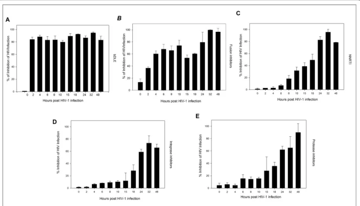
To further determine the antiviral target of bDLE, a time-of-addition experiment was performed using a single cycle infection assay. The time-of-addition experiment was used to determine the stage(s) of the viral life cycle that were blocked by bDLE. Several antiretroviral drugs were chosen as controls as they mark different stages of the viral cycle (i.e., fusion or entry, retrotranscription, protease activity, and integration into the genome). As seen in Figure 4(A-E), the antiviral activity of T-20, UC781, 118-D-24 and Amprenavir started to decline after the cycle stage that they targeted was passed. The fusion inhibitor’s activity declined after 2 h (Figure 4B), the RT inhibitor after 8 h (Figure 4C), the integrase inhi-bitor after 18 h (Figure 4D) and the protease inhiinhi-bitor after 15-18 h (Figure 4E). In contrast, bDLE retained its antiviral activity up to 48 h (Figure 4A) after the HIV
inoculation, inhibiting HIV-1 infection in 80% of infected cells against the positive control.
Cell protection assays
HeLa-CD4-LTR-b-gal cells were pretreated with bDLE for 30 minutes, which was then removed from the extracellu-lar compartment with three washes, and subsequently the HeLa-CD4-LTR-b-gal cells were exposed to HIV-1IIIBfor different pretreatment times (1, 5, 7, 10, 24 and 48 hours). As shown in Figure 5, HIV-1 infection was inhibited, as host cells were protected even after 7 hours from the bDLE removal.
Discussion
Due to the DLE immunomodulatory properties dis-cussed before, progression to AIDS in asymptomatic HIV-1 infected individuals treated with conventional
Figure 3Inhibition of Env/CD4-mediated membrane fusion.b-gal activity was measured after CD4 cells and Env cells were co-cultured after exposure toA) bDLE,B) UC781 andC) T-20 under different circumstances: (■) CD4 cells were exposed to the compound and co-cultured with Env cells for 24 hours; (▲) CD4 cells were exposed to the compound for 30 minutes, washed, and co-cultured with Env cells for 24 hours; (✘) Env cells were exposed to the compound for 30 minutes, washed, and co-cultured with CD4 cells for 24 hours. Percentage values are relative to the positive control (cell-to-cell fusion without pretreatment with drug). The data represent the means ± standard deviations from three separate experiments, each of which was carried out in duplicate.
Laraet al.BMC Research Notes2011,4:474 http://www.biomedcentral.com/1756-0500/4/474
anti-retrovirals, has shown a retarded progression to AIDS under adjuvant treatment with dialyzable leuko-cyte extract (DLE), demonstrated by lower incidences of opportunistic infections and improved cellular immunity
[9,38,44]. DLE simultaneously shows anti-HIV activity [44,47,48], modulates different types of immune effec-tors (e.g., cytokines and transcription factors)[11,47,49] and restores leukocyte subsets in treated patients [16,29,35,37]. All these properties make DLE a potential drug to be used in a therapeutic combination with anti-retrovirals to improve immune and clinical responses.
Bovine dialyzable leukocyte extract (bDLE) is defined as the dialyzate of a heterogeneous mixture of low mole-cular weight substances released from disintegrated blood leukocytes or lymphoid tissue obtained from homogenized bovine spleen. Previous studies have shown inhibition of HIV-1 infection by suppression of the activity of essential transcription factors [42-44] and cytokines by DLE. The purpose of this study was to demonstrate the mechanism of antiviral action of bDLE in vitroin the inhibition of HIV-1 and the protection of host cells from infection.
First, we compared the half cytotoxic concentration of bDLE when exposed to HeLa-CD4-LTR-b-gal cells (CC50 = 3.41 IU) (Figure 1A) with the concentration of bDLE at which HIV-1IIIB infectivity was inhibited by 50% (IC50= 1.33 IU) (Figure 1B) [48]. Then, the thera-peutic index was determined (TI = 2.56) and used as an indicator of bDLE overall efficacy and safety. Despite
Figure 4Time of intervention in HIV-1 life cycle. HeLa/CD4-LTR-b-gal cells were infected with HIV-1IIIBcell-free virus beforeA) bDLE (2 IU),B) T-20 (100
μM),C) UC781 (70 nM),D) 118-D-24 (120μM) andE) Amprenavir (0.1 mM), were added upon HIV-1 inoculation (time zero) or at various time points post-infection.b-gal activity was measured following 24 hr of incubation. Percentage values are relative to the positive control (infected cells without drug pretreatment). The data represent the means ± standard deviations from three separate experiments, each of which was carried out in duplicate.
Figure 5Cell protection against HIV-1 infection.b-gal activity was measured after HeLa/CD4-LTR-b-gal cells were exposed to bDLE (2 IU) for 30 minutes, washed and exposed to HIV-1IIIBcell-free virus at
the fact that TI was lower than expected (< 10), bDLE is a compound that has been used in clinical assays for more than fifty years without adverse reactions [10]. Better understanding of its inhibition mechanism can contribute to the development of new and improved anti-HIV-1 agents, which could be more efficient and have lower cytotoxicity. Furthermore, the residual infec-tivity [50,51] of cell-free viruses after bDLE treatment showed no inhibitory activity (Figure 2), which suggests that bDLE does not act as a virucide on the viral mem-brane to inhibit infection. bDLE showed inhibition of fusogenic cell-cell interactions in a dose-dependent manner when bDLE was exposed to CD4 cells, bDLE was then removed and the CD4 cells were mixed with Env cells. However, after 30 minutes pretreatment of Env cells with bDLE and then transfer to CD4 cells, there was no inhibition of Env-CD4 cell fusion. This observation further supported our previous results that bDLE acted on CD4 expressing cells and not on the Env of the HIV-1 virus. Furthermore, when exposed to CD4 cells for only 30 minutes, which is the time required for conformational changes in gp120 after CD4 binding [52], bDLE again showed inhibition in a dose-dependent manner (Figure 3A). These data suggested that bDLE acted on CD4 cells to inhibit HIV-1 infection. Antiviral results reported by Fernandez-Ortegaet al. [53,54] have also contributed to the knowledge of the molecular mechanisms responsible for the effectiveness of bDLE against HIV infection.
To further determine the antiviral target of bDLE, a time-of-addition experiment was used to define the stage (s) of the viral life cycle that are blocked by these com-pounds. These results were compared with several antire-troviral drugs as controls that marked different stages of the viral cycle [55-61] (Figure 4B-E). Our findings sug-gested that bDLE highly inhibited HIV-1 infection at all stages (Figure 4A), possibly due to viral Tat protein down-regulation (Tat activatesb-galactosidase indicator gene expression in HeLa-CD4-LTR-b-gal cells). Inhibi-tion of HIV-1 Tat activity correlates with down-regula-tion ofbcl-2 [62], but the action of bDLE onbcl-2 has not yet been determined in HIV-1 studies. Previously, bcl-2 was found to be reduced in breast cancer cell lines when treated with bDLE [32]. Furthermore, DLE inacti-vated the NF-B signaling pathway by reducing the secretion of cytokines, such as IL-1 and TNF-a, which are effective inducers of NF-B activity [63]. In HIV-infected T cells, NF-B-dependent transactivation is essential for HIV-LTR induction. Interestingly, even the function of HIV Tat in resting CD4 T lymphocytes depends onB responsive elements in the LTR [46]. Based on these interesting findings, it will be necessary to focus specifically on the transcriptional factors (NF-B
and SP1) and pro-apoptotic genes (bcl-2) in future research on bDLE as an antiviral against HIV-1 infection. Lastly, bDLE was capable of rendering CD4 expressing cells resistant against HIV-1 infection by residual active virus for several hours [64]. Previous results indicated that, although pretreatment of cells (MT-4) with DLE for 3 hours had no effect, inhibition of HIV-1 production was observed when cells were pre-treated for a longer period of time (from 1 to 7 days), an effect that was characterized by the decline in TNFaand TGFb1 gene expression and inhibition of transcriptional factors [9]. In our assays, after pretreatment of the HeLa-CD4-LTR-b-gal cells and bDLE removal prior to viral challenge, protection against infec-tion lasted 7 hours after bDLE was removed from the extracellular compartment (Figure 5). These results indi-cated that bDLE could induce long-term viral inhibition through cell protection, as well as modulate cell suscept-ibility to viral infectionin vitro, in agreement with pre-viously reported data on DLE obtained from human donors by molecular methods [9].
Conclusion
The data presented here were novel in that they proved that bDLE acted by inhibiting HIV-1 infection through protection of the host target CD4 cells at noncytotoxic levels. This effect was found to be modulated through transcriptional factors (NF-B and SP1) necessary for HIV-1 replication. In addition, bDLE was discovered to act through all viral cycle stages to protect cells from HIV-1 infection for hours without affecting the HIV membrane. Based on our results obtained above, bDLE should be further studied to determine its potential use as a thera-peutic agent in HIV-1 infection, especially due to its long-lasting cell protection against HIV-1 infection and lack of negative side effects.
Methods
Reagents, cells and HIV-1 isolates
The following reagents were obtained through the AIDS Research and Reference Reagent Program (NIH): HeLa-CD4-LTR-b-gal cells from Dr. Michael Emerman; HL2/3 cells from Dr. Barbara K. Felber and Dr. George N. Pavla-kis; HIV-1IIIB, fusion inhibitor T-20, integrase inhibitor 118-D-24 and protease inhibitor Amprenavir from Dr. Suzanne Gartner, Dr. Mikulas Popovic and Dr. Robert Gallo. UC781, a no nucleoside reverse-transcriptase inhi-bitor (NNRTI), was kindly donated by Dr. Gadi Borkow. The bDLE used in our study was produced by the Laboratory of Immunology and Virology at the Universi-dad Autonoma de Nuevo Leon, Mexico, following a mod-ified process described by Lawrenceet al. [9]. The bDLE was lyophilized, tested for endogenous pyrogens using the Limulus amebocyte lysate assay (MP Biomedicals,
Laraet al.BMC Research Notes2011,4:474 http://www.biomedcentral.com/1756-0500/4/474
Inc.), and determined to be free of bacterial contamina-tion by culturing in media andin vivomice inoculations. The bDLE obtained from 15 × 108 leukocytes was defined as one unit (1 Unit)[37].
Cytotoxicity Assays
A stock solution of bDLE was diluted two-fold diluted in growth medium and subsequently added into wells con-taining 5 × 10⁴ HeLa-CD4-LTR-b-gal cells. Microtiter plates were incubated at 37°C in a 5% CO2air humidified atmosphere for 24 hours. Assessments of cell viability were carried out using a CellTiter-Glo®Luminescent Cell Viability Assay (Promega). Cytotoxicity was evaluated based on the percentage cell survival relative to the result obtained in the absence of any compound.
HIV-1 Infection Inhibition Assays
Serial two-fold dilutions of bDLE were mixed with 105 TCID50of HIV-1IIIBand added to the wells containing 5 × 10⁴HeLa-CD4-LTR-b-gal cells with a multiplicity of infec-tion (MOI) of 0.2 - 0.5. HIV-1 infecinfec-tion was assessed after 24 hours of incubation by quantifying the activity of theb -galactosidase produced after infection with the Beta-Glo Assay System (Promega). The 50% inhibitory concentra-tion (IC50) was defined according to the percentage of infection inhibited relative to the positive control.
Virucidal Activity Assay
Serial two-fold dilutions of bDLE were added to HIV-1IIIB (T tropic virus) and HIV-1Ba-L(M Tropic) cell-free virus. After incubation for 5 min at room temperature, the mix-tures were centrifuged three times at 10, 000 rpm, the supernatant fluids removed, and the pellets washed three times. The final pellets were resuspended in Dulbecco’s Modified Eagle Medium (DMEM) and placed into 96-well plates with HeLa-CD4-LTR-b-gal cells. The cells were incubated in a 5% CO2humidified incubator at 37°C for 24 h. Assessment of HIV-1 infection was made with the Beta-Glo Assay System. The percentage of residual infec-tivity after bDLE treatment was then calculated with respect to the positive control of untreated virus.
Cell-based Fusion Assay
HeLa-derived HL2/3 cells (Env cells), which express the HIV-1HXB2Env, Tat, Gag, Rev, and Nef proteins, were co-cultured with HeLa-CD4-LTR-b-gal cells (CD4 cells) at a 1:1 cell density ratio (5 × 10⁴cells/well each) for 24 h in the absence or presence of two-fold dilutions of bDLE, UC781, and T-20 in order to examine whether the com-pounds interfered with the binding process of HIV-1 Env and the CD4 receptor. Also, both HeLa-CD4-LTR-b-gal and HL2/3 cells were exposed to the aforementioned compounds for only 30 minutes and then washed twice to eliminate residual compound before co-cultivating
with the other cell line. Upon fusion of both cell lines, the Tat protein from HL2/3 cells activatedb -galactosi-dase (b-gal) indicator gene expression in HeLa-CD4-LTR-b-gal cells [41].b-gal activity was quantified with the Beta-Glo Assay System (Promega). The percentage of inhibition of HL2/3-HeLa CD4 cell fusion was calculated with respect to the positive control of untreated cells.
Time of Addition Experiments
HeLa-CD4-LTR-b-gal cells were infected with 105TCID50 of HIV-1IIIBcell-free virus with a 0.2-0.5 MOI. bDLE (2 IU), T-20 (100μM), UC781 (70 nM), 118-D-24 (120μM) and Amprenavir (0.1 mM) were then added upon HIV-1 inoculation (time zero) or at various time points post-inoculation. The reference compounds were added at a concentration several times their EC50for infectivity of HIV-1IIIB. Infection inhibition was quantified after 24 h by measuringb-gal activity with the Beta-Glo Assay System.
Cell Protection Assays
HeLa-CD4-LTR-b-gal cells were incubated with bDLE (2 Units) for 30 minutes and subsequently washed with PBS three times. Then, the cells were exposed to 105 TCID50 of HIV-1IIIB cell-free virus with a 0.2-0.5 MOI for different times (1, 5, 7, 10, 24 and 48 h). Infection inhibition was quantified after 24 h by measuringb-gal activity with the Beta-Glo Assay System.
Statistical analysis
Graphs were done withSigmaPlot 10.0software and the values shown are means ± standard deviations from three separate experiments, each of which was carried out in duplicate. Cytotoxicity and inhibition assessment graphs are linear regression curves done withSigmaPlot 10.0 software.
Acknowledgements
The following funding sources supported our experiments: thePrograma de Apoyo a la Investigacion en Ciencia y Tecnologia(PAICyT) of the Universidad Autonoma de Nuevo Leon, Mexico, and theConsejo Nacional de Ciencia y Tecnologia(CONACyT) of Mexico.
Authors’contributions
All authors read and approved the final manuscript. HHL participated in the conception and experimental design of thein vitroHIV-1 manipulation and infection assays, in the analysis and interpretation of the data, and in the writing and revision of this report. LI-T participated in the analysis and interpretation the results. HHL and LI-T made equal contributions to this study. EN-GT participated in the analysis and interpretation of the data and in writing and revising this report. SM-FT participated in the analysis and writing of the report. JIB participated in revising this report. CR-P participated in the experimental design of this research.
Competing interests
The authors declare that they have no competing interests.
References
1. Fauci AS:The AIDS epidemic–considerations for the 21st century.N Engl J Med1999,341:1046-1050.
2. Ganguly N:State of the Globe: The Immunological Quest for an HIV/AIDS Vaccine Continues.J Glob Infect Dis2011,3:209-210.
3. Palella FJ Jr, Delaney KM, Moorman AC, Loveless MO, Fuhrer J, Satten GA, et al:Declining morbidity and mortality among patients with advanced human immunodeficiency virus infection. HIV Outpatient Study Investigators.N Engl J Med1998,338:853-860.
4. Sterne JA, May M, Costagliola D, de WF, Phillips AN, Harris R,et al:Timing of initiation of antiretroviral therapy in AIDS-free HIV-1-infected patients: a collaborative analysis of 18 HIV cohort studies.Lancet2009,
373:1352-1363.
5. Johnson VA, Brun-Vezinet F, Clotet B, Gunthard HF, Kuritzkes DR, Pillay D, et al:Update of the drug resistance mutations in HIV-1: December 2010.
Top HIV Med2010,18:156-163.
6. Lara HH, Garza-Trevino EN, Ixtepan-Turrent L, Singh DK:Silver nanoparticles are broad-spectrum bactericidal and virucidal compounds.J
Nanobiotechnology2011,9:30.
7. Caffrey M:HIV envelope: challenges and opportunities for development of entry inhibitors.Trends Microbiol2011,19:191-197.
8. Kirkpatrick CH:Transfer factors: identification of conserved sequences in transfer factor molecules.Mol Med2000,6:332-341.
9. Fernandez-Ortega C, Dubed M, Ruibal O, Vilarrubia OL, Menendez de San Pedro JC, Navea L,et al:Inhibition of in vitro HIV infection by dialysable leucocyte extracts.Biotherapy1996,9:33-40.
10. Lawrence HS, Borkowsky W:Transfer factor–current status and future prospects.Biotherapy1996,9:1-5.
11. Franco-Molina MA, Mendoza-Gamboa E, Castillo-Leon L, Tamez-Guerra RS, Rodriguez-Padilla C:Bovine dialyzable leukocyte extract modulates the nitric oxide and pro-inflammatory cytokine production in
lipopolysaccharide-stimulated murine peritoneal macrophages in vitro.J Med Food2005,8:20-26.
12. Franco-Molina MA, Mendoza-Gamboa E, Castillo-Tello P, Isaza-Brando CE, Garcia ME, Castillo-Leon L,et al:Bovine dialyzable leukocyte extract modulates cytokines and nitric oxide production in lipopolysaccharide-stimulated human blood cells.Cytotherapy2007,9:379-385.
13. Kirkpatrick CH, Rich RR, Smith TK:Effect of transfer factor on lymphocyte function in anergic patients.J Clin Invest1972,51:2948-2958.
14. Lang I, Nekam K, Gergely P, Petranyi G:Effect in vivo and in vitro treatment with dialyzable leukocyte extracts on human natural killer cell activity.Clin Immunol Immunopathol1982,25:139-144.
15. Fudenberg HH, Pizza G:Transfer factor 1993: new frontiers.Prog Drug Res 1994,42:309-400.
16. Lara HH, Ixtepan Turrent L, Garza Treviño EN, Tamez-Guerra RS, Rodriguez-Padilla C, (Eds):Clinical and Immunological assessment in breast cancer patients receiving anticancer therapy and bovine dialyzable extract as an adjuvant.Experimental and Therapeutic Medicine2010,1:425-431. 17. Estrada-Parra S, Nagaya A, Serrano E, Rodriguez O, Santamaria V, Ondarza R,
et al:Comparative study of transfer factor and acyclovir in the treatment of herpes zoster.Int J Immunopharmacol1998,20:521-535.
18. Mazzella G, Ronchi M, Villanova N, Mohamed AA, Pizza G, De VC,et al:
Treatment of chronic B virus hepatitis with specific transfer factor.
Journal Exp Pathol1987,1:421-423.
19. Fabre RA, Perez TM, Aguilar LD, Rangel MJ, Estrada-Garcia I, Hernandez-Pando R,et al:Transfer factors as immunotherapy and supplement of chemotherapy in experimental pulmonary tuberculosis.Clin Exp Immunol 2004,136:215-223.
20. Hastings RC, Morales MJ, Shannon EJ, Jacobson RR:Preliminary results on the safety and efficacy of transfer factor in leprosy.Int J Lepr Other Mycobact Dis1976,44:275.
21. Delgado O, Romano EL, Belfort E, Pifano F, Scorza JV, Rojas Z:Dialyzable leukocyte extract therapy in immunodepressed patients with cutaneous leishmaniasis.Clin Immunol Immunopathol1981,19:351-359.
22. Louie E, Borkowsky W, Klesius PH, Haynes TB, Gordon S, Bonk S,et al:
Treatment of cryptosporidiosis with oral bovine transfer factor.Clin Immunol Immunopathol1987,44:329-334.
23. Masi M, De VC, Baricordi OR:Transfer factor in chronic mucocutaneous candidiasis.Biotherapy1996,9:97-103.
24. Levin AS, Spitler LE, Stites DP, Fudenberg HH:Wiskott-Aldrich syndrome, a genetically determined cellular immunologic deficiency: clinical and
laboratory responses to therapy with transfer factor.Proc Natl Acad Sci USA1970,67:821-828.
25. Wolf RE, Fudenberg HH, Welch TM, Spitler LE, Ziff M:Treatment of Bechcet’s syndrome with transfer factor.JAMA1977,238:869-871. 26. Valdes Sanchez AF, Martin Rodriguez OL, Lastra AG:[Treatment of extrinsic
bronchial asthma with transfer factor].Rev Alerg Mex1993,40:124-131. 27. Kaminkova J, Lange CF:Transfer factor and repeated otitis media.Cell
Immunol1984,89:259-264.
28. Abramson A, Khan A, Tate GW Jr, Martin RG, Hill NO:Immunocompetence and transfer factor therapy in uveitis.Br J Ophthalmol1980,64:332-338. 29. Franco-Molina MA, Mendoza-Gamboa E, Zapata-Benavides P,
Vera-Garcia ME, Castillo-Tello P, Vera-Garcia dlF,et al:IMMUNEPOTENT CRP (bovine dialyzable leukocyte extract) adjuvant immunotherapy: a phase I study in non-small cell lung cancer patients.Cytotherapy2008,10:490-496. 30. Whyte RI, Schork MA, Sloan H, Orringer MB, Kirsh MM:Adjuvant treatment
using transfer factor for bronchogenic carcinoma: long-term follow-up.
Ann Thorac Surg1992,53:391-396.
31. Franco-Molina MA, Mendoza-Gamboa E, Miranda-Hernandez D, Zapata-Benavides P, Castillo-Leon L, Isaza-Brando C,et al:In vitro effects of bovine dialyzable leukocyte extract (bDLE) in cancer cells.Cytotherapy2006,
8:408-414.
32. Mendoza-Gamboa E, Franco-Molina MA, Zapata-Benavides P, Castillo-Tello P, Vera-Garcia ME, Tamez-Guerra RS,et al:Bovine dialyzable leukocyte extract modulates AP-1 DNA-binding activity and nuclear transcription factor expression in MCF-7 breast cancer cells.Cytotherapy2008,
10:212-219.
33. Franco-Molina MA, Mendoza-Gamboa E, Castillo-Leon L, Tamez-Guerra RS, Rodriguez-Padilla C:Bovine dialyzable leukocyte extract protects against LPS-induced, murine endotoxic shock.Int Immunopharmacol2004,
4:1577-1586.
34. Roberts L, Passmore JA, Williamson C, Little F, Bebell LM, Mlisana K,et al:
Plasma cytokine levels during acute HIV-1 infection predict HIV disease progression.AIDS2010,24:819-831.
35. Gottlieb AA, Sizemore RC, Gottlieb MS, Kern CH:Rationale and clinical results of using leucocyte-derived immunosupportive therapies in HIV disease.Biotherapy1996,9:27-31.
36. Raise E, Guerra L, Viza D, Pizza G, De VC, Schiattone ML,et al:Preliminary results in HIV-1-infected patients treated with transfer factor (TF) and zidovudine (ZDV).Biotherapy1996,9:49-54.
37. McMeeking A, Borkowsky W, Klesius PH, Bonk S, Holzman RS, Lawrence HS:
A controlled trial of bovine dialyzable leukocyte extract for cryptosporidiosis in patients with AIDS.J Infect Dis1990,161:108-112. 38. Pizza G, Chiodo F, Colangeli V, Gritti F, Raise E, Fudenberg HH,et al:
Preliminary observations using HIV-specific transfer factor in AIDS.
Biotherapy1996,9:41-47.
39. Li JC, Yim HC, Lau AS:Role of HIV-1 Tat in AIDS pathogenesis: its effects on cytokine dysregulation and contributions to the pathogenesis of opportunistic infection.AIDS2010,24:1609-1623.
40. Lim SP, Garzino-Demo A:The human immunodeficiency virus type 1 Tat protein up-regulates the promoter activity of the beta-chemokine monocyte chemoattractant protein 1 in the human astrocytoma cell line U-87 MG: role of SP-1, AP-1, and NF-kappaB consensus sites.J Virol2000,
74:1632-1640.
41. Lalonde MS, Lobritz MA, Ratcliff A, Chamanian M, Athanassiou Z, Tyagi M, et al:Inhibition of both HIV-1 reverse transcription and gene expression by a cyclic peptide that binds the Tat-transactivating response element (TAR) RNA.PLoS Pathog2011,7:e1002038.
42. Fernandez-Ortega C, Dubed M, Ramos Y, Navea L, Alvarez G, Lobaina L, et al:Non-induced leukocyte extract reduces HIV replication and TNF secretion.Biochem Biophys Res Commun2004,325:1075-1081.
43. Ojeda MO, Fernandez-Ortega C, Rosainz MJ:Dialyzable leukocyte extract suppresses the activity of essential transcription factors for HIV-1 gene expression in unstimulated MT-4 cells.Biochem Biophys Res Commun 2000,273:1099-1103.
44. Ojeda MO, van’t Veer C, Fernandez Ortega CB, Arana Rosainz MJ, Buurman WA:Dialyzable leukocyte extract differentially regulates the production of TNFalpha, IL-6, and IL-8 in bacterial component-activated leukocytes and endothelial cells.Inflamm Res2005,54:74-81.
45. Elrefaei M, Burke CM, Baker CA, Jones NG, Bousheri S, Bangsberg DR,et al:
HIV-specific TGF-beta-positive CD4+ T cells do not express regulatory Laraet al.BMC Research Notes2011,4:474
http://www.biomedcentral.com/1756-0500/4/474
surface markers and are regulated by CTLA-4.AIDS Res Hum Retroviruses 2010,26:329-337.
46. Hiscott J, Kwon H, Genin P:Hostile takeovers: viral appropriation of the NF-kappaB pathway.J Clin Invest2001,107:143-151.
47. Carey JT, Lederman MM:Treatment of AIDS with transfer factor.JAMA 1987,258:3515-3516.
48. Flory E, Weber CK, Chen P, Hoffmeyer A, Jassoy C, Rapp UR:Plasma membrane-targeted Raf kinase activates NF-kappaB and human immunodeficiency virus type 1 replication in T lymphocytes.J Virol1998,
72:2788-2794.
49. Alvarez-Thull L, Kirkpatrick CH:Profiles of cytokine production in recipients of transfer factors.Biotherapy1996,9:55-59.
50. Lara HH, Ayala-Nunez NV, Ixtepan-Turrent L, Rodriguez-Padilla C:Mode of antiviral action of silver nanoparticles against HIV-1.J Nanobiotechnology 2010,8:1.
51. Yang QE, Stephen AG, Adelsberger JW, Roberts PE, Zhu W, Currens MJ, et al:Discovery of small-molecule human immunodeficiency virus type 1 entry inhibitors that target the gp120-binding domain of CD4.J Virol 2005,79:6122-6133.
52. Jones PL, Korte T, Blumenthal R:Conformational changes in cell surface HIV-1 envelope glycoproteins are triggered by cooperation between cell surface CD4 and co-receptors.J Biol Chem1998,273:404-409.
53. Demarchi F, d’Adda di FF, Falaschi A, Giacca M:Activation of transcription factor NF-kappaB by the Tat protein of human immunodeficiency virus type 1.J Virol1996,70:4427-4437.
54. Gaynor R:Cellular transcription factors involved in the regulation of HIV-1 gene expression.AIDS1992,6:347-363.
55. Auwerx J, Stevens M, Van Rompay AR, Bird LE, Ren J, De CE,et al:The phenylmethylthiazolylthiourea nonnucleoside reverse transcriptase (RT) inhibitor MSK-076 selects for a resistance mutation in the active site of human immunodeficiency virus type 2 RT.J Virol2004,78:7427-7437. 56. Hombrouck A, Van RB, Michiels M, Noppe W, Christ F, Eneroth A,et al:
Preclinical evaluation of 1H-benzylindole derivatives as novel human immunodeficiency virus integrase strand transfer inhibitors.Antimicrob Agents Chemother2008,52:2861-2869.
57. Stevens M, Pannecouque C, De CE, Balzarini J:Novel human
immunodeficiency virus (HIV) inhibitors that have a dual mode of anti-HIV action.Antimicrob Agents Chemother2003,47:3109-3116.
58. Svarovskaia ES, Barr R, Zhang X, Pais GC, Marchand C, Pommier Y,et al:
Azido-containing diketo acid derivatives inhibit human
immunodeficiency virus type 1 integrase in vivo and influence the frequency of deletions at two-long-terminal-repeat-circle junctions.J Virol2004,78:3210-3222.
59. Witvrouw M, Balzarini J, Pannecouque C, Jhaumeer-Laulloo S, Este JA, Schols D,et al:SRR-SB3, a disulfide-containing macrolide that inhibits a late stage of the replicative cycle of human immunodeficiency virus.
Antimicrob Agents Chemother1997,41:262-268.
60. Witvrouw M, Fikkert V, Pluymers W, Matthews B, Mardel K, Schols D,et al:
Polyanionic (i.e., polysulfonate) dendrimers can inhibit the replication of human immunodeficiency virus by interfering with both virus adsorption and later steps (reverse transcriptase/integrase) in the virus replicative cycle.Mol Pharmacol2000,58:1100-1108.
61. Zhang X, Pais GC, Svarovskaia ES, Marchand C, Johnson AA, Karki RG,et al:
Azido-containing aryl beta-diketo acid HIV-1 integrase inhibitors.Bioorg Med Chem Lett2003,13:1215-1219.
62. Corallini A, Sampaolesi R, Possati L, Merlin M, Bagnarelli P, Piola C,et al:
Inhibition of HIV-1 Tat activity correlates with down-regulation of bcl-2 and results in reduction of angiogenesis and oncogenicity.Virology2002,
299:1-7.
63. Duh EJ, Maury WJ, Folks TM, Fauci AS, Rabson AB:Tumor necrosis factor alpha activates human immunodeficiency virus type 1 through induction of nuclear factor binding to the NF-kappa B sites in the long terminal repeat.Proc Natl Acad Sci USA1989,86:5974-5978.
64. Zussman A, Lara L, Lara HH, Bentwich Z, Borkow G:Blocking of cell-free and cell-associated HIV-1 transmission through human cervix organ culture with UC781.AIDS2003,17:653-661.
doi:10.1186/1756-0500-4-474
Cite this article as:Laraet al.:Antiviral mode of action of bovine dialyzable leukocyte extract against human immunodeficiency virus type 1 infection.BMC Research Notes20114:474.
Submit your next manuscript to BioMed Central and take full advantage of:
• Convenient online submission
• Thorough peer review
• No space constraints or color figure charges
• Immediate publication on acceptance
• Inclusion in PubMed, CAS, Scopus and Google Scholar
• Research which is freely available for redistribution