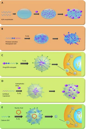
Elastin-like polypeptides in drug delivery
Contents
1. Introduction
2. Macroscopic devices for drug delivery
2.1ELR-based depots and hydrogels
2.1.1 ELR-based depots and hydrogels for reducing systemic toxicity
2.1.2 ELR-based depots and hydrogels for improving therapeutic
efficiency
3. Nanoparticle-based devices for drug delivery
3.1 Nanocarriers derived from ELRs
3.1.1 ELR block copolymers
3.1.2 ELR monomers
3.2 Nanocarriers derived from SELRs
3.3 Hybrid nanocarriers containing ELRs
4. Drug-ELR conjugates for drug delivery.
4.1 Drug-ELR conjugates as chemotherapeutic agents
4.2Drug-ELR conjugates with self-assembling capabilities upon conjugation
5. Conclusions
Acknowledgements
References
Keywords: Elastin-like recombinamers (ELRs), drug delivery, depots, nanoparticles,
smart materials, self-assembling, chemotherapeutic agents.
Abstract
The use of recombinant elastin-like materials, or elastin-like recombinamers (ELRs), in
drug-delivery applications is reviewed in this work. Although ELRs were initially used
in similar ways to other, more conventional kinds of polymeric carriers, their unique
properties soon gave rise to systems of unparalleled functionality and efficiency, with
the stimuli responsiveness of ELRs and their ability to self-assemble readily allowing
important factor that has driven the current breakthrough properties of ELR-based
delivery systems. Recombinant technology allows an unprecedented degree of
complexity in macromolecular design and synthesis. In addition, recombinant materials
easily incorporate any functional domain present in natural proteins. Therefore,
ELR-based delivery systems can exhibit complex interactions with both their drug load and
the tissues and cells towards which this load is directed. Selected examples, ranging
from highly functional nanocarriers to macrodepots, will be presented.
1. Introduction
From a classical point of view, controlled drug-delivery systems have well-defined
goals, including the maintenance of adequate drug levels for sufficiently prolonged
periods of time. To that end, drug-delivery systems act by increasing drug solubility and
availability as well as modifying the pharmacokinetics of the drug to achieve its
sustained and continuous presence. The materials and formulations used for this initial
principle have been extensively reviewed elsewhere [1].
The role of the drug-delivery system can, however, be more ambitious, with properties
such as targeting being pursued in even the very earliest designs. In this sense, the initial
goal of maintaining adequate drug levels is combined with spatial control of action. In
this approach, the drug-delivery system should be able to find the sites where the drug is
needed and release its cargo at these sites, thereby avoiding the exposure of healthy
tissues and regions to the drug. Although this property of targeting is always relevant as
a general ideal behaviour of any drug-delivery system, it is particularly relevant in areas
such as anticancer drugs as anticancer therapies are always accompanied by severe side
effects that can be substantially reduced by using such targeted drug-delivery systems.
Evidently, the possibilities for improving the efficiency of drug-delivery systems do not
end at this level and, once again, novel approaches continue to be developed. Some
examples of more sophisticated drug-delivery systems include intracellular drug
delivery or spatial-temporal control of the delivery of combined drugs [2], or tailored
devices suitable for a particular drug and disease combination. The ideal system is one
that combines sensing with delivery in the sense that the system is inactive until a
certain pathological condition is detected, at which point the drug is released to correct
The evolutionary progress in the development of advanced drug-delivery systems has
led, in turn, to the development of advanced materials. As the complex behaviour of
such systems can is only based on the complex functionality of the materials used to
build them, there is always a direct relationship between the complexity of the system
and the complexity of the materials used to create the system. In essence, our ability to
create complex and sophisticated materials limits the degree of complexity and
functionality that can be achieved when developing such advanced drug-delivery
systems. However, generally speaking, the synthesis of complex materials is time- and
resource-consuming. Chemical synthesis has an important drawback, namely that the
production costs of a synthetic material increase exponentially with its molecular
complexity. Nowadays there is only one exception in which increasing complexity is
not linked to increasing production costs – the recombinant materials also known as
“recombinamers” [4], which are based on synthetic genes. Taking advantage of the
current development of genetic engineering, it is now possible, for the first time, to
create a synthetic gene with a composition that is controlled at the single base-pair level.
The gene sequence is therefore essentially unlimited as it is not restricted to those genes
related to natural proteins; although, evidently, natural proteins are an immense source
of inspiration in the search for functional domains. Finally, these genes can be inserted
into the genome of a producing cell, typically a microorganism, for expression, and this
modified microorganism becomes a nearly infinite and cheap source of the desired
material. Although creation of the gene may require an initial investment, this work
only has to be performed once. After that, cell-based production rapidly compensates
these initial costs related to gene construction. As such, recombinamers are
proteinaceous materials based on a synthetic gene with a well-defined and -engineered
design. They usually have a high molecular weight, thus meaning that they are truly
macromolecules, and can be particularly complex, much more complex than any other
macromolecule produced by chemical synthesis. In addition, the composition of these
systems is strictly controlled, thus meaning that complexity and control form a binomial
that can expand functionality to unseen levels. Their useful properties have motivated
their use in a wide variety of materials and biological applications and, indeed, they
show a huge potential as breakthrough materials in the development of advanced
drug-delivery systems. In this regard, in addition to elastin-like recombinamers, several
classes of recombinant polymers, such as silk-like, and silk-elastin-, collagen- and
example, given the benefits of combining semi-crystalline silk-blocks and elastomeric
elastin-blocks, SELRs possess multi-stimuli-responsive properties and tunability,
thereby becoming promising candidates for the targeted delivery of anticancer drugs
and controlled gene release [9]. Similarly, resilin-like-recombinamer-based
nanoparticles could also find potential uses as responsive components in drug-delivery
applications. Thus, Li et al. have demonstrated that the transition temperature and sizes
of RLR-based nanoparticles can be modulated by varying the polypeptide
concentration, salt identity, ionic strength, pH, and denaturing agents [10]. Additionally,
thermoresponsive self-assembly of well-defined nanovesicles from a collagen-like
peptide has also been demonstrated [11].
Natural elastin, which is one of the most abundant fibrous proteins in the
vertebrate extracellular matrix, provides elasticity to flexible tissues and is the
prevailing constituent of mature elastic fibers [12]. In addition to performing a
mechanical and structural role, elastin acts as a signalling molecule to modulate
cell-matrix interactions. Along the elastin sequence in fact have been identified motifs that
control the cell behaviour as well as the “matrikine” hexapeptide VGVAPG [13], the
consensus sequence GXXVP that interact with the elastin/laminin receptor [14], or the
integrin-mediate cell adhesion motif GRKRK [15]. These bioactive motifs are able to
induce and regulate, the cell adhesion, proliferation or differentiation and possess
chemotactic properties on keratinocytes, fibroblasts, neutrophils and monocytes in
addition to drive the extracellular matrix remodelling[16].
ELR compositions are inspired by the natural elastin precursor tropoelastin, especially
the recurrent tetra- and pentapeptides found in its composition of the elastomeric
domains [17]. Of these, the most widely used repetitive peptide in ELRs is the
pentapeptide VPGXG, and some equivalent variations, where X represents any amino
acid except L-proline. The choice of guest amino acid in the pentapeptide sequence can
dramatically alter the physicochemical properties of the final recombinamer, therefore
balancing the composition depending on the choice of this amino acid, depending on the
final properties desired, is a key parameter when designing the initial basic composition
of the ELR. Although detailed information regarding the dependence of these properties
on the choice of the guest amino acid can be found elsewhere [17], in short, and in
ELRs are stimuli-responsive materials. The mechanism of self-assembly of ELRs and
their sensitivity to different stimuli has been described extensively in the literature.
Their most striking property is perhaps the acute smart nature of these polymers. This
smart nature is based upon a molecular transition of the polymer chain that takes place
in the presence of water. This transition, known as the inverse temperature transition
(“ITT”) and first described for ELPs, has become the key issue in the development of
peptide-based elastin. Indeed, all functional ELRs exhibit this reversible phase
transition behavior [18]. In aqueous solution, and below a certain critical temperature
(Tt), the free polymer chains remain as disordered, random coils in solution [19] that are
fully hydrated, mainly by hydrophobic hydration. This hydration is characterized by the
existence of ordered clathrate-like water structures surrounding the apolar moieties of
the polymer[20-22] with a structure resembling that described for crystalline gas
hydrates[22, 23]. In contrast, above Tt, poly(GVGVP) folds hydrophobically and
assembles to form a phase-separated state containing 63% water and 37% polymer by
weight[24] in which the polymer chains adopt a dynamic, regular, non-random structure
known as a β-spiral, involving one type II β-turn per pentamer, and stabilized by
intra-spiral inter-turn and inter-intra-spiral hydrophobic contacts[18]. This is the product of the
ITT. In this folded and associated state, the chain loses essentially all of the ordered
water structures resulting from hydrophobic hydration [20]. During the initial stages of
polymer dehydration, hydrophobic association of β-spirals takes on fibrillar form that
grows into a particle several hundred nanometres in size before settling into the visible
phase-separated state [18, 25]. This folding is completely reversible on lowering the
sample temperature below Tt [18]. The ITT, and its associated Tt, is in many respects
similar to a lower critical solution temperature (LCST) and many studies in this field
have considered Tt and LCST to be equivalent even though the existence of a regularly
folded structure—the beta spiral—above Tt differentiates the behaviour of an ELR from
many other macromolecules exhibiting an LCST.
In ELRs, Tt depends on the mean polarity of the polymer, increasing as the
hydrophobicity decreases. This is the origin of the so-called “ΔTt mechanism” [18] and
“amplified ΔTt mechanism” [26]; i.e., if a chemical group that can be present in two
different states of polarity is present in the polymer chain, and these states are reversibly
convertible by the action of an external stimulus, the polymer will show two different Tt
values. This change in Tt (“ΔTt”) opens a working temperature window in which the
following changes in the environmental stimulus. These ΔTt mechanisms have been
exploited to obtain a large number of smart elastin-like derivatives [18, 26-28]. In this
sense, certain guest amino acids, such as glutamic/aspartic acid, lysine and others,
confer pH sensitivity, a mechanism that is also exploited in the following model
pH-responsive polymer. The γ-carboxylic function of the glutamic acid (E) residue in the
ELR [(VPGVG)2-VPGEG-(VPGVG)2]n suffers strong polarity changes between its
protonated and deprotonated states as a consequence of pH changes around its effective
pKa [18].
The list of external stimuli that can be used to trigger such smart behaviour is nowadays
quite extensive and, in addition to thermal and pH responsiveness, includes pressure,
light, redox state (electric current) and a variety of chemical potentials such as calcium
or glucose concentration. A good example of this is the photo-responsive ELPs, which
carry photochromic side chains either coupled to functionalized side chains in the
previously formed polymer (chemically or genetically engineered) or by using
non-natural amino acids that are already photochromic prior to chemical polymerization [27,
29] .
The biocompatibility of the material is a key parameter for its use in drug-delivery
systems. Thus, the lack of immunogenicity already described for ELRs, along with their
biodegradability and biocompatibility for human tissue, tissue fluids, and blood, make
these polymers exceptional candidates as carriers in delivery systems [30-32]. The
inflammatory response of ELRs has been studied by different authors using in vivo
assays. For example, Sallach et al. have developed a recombinant elastin-mimetic
triblock copolymer that shows a minimal inflammatory response [33]. Moreover R.
Herrero-Vanrell et al., also observed a poor inflammatory response when they used
poly(VPAVG) as a vehicle for intraocular drug-delivery systems [34].
This manuscript will review the use of recombinamers, in particular ELRs, in the field
of drug delivery which, although still incipient, has already shown its ability and ample
potential. The use of recombinamers as drug depots will be reviewed in the first part of
this manuscript. This includes those systems in which the drugs are either adsorbed,
chemically bound or fused to them using genetic engineering techniques. The design of
selected nanometric drug-delivery systems will subsequently be discussed. Finally, the
paper ends with a brief look at the developments that can be envisaged for this field in
the near future in order to gain a better perspective of what this approach could
different ELR-devices for drug delivery described in the literature. Varying the
composition of the ELR or ELR conjugate may result in more or less complex systems.
2. Macroscopic devices for drug delivery
Nowadays, even in cases where the patient suffers localized disease or pain (of a single
organ or part of it), the treatments that are usually available to physicians involve
systemic drug administration. This kind of administration is particularly suitable for
acute treatments as it requires minimal expertise, although it also presents several
disadvantages for long-term therapies, especially the fact that the drugs administered are
distributed throughout body, including the site of action, thus meaning that a higher
dosage is required to achieve the desired efficacy and consequently increasing the risk
of adverse systemic side effects. Additionally, restrictions related to the molecular
structure of drugs exist as very few molecules are able to overcome the physiological
barriers required to reach their objectives [35]. Moreover, the rapid body clearance
observed when drugs are dosed systematically (enteral or parenteral administration)
means that frequent and repeated administrations are required to achieve sustainable
drug delivery.
In recent years, alternative strategies have been developed to refine and improve drug
delivery and therefore therapeutic efficacy. In this regard, nano- and
microparticle-based delivery devices have demonstrated an ability to provide the controlled release of
small molecules for periods of several weeks [36]. The local implantation of depots is
one the most common methods used for drug administration, especially when long-term
delivery is required. Indeed, the use of this kind of drug reservoir allows local drug
concentration to be increased and sustained release to be achieved for both small and
large drug molecules [36, 37]. Several drug depots have been produced in the form of a
continuous solid mass or loaded hydrogels, and new approaches have been proposed to
obtain less invasive implantation/removal procedures for these devices, such as the
development of injectable biodegradable hydrogels that form the required depot in situ
[38].
Hydrogels are defined as three-dimensional networks of polymeric chains that are able
to swell in water but do not dissolve. The formation of 3D polymeric hydrogels was
originally obtained by chemical synthesis of monomers, although this meant that no
progress in this field has evolved towards smart devices produced using a new
generation of biomaterials whose designed molecules are able to self-assemble or
cross-link under physiologically friendly conditions and whose properties can be fine-tuned
[39]. The properties that make this new generation of hydrogels particularly suitable for
use in medical devices include their biocompatibility and soft hydrophilic structure,
along with the fact that they exhibit controlled porosity, swelling behaviour, a wide
range of mechanical properties, degradability, and stimuli-responsiveness to changes in
their environment [40].
2.1. ELR-based depots and hydrogels
Elastin-like recombinamers are possibly the most promising candidates amongst the
smart biomaterials that are currently being investigated for this purpose as these
protein-based polymers can be synthesized using genetic- and protein-engineering techniques,
their macromolecular structure is well defined and their sequence can be designed to
calibrate their properties to better fit their required functions [41]. Moreover, most of
the key properties of ELR-based hydrogels can easily be tailored to improve their
performance. The porosity and swelling behaviour [42, 43] of such hydrogels is an
advantageous characteristic in drug-delivery applications as it allows the hydrogel to be
loaded with both small and large molecules such as DNA, growth factors or proteins
that can be released at a rate dependent on the diffusion coefficient but also in response
to a physiological stimulus. Indeed, their own porosity and pore size can be modulated
by varying simple parameters such as concentration [44], CO2 pressure [45] or by using
gas-foaming and salt-leaching techniques [46]. Likewise, the mechanical properties of
the hydrogels employed in drug-delivery applications should be adapted according to
the requirement of the site where they will be implanted in the body and its mechanical
stresses. Adaptation of their elastic/viscous behaviour can be achieved by adjusting the
polymer concentration and crosslinking conditions [47, 48]. As described previously, all
VPGXG-based ELRs present temperature responsiveness in aqueous solution
depending on the chemical properties of the guest amino acid [49], properties that may
also provide further responsiveness [50] to other stimuli such as pH [51], ion
concentration [52] or UV-vis light [26] and that are preserved when ELR-based
hydrogels are produced, thereby resulting in multi-stimuli-responsive hydrogels [53].
The tuneable stimuli-responsiveness of ELRs allows, among others, the possibility of
physiological conditions to form a local delivery depot [53]. The half-life of the
resulting ELR-based coacervate at the administration site is at least 25-fold longer than
for the soluble version [54]. Finally, ELRs are extremely non-inflammatory and
biocompatible materials [55] and the removal of ELR-based devices when their payload
is exhausted is not necessary as the biodegradation of these protein-based scaffolds
follows the same natural routes as those found for structural proteins[56-58], whose
degradation products, namely simple amino acids, do no present any toxicity or adverse
responses[33]. Although no systematic studies on the biodegradability of ELR-based
hydrogels are yet available, several parameters related to the requirements and
characteristics of the body site should be taken into consideration when designing a
drug-delivery device. As is the case in vitro [31], the in vivo half-life of the ELR-based
hydrogel mainly depends on both enzymatic digestion and dissolution, therefore factors
such as porosity, recombinamer sequence (in terms of presence of protease-sensitive
sequences), and the cross-linking rate can be modified to tune their stability. For
instance, the biodegradability of hydrogels obtained by coacervation may be markedly
lower than that for their crosslinked counterparts as a result of dissolution. Moreover, in
an environment in which ELR-based depots are exposed to a prolonged nonspecific
proteolytic action, the ELRs, like all protein molecules, will be digested in a short time,
whereas in less aggressive environment the half-life of such depots can exceed twelve
months [33].
ELR-based depots have been developed as therapeutic agent reservoirs for use in
intra-articular [31, 54] ocular [59] or infectious [60] disease, cancer [61, 62] and in diabetes
[63], with some of the most recent examples being discussed below (Figure 2).
2.1.1 ELR-based depots and hydrogels for reducing systemic toxicity
Unfortunately, the most efficient therapies to deal several important diseases may affect
the wellness and patient health due to their systemic side effects. The local
neuroinflammation of the disc herniation, for example, can be alleviated by aggressive
medications that cause moderate immunosuppression consequently, an alternative
strategy of confined drug delivery is especially required. Shamji et al. have described
tritium-labeled ELR depots that possess a local half-life eight times greater than their
soluble counterparts under physiological conditions and when placed in the perineural
healing effectiveness [64]. The same therapeutic strategy of utilizing injectable drug
gels for neuroinflammation treatment but changing the therapeutic agent was carried out
to produce, depots of conjugate curcumin [65] or necrosis factor alpha inhibitor
(sTNFRII) fused to an ELR carrier to invert the inflammatory response in dorsal root
ganglion explants [66].
Similarly, the clinical requirement of controlling post-surgical infections by way of the
local and continued release of antibiotics can benefit from the use of ELR-based depots.
Bacterial infections continue to be a major complication after surgical procedures,
especially in orthopaedic surgery, despite recent advances in parenteral antibiotic
prophylaxis, which mostly consists in long-term intravenous antibiotic administration
[67]. The delivery of antibiotics using depots can enhance the local concentration of the
drug and limit systemic toxicity during wound healing. In this work, drug loading was
performed by lyophilizing cross-linked hydrogels with a homogenous porous size and
then hydrating them in aqueous solutions containing drugs such as cefazolin or
vancomycin. The ELR hydrogels entrapped antibiotics and released them slowly and in
a sustained manner (25 hours for cefazolin and approximately 1170 hours for
vancomycin). The bioactivity of these devices has been assayed in an inhibition test of
Bacillus subtilis bacterial culture and found to be similar to that of the free antibiotics
during throughout the delivery time [68]. To solve the same clinical issue, Anderson et
al. tried to improve the mechanical properties of antibiotic-collagen depots, which are
unstable in aqueous solution, by mixing with an ELR in a proportion of 3:1
respectively. Non-crosslinked ELR-collagen hydrogels loaded with the antibiotic
doxycycline hyclate were obtained after incubation of a solution containing all the
components for 24 hours under physiological conditions [69]. The resulting
ELR-collagen hydrogels showed a significantly higher elastic modulus and sustained
doxycycline release over a period of 5 days. Moreover, the antibiotic released showed
antibacterial activity against at least four bacterial strains of clinical interest
(Escherichia coli, Pseudomonas aeruginosa, Streptococcus sanguinis, and
Staphylococcus aureus).
2.1.2. ELR-based depots and hydrogels for improving therapeutic efficiency
As stated above, the therapies used to date to alleviate the symptoms of chronic illness
ease with which the drug can reach the target tissue, the dosage required and its
turn-over. The drug depot approach offers the possibility of formulation in multiple dosage
forms to calibrate the delivery strategies and provide a suitable alternative for reducing
the frequency of administration and, consequently, patient discomfort. Dry eye disease
(DED) arises due to a chronic lack of lubrication and moisture on the surface of the eye,
thereby causing pain, loss of clear vision, and increasing the risk of infection. It is one
of the most common ocular diseases, affecting between 5% and 35% of the global
population [70]. Continuous topical administration of artificial tear solutions, which is
the common treatment for mild cases of the disease, is not appropriate for the most
severe cases. An alternative and efficient therapy for this syndrome involves
administration of the regulatory human tear protein lacritin, an abundant protein
component of human tears that increases both lacrimal gland secretory activity and
mitosis of the corneal epithelium [71]. To increase its retention on ocular surfaces in
which all proteins suffer a rapid clearance, Wang et al. designed and produced a
lacritin-ELR fusion protein, the ELR component of which was able to drive the
self-assembly and phase separation of the construct and thus to form depots. The
lacritin-mediated cell response in in vitro assays showed that this system was able to promote
secretion in specific lacrimal gland cells. Moreover, in vivo studies performed in an
NOD (non-obese diabetic) mouse model demonstrated the ability of the lacritin-ELR to
stimulate tear secretion in a similar manner to the soluble drug while enhancing the
local retention time in the lacrimal gland by at least six fold. The two components of the
construct preserved their properties, namely the pro-secretory function of lacritin and
the temperature-responsiveness of the ELR, which allowed coacervation of the fusion
ELR below physiological temperature. This allowed the authors to obtain an injectable
intralacrimal depot that afforded sustained delivery of the drug (Figure 3) [59].
Another ELR fusion protein has been designed to produce an injectable drug depot that
can easily be injected into the knee joint, one of the tissues in which systemic
administration is insufficient due to the paucity of vascularity. An increased level of
cytokines in synovial fluid after joint injury is related to the onset and progression of
different arthritis-type conditions [72], therefore the intra-articular administration of
cytokine antagonists is a promising therapy despite the fact that their rapid turnover
limits their effectiveness. With the aim of obtaining a sustained drug-eluting system, the
interleukin-1 receptor antagonist (IL-1RA) has been fused with an ELR and the final
the fused IL-1RA exhibited a reduced bioactivity than the free version, the gradual and
constant depot disaggregation achieved enhanced local drug concentrations [73],
thereby extending the residence time in the joint and consequently reducing cartilage
degeneration at both a macroscopic and a microscopic level [74]. Recently, a different
approach has been proposed to increase the persistence of cytokine antagonists without
reducing functionality. Thus, Kimmerling et al. synthesized ELR-based cross-linked
hydrogels to form a sponge-like device that was able to encapsulate both native IL-1RA
and soluble tumour necrosis factor receptor II (sTNFRII) [75]. In this work, the authors
compared the therapeutic effect of the continued delivery of IL-1RA and/or sTNFRII on
post-traumatic arthritis remission in a murine intra-articular defect model. The
prolonged release of IL-1Ra decreased post-traumatic arthritic disease by reducing the
degeneration of cartilage, facilitating bone healing and decreasing synovial
inflammation. Both ELR devices loaded with sTNFRII only or jointly with IL-1RA
achieved the sustained delivery of therapeutic agents, although no protective effects for
cartilage and bone regeneration were observed in this study due to the inhibition of
tumour necrosis factor .
Diabetes mellitus type II is a hyperglycaemia-related metabolic disorder that affects
approximately 6% of the adult population in industrialised nations and is therefore one
of the most common chronic diseases [77]. A promising alternative therapy for diabetes
type II involves treatment with glucagon-like peptide 1 (GLP-1), an incretin protein
hormone secreted by the gastrointestinal cells that stimulates insulin secretion and
mitosis signalling pathways in pancreatic and insulinoma cells [78]. However, the rapid
degradation suffered by this peptide in the bloodstream (its half-life is less than 2
minutes) limits its use in diabetes treatments.
With the aim of constructing improved, controlled-release glucose-delivery systems,
Chilkoti and co-workers designed and produced two genetically fused ELRs which final
products presented different GLP-1 drug cargo. The fusion constructs self-assembled to
form depots and could be used in two different delivery strategies. In the first one, the
ELR and the drug component are separated by a target sequence of the ubiquitous
exopeptidase DPPIV which, after cleavage, releases GLP-1 monomers from the depot
[63]. In order to limit the natural proteolysis of GLP-1, the endogenous DPPIV cleavage
sites were eliminated by site-directed mutagenesis. The mouse model used to evaluate
the in vivo effects confirmed that the ELR-GLP-1 construct was injectable and able to
thus meaning that lower glucose levels were maintained for at least five days [63]. In
the second approach, in vivo assays showed that a solution of GLP-1-ELR lacking the
proteolytic target easily formed subcutaneous insoluble coacervates, which provided
sustainable delivery of the GLP1-ELR. Sustained delivery of the GLP1-ELR was
compared with both GLP1 monomer and other GLP1-ELR versions soluble under
physiological conditions. Although the GLP1 released was genetically fused to the
ELR, it nevertheless preserved and stabilized glucose activity and, furthermore, was
able to extend the protein plasma half-life when delivered systemically. Indeed, a single
injection in mice produced a five-times longer effect with respect to the soluble version
and 120 times longer than that provided by the native peptide [79].
3. Nanoparticle-based devices for drug delivery
The challenge of delivering the correct concentration of a therapeutic agent at its site of
action, and for sufficient time to be efficient, can be overcome by using traditional
drug-loaded depots when the damage has well-localized targets. Unfortunately, many
important diseases cannot be treated with a local single application that provides a
prolonged and confined drug delivery. This is the case, for example, when the
simultaneous treatment of different organs is required, and this is especially important
in metastatic cancers. In order to reach widespread cancer cells, molecular drugs must
be specifically directed and protected from natural and tumor-related barriers while in
vascular circulation. To this end, over the last few years biomedicine has turned to the
use of nanomaterials as smart drug-carriers, although the clinical outcome in treating
these diseases is still far from optimal [80-82]. In this regard, a large number of
biomaterials that are considered to be effective drug-delivery systems on the nanoscale
can be found in the literature [83-85]. Several of these candidates comprise well-known
polymers adapted to the new requirements although, more recently, tailor-made
biopolymers with a structural design focused on each specific application are emerging
as the best solution.
The ideal nanocarrier should transport the correct amount of drug to the target cell
according to the criteria proposed by Lin and Cui [85]. Basically, these include
sufficient drug loading, good stability during circulation to reach the target safely,
selection and accumulation in the target cell and, finally, promotion of the interaction
Although traditional biomaterials can successfully meet the first two conditions, the last
ones (concerning specific interactions with the molecular components of the cell) are
rarely achieved by just one type of these materials. This is due to the fact that, in both
steps, nanocarriers have to recognize the natural molecules of the target cell and use
them to deliver their payload correctly. These interactions have been achieved by the
use of hybrid nanodevices but also with genetically engineered nanocarriers specifically
designed according to the task at hand [7, 8, 82]. These innovative biopolymers offer
numerous advantages in addition to those inherent to their recombinant nature (versatile
structure, environmentally friendly production, monodisperse sequence, ...). In addition,
they can be chemically modified and adapted as traditional materials and can interact
with the cellular components in a similar manner to a natural protein. Genetically
engineered carriers can be polymeric, such as polypeptides derived from elastin or silk,
or non-polymeric, such as the vault and virus proteins recently reviewed by Shi et al.
[8]. In the first case, another important characteristic turns out to be essential for their
use in the synthesis of drug nanocarriers. Thus, in addition to their low immunogenicity
and good biocompatibility, these polymers self-assemble, by way of a
stimulus-responsive mechanism that is highly tunable in ELRs.
3.1. Nanocarriers derived from ELRs
3.1.1. ELR block copolymers
ELR-based nanoparticles were initially developed by the synthesis of tailor made
di-block copolymers and the main genetic strategies used have been extensively reviewed
elsewhere [4, 7, 86]. The selection of the appropriate guest residue in the amino acid
sequence VPGXG and the combination of diverse blocks, with the resulting differences
in their respective transition temperatures, results in ELR-based amphiphilic block
copolymers. When the solution temperature is increased above the lower Tt the
hydrophobic block suffers its typical transition whereas the hydrophilic block (higher
Tt) remains soluble. This means that the hydrophobic moiety folds and segregates from
the aqueous solution whereas the hydrophilic block remains fully hydrated in contact
with the surrounding water, forming the corona of a micellar structure [87]. This means
that non-polar drugs can be entrapped in the hydrophobic core, whereas the hydrophilic
In addition, the number of repeats and the architecture of the ELR blocks has been
shown to be useful for both tuning the Tt and selecting the type of nanoparticles [89].
This study demonstrated how the common micellar nanostructure derived from the
self-assembly of ELR block copolymers can be shifted to a hollow vesicle (Dh ≈ 200nm) by
changing the block lengths and arrangements. Therefore, close control over the
aggregate morphology could allow the quantity and quality of the loaded drug to be
varied. In contrast, more recent studies [90, 91], have reported the synthesis of
cylindrical micelles or vesicles containing non-elastin assembly domains or
complementary leucine zipper motifs, respectively. In the latter case, the average
diameters were 1.26 and 1.82 μm while the thickness of the vesicle membrane was
about 20 nm. As a proof of their suitability as drug carriers, the microvesicles were used
to efficiently encapsulate both fluorescein and fluorescent polystyrene nanoparticles
(diameter ≈ 500 nm), although their application in vivo will require a reduction in the
critical salt concentration values required to form the vesicles to physiological levels
[91].
Another example is the ELR diblock copolymer with triple stimulus responsiveness
synthesized by Chilkoti’s group [92], which responds to temperature, pH and transition
metal ions in a physiologically relevant range of 32-40°C, pH 7.4-6.4 and Zn2+
concentration (0-25 M). This [VG7A8]-80/[VH4]-100 diblock forms stable 60-nm
micelles under physiological conditions with the histidine-rich block (VH4) at their
core. pH subsequently stimulates their disassembly in the extracellular tumor pH range
(≈6.8), whereas physiological Zn2+ concentrations stabilize them without affecting their
pH sensitivity. The in vivo behavior of this biopolymer was examined by i.v. injection
in nude mice bearing tumor xenografts. The pH-sensitive nanocarriers demonstrated a
prolonged circulation half-life and around 4% of the injected dose reached the tumor
after 8 h. Additionally, they presented a homogeneous intratumoral distribution and
deeper penetration than pH-insensitive nanoparticles, which remain in the perivascular
space because the tumor pH cannot disassemble them into the individual polymer
chains. These pH-sensitive nanocarriers appear to be excellent candidates for carrying
radio- and chemotherapeutical agents for which tumor penetration is a critical success
factor [92].
A different strategy has been developed to increase the drug loading of the toxic and
studies, a serine-based ELR block was flanked by an isoleucine-based block and the
FK506 binding protein (12KDa), the human receptor for Rapa. Due to its low water
solubility and use of the two-phase solvent encapsulation method, the drug was
entrapped in the core of the nanoparticle in addition to being retained in the corona. This
dual strategy increase the drug loading to 75%, the solubility at least 10-fold and the in
vitro half-life release from 2 h to nearly 60 h. The Rapa-ELR nanoparticles were
successfully tested in two mouse models. Thus, i.v. injection in a human breast cancer
model produced a drastic reduction in tumor growth and lower toxicity than free Rapa
[94]. Similarly, their use in a mouse model of Sjögren's syndrome suppressed
lymphocytic infiltration in the lacrimal gland and reduced cathepsin S levels [93].
In a recent paper, the same research group used a previously described chimeric ELR
[95] to target a completely different tissue [96]. In this case the ELR diblock copolymer
contained a fiber capsid protein of adenovirus serotype 5 directed to the coxsackievirus
and adenovirus receptor (CAR), a cell adhesion protein present in liver and lacrimal
gland cells. The in vitro internalization of ELR nanoparticles was successfully
demonstrated in liver cells in the former study, whereas in the latter they were
internalized and transported from basolateral to apical membranes of the lacrimal gland
acinar cells after intra-lacrimal injection in a mouse model. This transcytosing property
could be used in future for sustained delivery of drugs to the ocular surface by i.v. or
s.c. nanoparticle application.
3.1.2. ELR monomers
The synthesis of fusion proteins comprising ELRs in E. coli can include therapeutic
agents when they are polypeptides with simple structures (e.g. toxic peptides or
monomeric growth factors); otherwise, physical encapsulation is the preferred option
[97-100]. When this is the case two aspects become critical: the polypeptide must
conserve its biological function in the fusion protein and the ELR block must retain its
ability to self-assemble. The specific fusion protein comprising monomeric keratinocyte
growth factor (KGF – Mw 22.5 kDa) and a hydrophobic ELR (Tt 28°C) produced stable
nanoparticles (Dh 530nm) under physiological conditions [101]. When the activity of
the recombinant growth factor was assessed, the in vitro cellular test showed a similar
contrast, the molecular activity (measured as substrate phosphorylation) was clearly
reduced when the KGF was fused, thus indicating that its aggregation hinders
interaction with the receptor without compromising the overall cellular effect. The in
vivo healing results in genetically diabetic mice showed that the KGF-ELR nanoparticle
enhanced granulation tissue and improved re-epithelialization when compared with free
factor. An alternative to the fusion proteins with monomeric growth factors using the
single-chain vascular endothelial growth factor was described more recently [102]. In
this work, both molecules were non-covalently linked by a coiled-coil association and
the nanoparticle was found to be effective against cancer cells by inducing cell
apoptosis.
The advantages of fusing therapeutics to ELR-based carriers are numerous. Thus, these
polypeptides can be easily expressed and purified from E. coli sources, which makes it
possible to produce huge amounts of the fused peptide while avoiding any putative
toxicity [103-110]. Additionally, as mentioned above, the intrinsic self-aggregation
property of ELRs can be exploited for several purposes [32, 111, 112]. Finally, yet
perhaps more importantly, ELR polypeptides are macromolecules with a long plasma
half-life [94, 113], which stabilizes circulation of the fused peptide, and a remarkable in
vivo biocompatibility [33, 114] and biodegradability [115] that allow their use as
long-term depots with a minimal inflammatory response.
Recently, a combined strategy has demonstrated promising results against pancreatic
cancer [108]. The authors fused two therapeutic peptides to the ELR for a local therapy
by mild hyperthermia, and combined it with a systemic treatment with Gemcitabine, a
pyrimidine nucleoside analogue of cytarabine. The ELR fragment was flanked by a Bac
cell-penetrating peptide, which mediates intracellular uptake of the nanoparticles, and
by a cell cycle inhibitory p21-derived peptide that leads to arrest at the G1 and G2 or S
phase of the cancer cell cycle. The chimeric polypeptide efficiently inhibited the growth
of three pancreatic cancer cell lines and its activity was enhanced by combination with
gemcitabine. However, this synergism in inhibiting tumor growth with the combination
treatment was not achieved in the xenograft mouse model, probably because additional
studies to adjust the dose ratios between two drugs are required.
The inclusion of bioactive peptides has also been used for the synthesis of ELR-based
nanoparticles for DNA encapsulation. Plasmid condensation was initially accomplished
copolymer was incubated with a plasmid containing the Green Fluorescent Protein
gene, the resulting polyplexes (Rh ≈ 100 nm) were able to transfect cells and its
cytotoxicity was clearly reduced. Therefore, the ELR moiety induced formation of the
nanoparticles as well as neutralization of the intrinsic cytotoxicity of the oligolysine
peptides. In contrast, in a recent paper, a plasmid-condensing peptide (RH3) was fused
to four different and short ELRs, varying their guest residue [93]. All biopolymers were
able to condense pDNA into nanoparticles (Rh <200 nm) and their immunogenicity was
analysed by repetitive injections into immunocompetent mice. The IgG response
showed that, in contrast to those containing K and E, the less immunogenic ELRs were
those including non-charged guest amino acids (S and A). Finally, functional peptides
(penetratin and LAEL fusogenic peptides) were fused to a positively charged ELR and
their cellular transfection ability tested [117]. The resulting biopolymers condensed the
plasmid DNA and the polyplexes (Dh 150-300nm) were internalized by cancer cells via
endocytic pathways and were able to transfect them, especially when both fusogenic
peptides were present in the same molecule (Figure 4). Interestingly, all the ELR
polyplexes showed better biocompatibility when compared with the cationic polymer
PEI. These results demonstrate the utility of ELRs as biocompatible non-viral systems
for gene-therapy applications.
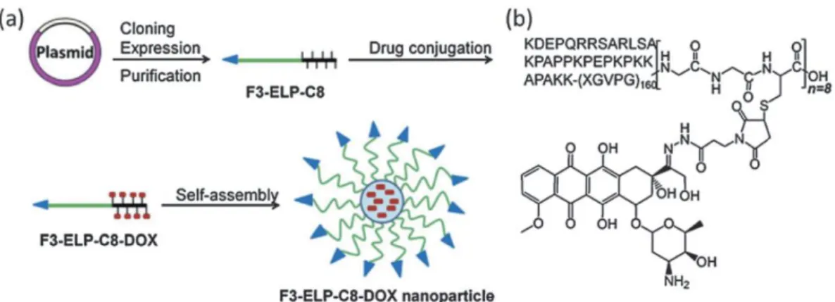
Recently, alternative approaches to tuning the polymer transition, and thus nanoparticle
assembly, have been studied [118]. One successful strategy for overcoming this
limitation is the chemical conjugation of therapeutic non-protein molecules
(doxorubicin, paclitaxel, salinomycin, etc.) to the ELRs [119-121]. In these examples,
the hydrophobic drug triggers assembly of the chimeric molecules into micelles and the
ELR polymer protects the therapeutic load until it reaches its cellular targets. The
roadmap described by Chilkoti should be consulted for a rational design of thermally
responsive drug-loaded nanocarriers [122]. This work provides a simple predictive
model for selecting drugs suitable for inducing conjugation-triggered self-assembly and
for entrapment in the nanoparticle core. Recently, a new model has been described in
order to design drug-loaded ELR nanocarriers for an alternative targeting strategy:
so-called “heat seeking” nanoparticles, which thermally target an externally heated tumor
[123]. As the approved temperatures for mild hyperthermia of solid tumors range from
40 to 45ºC, this work presents a mathematical model for predicting the Tt of the
model has been successfully tested in a colon carcinoma mouse model with
Doxorubicin-conjugated ELRs.
A combined encapsulation strategy was assessed by Zhao et al. with the aim of
increasing drug loading in ELR-based nanocarriers [120] Thus, salinomycin was
chemically conjugated to cysteine residues and free drug was also physically entrapped.
The covalently attached drug induced the synthesis of stable nanoparticles surrounded
by ELRs, whereas the entrapped drug increased the drug loading up to 25%.
Additionally, two additives were added to increase the load up to 75%, with the first
neutralizing the negative charge present in the drug and the second increasing the
hydrophobicity of the nanoparticle core. This complex nanocarrier was found to be
cytotoxic in a cancer cell line, with an extended in vitro drug delivery half-life of 4 h, a
5.2-times longer in vivo plasma half-life and higher accumulation in the tumor.
However, this nanodevice did not inhibit tumor growth more effectively than free
salinomycin, probably due to the intrinsic characteristics of the breast tumor selected.
The performance of ELR-derived nanocarriers in cancer treatment has recently been
compared with a nanoformulation of the highly hydrophobic drug placitaxel (PTX)
approved by the Food and Drug Administration [121]. This nanomedicine (Abraxane)
consists in a 130-nm diameter particle of PTX physically bound to human serum
albumin. In contrast, PTX was chemically conjugated in the ELR-PTX nanoparticle via
a pH-sensitive linker and the Rh in PBS was around 32 nm. Both nanoparticles were
tested in terms of their in vitro and in vivo anticancer efficacy and compared with free
PTX in human prostate and triple-negative breast cancer models. The in vivo plasma
exposure of ELR-PTX nanoparticles was seven-fold higher than free drug and two-fold
higher than Abraxane, thus meaning that the ELR-PTX concentration in the tumor was
2.5-fold higher than for free PTX, whereas ELR-PTX accumulation in healthy organs
was clearly lower than for Abraxane, particularly in muscle and liver. When the
different formulations were compared in the cancer models, a single intravenous
injection of ELR-PTX nanoparticles showed better tumor regression than Abraxane in
the breast model and, while the Abraxane-treated mice bearing the prostate tumor
survived ≤60 days, 100% of the mice injected with ELR-PTX survived for >70 days.
These results, along with the inherent features of the genetically engineered
nanocarriers, provide new reasons for testing their prompt translation into clinical
3.2. Nanocarriers derived from SELRs
Silk-like proteins (SLPs) are another type of recombinant material with demonstrated
success in biomedical applications [124, 125]. These biopolymers are designed taking
into account the repetitive peptide sequences found in silkworm and spider silk. The
most common of all silk variants is probably the hexapeptide GAGAGS from Bombyx
mori fibroin, although recombinant spider silk has also been used in nanoparticles for
gene and drug delivery [126, 127]. In aqueous solution, these silk-derived polymers
undergo an essentially irreversible conformation transition from random coil to beta
sheet and a subsequent beta sheet aggregation growth accelerated by an increase in
temperature. The good biocompatibility and biodegradability shown by these materials,
and particularly the mechanical strength of the resulting aggregation products,
stimulated the design of chimeric materials such as silk-elastin like recombinamers
(SELR). There are many examples of the use of SELRs in drug delivery and these have
been extensively reviewed in the last few years [9, 128, 129]. However, very few
studies reporting the synthesis of SELR-derived nanoparticles have been published
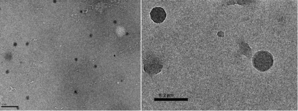
[130-133]. These studies demonstrated that the self-assembly of SELR into spherical
nanoparticles is a process in which the length of the silk block determines both the
kinetics and the size of the aggregates. Thus, an initial temperature-driven aggregation
mediated by the elastin block forms nanoparticles (around 40 nm in diameter) that then
self-assemble into a nanofibrillar morphology in an annealing time-dependent manner
(Figure 5). This coordinated and concomitant dual-gelation mechanism leads to the final
maturation into a resistant hydrogel made of the fibrous structures when the
concentration of the material and annealing time are appropriate [131, 133].
Consequently, the use of SELRs in nanocarrier synthesis for drug delivery has excellent
potential but requires more in-depth studies of the production procedures and choice of
the final target.
The first attempt to investigate the applicability of self-assembled SELR nanoparticles
in drug delivery was described recently by Xia et al. [132] with the antitumor drug
doxorubicin in an in vitro system. In this work, three previously described SELR
constructs [131], with different ratios of silk component, were incubated with DOX,
which triggered their self-assembly into micellar-like nanoparticles (with Rh ranging
drug-loading efficiency was higher for the shorter silk blocks in the SELR and the
nanoparticles were found to be stable for 48 h under physiological conditions, with a
minor increase in size and polydispersity. As regards in vitro studies with the HeLa
cancer cell line, the SELRs with no drug were found to be non-cytotoxic up to 0.2mg/ml
whereas the IC50 for drug loaded-nanoparticles was lower the shorter the silk block,
with a 1.8-fold higher cytotoxicity than the free drug. This indicates that DOX
internalization was modulated by the SELR and the importance of the selection of the
silk-block for appropriate drug delivery [132].
3.3. Hybrid nanocarriers containing ELRs
The versatile and exclusive characteristics of ELRs, along with their chemical
properties, have catalyzed the emergence of hybrid compositions with traditional
materials. These new BioHybrid materials are synthesized by conjugation of organic or
inorganic substances with the appropriate ELRs and present promising opportunities in
the drug-delivery field. Relevant examples of this innovative strategy involving
polysaccharides, lipids, silica and metals can be found in the literature from the last few
years. Thus, multicompartmental capsules were formed using the layer-by-layer method
by combining polysaccharides (chitosan and alginate) and an RGD-containing ELR that
confers both stimuli-responsiveness and cell adherence on them. Their application as
drug-delivery devices was demonstrated when the release profile of the entrapped
rhodamine was found to be temperature dependent [134].
ELRs containing thermosensitive liposomes (e-TSL) are composed of four different
components, namely a phospholipid, cholesterol, PEG and ELR-lipid conjugates, and
their potential use was studied using calcein and doxorubicin [135]. The optimized
liposome formulation (Dh 161 nm) encapsulated DOX and was tested using mild
hyperthermia as a stimulus for drug release. The blood half-life of DOX increased in
mice from 12 min (free DOX) to 2 h when entrapped in the e-TSL and showed superior
drug accumulation in tumors. Finally, the e-TSL exhibited anticancer properties in a
murine mammary tumor model with a hyperthermia-dependent effect.
Two hybrid organic-inorganic materials based on thermosensitive ELRs have been
synthesized recently. Thus, silica micelles are formed when an ELR diblock copolymer
additional condensation of the silica resulted in highly negative monodisperse
nanoparticles (Rh ≈ 35nm) that appear to be promising for encapsulation of therapeutic
or imaging moieties [136]. Similarly, the chemical conjugation of an ELR on the
periphery of a dendrimer produced thermally sensitive nanoparticles that were loaded
with photothermogenic AuNPs. These photothermal dendrimers bound to cells and
induced photocytotoxicity in a temperature-dependent manner [137].
4. Drug-ELR conjugates for drug delivery
A large number of studies have attempted to improve the bioavailability and
pharmacokinetics of small-molecule drugs by conjugation thereof to ELRs, with a
reduction in the therapeutic dose and an increased drug efficiency being the expected
benefits of such an approach. ELRs show certain intrinsic advantages for this purpose,
one of which is their biodegradability as they degrade into natural amino acids, thus
allowing the use of high MW ELRs, even above the renal clearance limit, without the
risk of accumulation.
Other interesting properties of ELRs that are often exploited to increase their delivery
efficiency are their temperature sensitivity and ability to self-assemble. The latter is
particularly relevant as regards spontaneous assembly into nanocarriers. Fortunately,
several studies have shown that conjugation of the drug is not detrimental to these
beneficial properties of ELRs. Indeed, in some cases, they can even be improved and
tuned as a consequence of drug conjugation. One example of this is hyperthermia
therapy for cancer, where it is already been demonstrated that a precise heating of
deep-seated tissues can be used to thermally target ELRs to internal organs by taking
advantage of the thermal sensitivity of ELRs.
The use of recombinant DNA techniques to produce these ELRs allows the easy
incorporation of peptide-based functional domains that greatly contribute to the final
goals of increasing the amount of drug delivered to the desired tissue or affected area,
while decreasing the drug exposure experienced by healthy tissues. In this sense,
although the molecular composition of ELRs tends to be quite complex, in contrast to
other chemically synthesized macromolecules, the complexity in these compounds is
4.1. Drug-ELR conjugates as chemotherapeutic agents
Different drugs have been conjugated to ELRs, with doxorubicin (Dox), a well-known
chemotherapeutic anticancer agent, being one of the most widely used. Indeed, the
conjugation of Dox to ELRs via acid-labile hydrazine bonds has been proven to be
highly efficient as, once the nanocarriers used as drug-delivery systems have been
internalized by the cells, the resulting systems are able to release their drug load in the
acidic environment of lysosomes, with negligible amounts of Dox being released into
the external medium of the cell [139].In this initial work, the conjugation of Dox to the
thiol group of a single cysteine in the ELR was carried out via different pH-sensitive,
maleimide-activated hydrazone linkers. The linker structure and length had little effect
on the Tt of the resulting ELR–Dox conjugates, all of which exhibited similar Tt’s to
that of the native ELR. However, ELR –Dox conjugates with longer linkers exhibited
slower transition kinetics compared to those with shorter linkers. The highest release
achieved upon cleavage of the hydrazone bond of the ELR –Dox conjugate at pH 4 was
nearly 80% over 72 h and was provided by the conjugate with the shortest linker.
In a different study, an ELR-Dox system linked, as above, via an acid-labile hydrazone
was shown to be endocytosed by squamous cell carcinoma cells (FaDu) and trafficked
into lysosomes. Both the ELR–Dox conjugate and the free drug exhibited almost
identical in vitro cytotoxicity, although their subcellular localization was differed
significantly. Thus, the free drug was largely concentrated in the nucleus, whereas the
conjugate was dispersed throughout the cytoplasm with limited nuclear accumulation.
These differences suggest a different mechanism of cytotoxicity for the conjugate as
compared with the free drug [140].
The ability of cancer cells to become simultaneously resistant to different drugs, a trait
known as multidrug resistance, remains a major obstacle for successful anticancer
therapy. One major mechanism of resistance involves cellular drug efflux by expression
of P-glycoprotein (P-gp). However, P-gp mediated resistance can often be overcome by
using P-gp inhibitors, synthesising novel analogs, or conjugating drugs to
macromolecular carriers in order to circumvent the efflux mechanism. Along these
macromolecular ELR-derived therapeutic agents (Tat- ELR -GFLG-Dox). These studies
showed that ELR-bound Dox was equally cytotoxic in both sensitive and resistant cell
lines (MES-SA/Dx5). Indeed, in contrast to free Dos, which was rapidly pumped out by
the P-gp transporter, ELR-bound Dox was shown to accumulate in MES-SA/Dx5 cells.
In these conjugates, the ELR was flanked with a Tat cell penetrating peptide at the
N-terminus and a GFLGC cathepsin cleavage sequence by cassette mutagenesis [141]. The
cell internalization mechanism observed for the ELR was shown to be mainly
endocytotic in nature [142]. This particular study tested two different versions of the
conjugates (Tat- ELR -GFLG-Dox), one containing ELR1, which showed thermal
responsiveness at near physiological temperatures (Tt=40°C), and one containing ELR2,
which does not aggregate at the hyperthermia temperature (Tt=65°C). Focused
hyperthermia above a specific transition temperature at the tumour site caused the ELR
to aggregate and accumulate, thereby increasing the local concentration of the drug
load. The ability of Tat- ELR-GFLG-Dox to be thermally targeted in the resistant
MES-SA/Dx5 cell line was also confirmed. Application of hyperthermia to Tat-
ELR1-GFLG-Dox increased the drug’s toxicity (nearly three-fold) and apoptosis, whereas no
toxicity increase was seen with the non-thermally sensitive control construct Tat-
ELR2-GFLG-Dox, thus indicating that the effect observed was due to aggregation of
the polypeptide. The toxicity enhancement was 20-fold higher in the sensitive cell line.
Paclitaxel (Ptx) has also been used in conjugation with an ELR. As above, Ptx shows
poor aqueous solubility, and was therefore grafted to the ELR to obtain an
acid-sensitive paclitaxel prodrug for the potential treatment of breast cancer [143]. This work
showed that free Ptx is more effective than the macromolecular conjugate SynB1-
ELR1-Ptx in inhibiting the proliferation of sensitive MCF-7 cells. However, for
resistant MCF-7Tax cells, although the difference decreases substantially, the toxicity of
free Ptx is much higher, thereby suggesting the existence of alternative resistance
factors to the P-gp pump that cannot be overcome by polymer-based delivery. Finally,
under hyperthermia conditions, SynB1- ELR1-Ptx induced apoptosis in a manner
similar to conventional paclitaxel in MCF-7TAX cells. This finding can be attributed to
an increase in the intracellular concentration of SynB1- ELR1-Ptx. It is well known that
the use of SynB1 as the CPP promotes internalization via adsorptive-mediated
endocytosis [144].
The demonstration of the efficacy of ELR-Dox conjugates in vivo was a remarkable
of the thermal targeting of an ELR-Dox compound was reported by Shama Moktan et
al. [145]. Thus, the thermal targeting of SynB1- ELR1-Dox (consisting of a
cell-penetrating peptide at the N-terminus and the 6-maleimidocaproyl hydrazone derivative
of Dox at the C-terminus of the ELR), in combination with induced hyperthermia,
resulted in complete inhibition of tumour growth and a substantially higher therapeutic
benefit of the drug in an animal model (E0771 syngeneic mouse breast cancer model).
This cancer model is otherwise only partially responsive to standard Dox treatment. The
advantage of combining hyperthermia with ELR in vivo is that hyperthermia increases
the permeability of tumour vasculature compared with normal vasculature, thereby
resulting in enhanced extravasation of macromolecules. Another remarkable conclusion
of this work was that Dox was found in the heart of animals treated with free
doxorubicin, whereas no detectable levels of Dox were found in animals treated with
ELR-Dox, thereby indicating a good correlation between tumour targeting and a
reduction in the potential cardiac toxicity of ELR-Dox.
The final example in this section is the conjugation of radioactive Iodine (131I) to
different ELRs to use such conjugates as radiotherapeutic agents. Thermal responsive
ELRs have been conjugated to such isotope to create injectable depots. That allowed the
location, by injection, of the radionuclei in the bulk of advanced-stage cancers which
reduced the bulk of those inoperable tumours, enabling surgical removal of de-bulked
tumours [61, 62].
4.2. Drug-ELR conjugates with self-assembling capabilities upon conjugation
The packaging of drugs into nanoscale delivery vehicles (diameter of 10-100 nm) is of
particular interest for cancer therapy, especially as numerous studies have shown that
objects within this size range accumulate within solid tumours due to the enhanced
permeability thereof and the retention effect, which results from abnormalities in
tumour blood and lymphatic vasculature. In this sense, different studies have focussed
on the influence of the size and heterogeneity of ELR-Drug nanocarriers on their
therapeutic efficiency. In the case of ELRs, the formation of such nanocarriers relies
exclusively on their temperature-triggered self-assembly, thus meaning that handling
and fabrication of the delivery system is greatly facilitated.
Numerous attempts to discover the rules that drive self-assembly in ELR-Drug
![Figure 3: Schematic representation of the injectable lacritin-ELR depots formation in the lacrimal gland[59]](https://thumb-us.123doks.com/thumbv2/123dok_es/6170293.183052/33.892.130.759.675.1065/figure-schematic-representation-injectable-lacritin-depots-formation-lacrimal.webp)