F Jaramillo-Juárez et al. Acute renal failure in cirrhotic rats 331
www.medigraphic.com
Annals of Hepatology 2008; 7(4): October-December: 331-338
Annals of Hepatology
Original Article
Acute renal failure induced by carbon
tetrachloride in rats with hepatic cirrhosis
Fernando Jaramillo-Juárez;1 María Luisa Rodríguez-Vázquez;1 Ana Rosa Rincón-Sánchez;2# María Consolación Martínez;1
Genaro G. Ortiz;3 Javier Llamas;1 Francisco Anibal Posadas;1 José L. Reyes4
1Universidad Autónoma de Aguascalientes, Aguascalientes,
México.
2IECD, CUCS, Universidad de Guadalajara, Guadalajara, Jalisco,
México.
3Centro de Investigación Biomédica de Occidente-IMSS,
Guadalajara, Jalisco, México.
4Centro de Investigación y de Estudios Avanzados. Instituto
Politécnico Nacional, México
Address for correspondence: Ana Rosa Rincón-Sánchez, PhD.
Instituto de Enfermedades Crónico-Degenerativas CUCS-Universidad de Guadalajara
Guadalajara, Jalisco, México
E-mail: rincon_ana@hotmail.com; arrincon@cencar.udg.mx Fax: 52 (33) 1058 5200 ext 3893
Phone: 52 (33) 1058 5200 ext 3891, 3892
Manuscript received and accepted: 22 July and 20 August 2008
Abstract
Relationship between cirrhosis and renal dysfunction is not yet fully understood. A model of cirrhosis with acute hepatic and renal damage (RF), produced by CCl4 in rats, with hemodynamic and renal functional alterations, similar to those observed in decompensated cirrhosis (DC) in man, was used to study chemical nephrotoxicity in animals. We performed in male Wistar rats hepatic and renal functional and hemody-namic studies in control, cirrhotic and decompensated cirrhotic (DC) groups. Cirrhosis was induced with car-bon tetrachloride by chronic administration. Associa-tion between liver and renal funcAssocia-tional alteraAssocia-tions was detected in rats with decompensated cirrhosis, showing fall in mean arterial pressure and reduction of glomer-ular filtration rate and filtration fraction. Renal hemo-dynamics did not change in cirrhotic rats, similarly to what occurs in compensated cirrhotic patients. Howev-er, DC rats exhibited increased sodium, glucose and phosphate urinary excretions and decreased ATP in re-nal cortex. DC animals had severe hypoglycemia. There was an extensive liver fibrosis. Glomeruli had hypercellularity and tubules showed extensive vacu-olization in cirrhotic and DC rats. The present study suggests that in this model, damage typical of acute tu-bular necrosis ensues in cirrhotic rats. We describe
functional and morphological damage in liver and kid-ney in a model of cirrhosis that might predispose to the development of acute renal failure when an individual with hepatic damage is exposed in acute way to chemi-cal toxicants.
Key words: Acute renal failure, kidney, liver, renal ATP, glomerular filtration rate, hypoglycemia, glyco-suria.
Introduction
Alterations in kidney structure and function are fre-quently found in severe liver disease and once liver func-tion falls below a critical threshold, sodium retenfunc-tion oc-curs followed by ascites, associated with profound distur-bances of splachnic and systemic hemodynamics which in turn may affect renal function.1,2 As disease progresses,
constriction of intrarenal vascular system favors marked sodium and water retention, leading to refractory ascites, a progressive rise in plasma creatinine levels and reduc-tion of renal clearances (decompensated cirrhosis). Per-sistent renal hypoxia may also induce tubular damage.3
Development of renal failure in patients with liver failure is frequent, it occurs in approximately the 55% of patients. This complication may result when the cirrhotic individual is exposed to xenobiotics, either therapeutic drugs or environmental pollutants, especially heavy met-als. Renal function is rarely restored in the absence of he-patic recovery.4 Administration of carbon tetrachloride
(CCl4) to rats results in a reproducible experimental mod-el of cirrhosis, that resembles the disease in humans and provides a tool to study liver-kidney interrelationships.5,6
Previously we have performed an experimental model of acute liver and renal damage, produced by intragastric administration of a single dose of CCl4 to cirrhotic rats. In this experimental model there are hemodynamic and renal functional alterations similar to those observed in the human with decompensated cirrhosis. This model is useful to study the pathogenesis of renal failure associat-ed with liver damage when the hepatic function decrease after an acute liver damage.7
In our present work, we used our model to further characterize the systemic and renal hemodynamics, his-topathological and functional changes of proximal
tubu-Artemisa
www.medigraphic.com
lar cells (renal sodium handling, phosphate and glucose excretion rates) as well as the function of cells from the distal nephron (renal water handling), in cirrhotic and in cirrhotic rats with acute liver damage induced by carbon tetrachloride.
Material and methods
Animals:
Male Wistar rats were maintained under 12 h day/ night light cycles fed ad libitum with rodent lab diet (Ralston Rations-Kansas, KA). Animals were housed in the animal facility of University Autonomous of Aguas-calientes and all animal studies were conducted in accor-dance with the principles and procedures outlined in the National Institutes of Health’s Guide for the Care and Use of Laboratory Animals.
Induction of liver cirrhosis: Liver cirrhosis was achieved in 100 male Wistar rats (40-60 g initial weight; 250 – 350 g final weight) by intraperitoneal injection of 0.2 ml of a mixture of CCl4 (J.T. Baker USA) and mineral oil (Sigma Chemical Company, St. Louis MO) 3 times per week during 8 weeks, progres-sively increasing CCl4 concentration until the fourth week of treatment.7 The percentages of CCl
4 in mineral
oil (v/v) were as follows: week 1, 13%; week 2, 16%; week 3, 20% and weeks 4 to 8, 25%. Age and sex matched control animals received the same volume of mineral oil.7
Induction of acute renal damage: At the end of eight weeks of chronic CCl4 treatment and after 6 days of the last chronic CCl4 dose, cirrhotic rats received a single in-tragastric dose of a 1:1 (v/v) mixture of CCl4 /corn oil (0.5 mL per 100 g body weight). This acute treatment change cirrhotic by decompensated cirrosis rats and in-duce acute renal failure in the animals.7-9
Liver function tests: Liver damage was assessed by measurements of: a) serum albumin,10,11 b) aspartate
ami-notransferase (AST) and alanine amiami-notransferase (ALT) activities12 and c) total proteins. The method of Lowry et
al. modified by Peterson13 was used for protein
determina-tions using bovine albumin as standard.
Renal function tests: Animals were divided into three groups: control, cirrhotic and cirrhotic plus acute renal damage (decompensated cirrhosis). In those groups, we studied: (A) glomerular filtration rate (GFR) and plasma renal flow (PRF); (B) renal sodium handling (C) renal cor-tical Na+-K+- ATPase activity and tissue ATP
concentra-tions, (D) renal glucose and phosphate excretion rates and (E) renal water handling.
Histological samples examination: Four animals per group (control and CCl4-treated) were anesthetized with sodium pentobarbital and intravascularly perfused with phosphate buffer solution (PBS), pH 7.4, containing hep-arine (1,000 UI/L) and procaine (1 g/L) for rinsing of
liv-ers and kidneys. For in situ fixation, tissue was perfused with PBS, pH 7.4, containing 10% formaldehyde (v/v). Livers and kidneys were removed and fixed for 24 h in the same fixing solution. 5-μm sections were stained with hematoxylin and eosin. Tissue sections were evaluated by optical microscopy. Examiner was unaware of the group to which samples belong.
Renal hemodynamic and sodium handling assays.
Glomerular filtration rate (GFR) and renal plasma flow (RPF) were estimated by inulin and p-aminohippurate clearances, as previously described.14 Arterial blood
pres-sure (BP) was meapres-sured with a Statham P-23A transducer and recorded in a RP Beckman Dynograph. Sodium, inu-lin and PAH were measured by flame photometry, an-throne method and spectrophotometry, as previously de-scribed.14,15
Renal cortex ATPase activities and tissue ATP con-centrations: Renal cortex was separated from medulla and homogenized in solution containing (mM): 240 su-crose, 1 EDTA and 10 Tris-HCl, pH 7.5 (20 % w/v homo-genate). ATPase activities were measured according to Quigley and Gotterer 16 ATP concentration was
deter-mined from renal cortex, according to the method of Bucher, modified by Adams.
Phosphate and glucose excretion: Cirrhotic, DC and age-matched control rats were kept in individual meta-bolic cages. Twenty four hour urine samples were col-lected and urinary volume was recorded. Urinary and plasma phosphate and glucose were measured by Sumner method adapted for microsamples and by the Trinder method, respectively. The urinary/plasma phosphate and the urinary/plasma glucose ratios were calculated.
Renal water handling: Water handling was estimated by osmolal and free-water clearances. Urine samples were collected and total urinary volume of each rat was record-ed. Plasma and urine osmolalities were measured by cryoscopy (OSMETTE, Model 5004). Osmolal (Cosm) and free-water (CH2O) clearances were calculated as follows:
Cosm = [U]osm X Urine flow/[P]osm
where [U]osm and [P]osm are urine and plasmatic-osmo-lalities, respectively, and urine flow is expressed in mL/ min. Free-water clearance was calculated as follows:
CH2O = Urine flow - Cosm
Statistical analyses: Results show means ± SD. Data were analyzed using ANOVA followed by Tukey Kramer test. Differences were considered significant when p < 0. 05.
Results
F Jaramillo-Juárez et al. Acute renal failure in cirrhotic rats
www.medigraphic.com
Morphological studies
Morphology was normal in livers and kidneys of con-trol animals (healthy animals) (Figures 1a and 1d). In contrast, cirrhotic and decompensated rats with renal fail-ure (RF) showed extensive liver fibrosis confirming that hepatic cirrhosis was attained (Figures 1b and 1c). Kid-neys of cirrhotic and DC with RF-rats showed degenera-tive changes (Figures 1e and 1f). Glomeruli were denser in both cirrhotic and DC with RF-rats, than in control group. In the cirrhotic group, there was extensive mesan-gial hypercellularity and podocytes showed hyperchro-matic nuclei. Tubules exhibited fine and extensive vacu-olization. In DC with RF-group, there was severe glomer-ular damage with increased mesangial space and hyperchromatic nuclei of podocytes; tubules showed cloudy swelling and degeneration (Figure 1f).
Blood proteins were reduced and enzymes increased in cirrhotic and DC with RF-rats
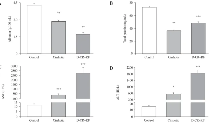
As expected, in cirrhotic and DC with RF rats, serum albumin concentrations were lower (33.6% and 60%, re-spectively) than in controls. In DC with RF group serum albumin further decreased 39.8 %, in relation to cirrhotic rats (Figure 2, panel A). Total protein decreased 50 % and 33.6 % in the cirrhotic and DC with RF rats, respec-tively, as compared to control values (Figure 2, panel B). AST and ALT in DC with RF group and ALT in cirrhotic rats increased significantly, compared to controls, and in the DC with RF group AST and ALT serum activities fur-ther increased 255 % and 238 %, as compared with cir-rhotic rats (Figure 2, panel C and D).
Arterial blood pressure and glomerular filtration decreased in DC with RF rats
Mean arterial blood pressure (MAP) was similar in con-trol and cirrhotic rats. By contrast, we also observed a fall in the MAP in the DC rats with RF, suggesting reduction in re-nal perfusion pressure (Table I). GFR also fell by 52 % in DC with RF group, as compared to control and cirrhotic ani-mals, while no differences were observed between these two groups. Moreover, filtration fraction fell by 43% in group of DC with RF and did not change in cirrhotic rats (Table I). Renal plasma flow (RPF) did not show differences.
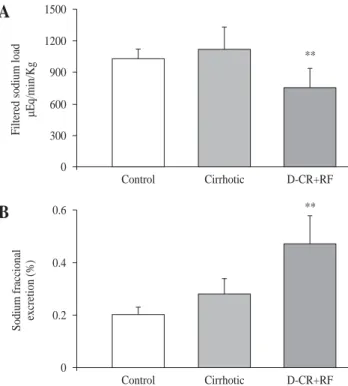
Excretion of sodium was strikingly increased in DC rats with RF
Probably related to decrement in MAP, filtered sodium load decreased in DC rats with RF (756 ± 184 μEq/min/kg) as compared to control and cirrhotic rats (1032 ± 94 and 1117 ± 214 μEq/min/kg, respectively). In spite of dimin-ished filtered sodium load, there was an increment in the fractional excretion of sodium (FeNa), in DC rats with RF.
FeNa increased by 135% in DC rats with RF as compared to control rats and, excretion was 68% higher when com-pared to the cirrhotic group (Figure 3), indicating severe alteration of the tubular handling of sodium.
Severe depletion of ATP renal concentrations in cirrhotic and DC with RF rats
Renal cortical ATP concentrations decreased in both CCl4-treated groups. We also observed fall of ATP signif-icantly, in tissue of cirrhotic DC rats with RF, 62% and 71%, respectively (Figure 4). By contrast, both groups, cirrhotic and DC rats with RF, showed renal cortical AT-Pases (Total, Mg++-dependent and Na+-K+-dependent)
that were significantly unaffected. Na+-K+-ATPase
activi-ty changed from a control value of 23 ± 3.3 to 32.5 ± 4.6 and 32.7 ± 10, nmol Pi min-1 mg-1, in cirrhotic and DC
rats with RF respectively. These findings suggest that low tissue concentration of ATP was probably related to nucleotide altered synthesis.
Increased glucose and phosphate urinary excretions in DC with RF rats
However, glucose urine/plasma ratio and phosphate urine/plasma ratio were markedly higher in both groups, cirrhotic and DC rats with RF, as compared to control val-ues, indicating marked urinary losses of glucose and phos-phate. Indeed, urine/plasma glucose ratio increased 7.5-fold in cirrhotic rats and 21.5-7.5-fold in DC rats with RF. Also, there was a significant difference in the urine/plasma glucose ratio values between DC rats and cirrhotic rats with RF (p < 0.001) (Table II). Related to phosphate urine/ plasma ratio, it increased 1.4-fold in cirrhotic rats (not sig-nificant) and 5.7-fold in DC rats group with RF (Table II). On the other hand, the glucose urinary concentration in-creased significantly in both cirrhotic (522 %) and RF rats (855 %) (Figure 5B). Because of these changes, serum glucose concentration markedly decreased in DC rats with RF group (54 %), without significant differences be-tween control and cirrhotic rats (Figure 5A).
Defective renal water handling in cirrhotic and DC with RF rats
www.medigraphic.com
Figure 1. Histopathologic evaluation of rat liver and kidney. (a) Control group. Hepatocyte (arrows), kupffer cells (arrowheads), and sinusoi-dal space (asterisk) disclose normal liver organization and structure. (b) Liver exhibited extensive damage with necrosis and fibrosis (arrows), hyperplasia of kupffer cells is observed (peak arrow) and most of liver morphology was altered by cirrhosis. (c) Extensive and diffuse dama-ge, with vesicular degeneration and necrosis (arrows) was observed in decompensated cirrhosis with renal failure group (dc+rf). (d) No mor-phological abnormalities were observed in the kidney of control animals: glomerular area (asterisk), mesangial space (small arrow), podocyte (arrowhead), proximal and distal tubules (arrows). (e) Cirrhotic animals exhibited hyperchromatic podocyte (arrowhead) and large mesangial spaces (small arrow); cloudy swelling (arrows) and hydropic degeneration (double arrow) were observed in the tubules. (f) In dc+rf group extensive mesangial hypercellularity with hyperchromatic nuclei (small and arrowhead), severe damage with structural modification in all the glomerular area were observed. tubules exhibited loss of brush border, cloudy swelling and cellular fragments inside the lumen (arrows).
Liver
Kidney
a
b
c
d
e
f
C
O
N
T
R
O
L
C
I
R
R
H
O
T
I
C
D
C
+
R
F
*
*
F Jaramillo-Juárez et al. Acute renal failure in cirrhotic rats
www.medigraphic.com
Discussion
Hemodynamic and functional renal alterations in DC rats with RF were more severe than in cirrhotic rats. When additional acute liver damage was induced in cirrhotic
rats, intense hemodynamic and renal functional alter-ations ensued. We observed fall in mean arterial pressure (MAP) probably due to diminished peripheral vascular resistance and significant reduction of GFR and filtration fraction in DC rats with RF, in a similar fashion as report-ed to cirrhotic rats 17. In contrast, renal hemodynamics
did not change in cirrhotic rats, similarly to what occurs in compensated cirrhosis in humans.
Renal response to decreased blood pressure is altered in severe liver disease with activation of sympathetic sys-tem and increased synthesis of several renal vasoconstric-tor compounds (endothelin, thromboxane A2 and F2 -iso-prostanes). Activated sympathetic nervous system, and presumably other renal vasoconstricting agents, cause a rightward shift in the autoregulatory curve, making renal Table I. Mean arterial blood pressure and renal hemodynamic data from control, cirrhotic and decompensed cirrhotic rats with renal failure, produced by CCl4.
Renal hemodynamics
Mean arterial (mL min -1 kg-1) Filtration
Group Weight (g) pressure (mmHg) C-In C-PAH fraction
Control (n = 12) 308 ± 6 110 ± 4 7.35 ± 0.7 13.8 ± 1.4 0.552 ± 0.05
Cirrhotic (n = 9) 340 ± 2 1 103 ± 8 7.47 ± 1.4 13.9 ± 2.0 0.551 ± 0.09
DC with RF (n = 8) 371 ± 1 6 7 5 ± 10 ** 3.54 ± 0.8* 10.7 ± 2.2 0.315 ± 0.03 *
DC with RF = decompensated cirrhosis with renal failure; C-In = clearance of inulin; C-PAH = clearance of para-amino-hippurate *p < 0.05 and **p < 0.01, when compared with the control group.
Figure 2. Hepatic function studies in control, cirrhotic and decompensated cirrhotic rats with renal failure (RF), produced by CCl4. Panel A) Serum albumin in control (empty bar), cirrhotic (hatched bar) and rats with acute hepatic and renal damage (fill bar).Panel B) Total plasma protein content, symbols as in panel A: C): Aspartate aminotransferase in the same groups of rats previously described; D): Alanine aminotransferase, ALT. Mean ± SEM are shown, *p < 0.05, **p < 0.01 and ***p < 0.001, when compared with control B) group.
4.5
3.0
1.5
0
Albumin
(g/100
mL)
**
**
Control Cirrhotic D-CR+RF
A
8040
20
0
Total
protein
(mg/mL) **
***
Control Cirrhotic D-CR+RF
B
60
3200
0
AST
(IU/L) ***
***
Control Cirrhotic D-CR+RF
C
2800 2400 2000 1600 1200 800 400 15 10 5
ALT
(IU/L)
*
***
Control Cirrhotic D-CR+RF
D
10 20 200 600 1000 1400 1800 2200
0
Table II. The urine/plasma ratio of glucose and phosphates from control, cirrhotic and decompensated cirrhotic rats with renal failure, produced by CCl4.
Group Urine/plasma Urine/plasma
www.medigraphic.com
blood flow more pressure-dependent. Thus, even modest decreases in MAP may result in a marked fall of renal blood flow.4 Hemodynamic alterations led to the use of
splanchnic vasoconstrictors, such as terlipressin.18 It has
been reported that GFR and RPF are unchanged in cir-rhotic rats.19 These findings are in agreement with our
re-nal hemodynamic data in cirrhotic and control rats. We observed increased excretions of sodium, glucose and phosphate in DC rats with RF. For glucose, the con-sequence was glycosuria, with marked hypoglycemia in
DC rats with RF. Moreover, both glucose urine/plasma ratio and phosphate urine/plasma ratio were also higher in DC rats with RF than in cirrhotic rats. Increased FeNa, intense glycosuria (with severe hypoglycemia) and phos-phaturia indicate severe functional alteration in proximal segments of the nephron. Morphological alterations in proximal tubules were in accordance with these func-tional alterations.
ATP tissue content was significantly reduced in DC rats with RF, reflecting scarcity of the main energetic source for sodium, glucose and phosphate transport. These severe metabolic alterations might partially ex-plain the high mortality observed in the RF experimental model. Relevance of our findings in human RF remains to be defined.
Proximal reabsorption of filtered sodium occurs in two steps: sodium enters the cell across the apical
Figure 3. Sodium renal handling in control, cirrhotic and decom-pensated cirrhotic rats with renal damage (RF), produced by CCl4. Hepatic cirrhosis was produced in Wistar male rats with CCl4 (ip,
for 8 weeks) and acute hepatic and renal damage was induced by an additional dose of CCl4 given orally to cirrhotic rats. Mean ±
SEM are shown, **p < 0.01, when compared with control group. 0
**
Control Cirrhotic D-CR+RF
A
300 600 900 1200 1500
Filtered sodium load
Eq/min/Kg
m
**
Control Cirrhotic D-CR+RF
B
0.60.4
0.2
0
Sodium fraccional excretion (%)
Figure 4. Renal cortical ATP from control, cirrhotic and decom-pensated cirrhotic rats with renal failure (RF), produced by CCl4.
Symbols as in figure 3. Mean ± SEM of the percent change are shown, ***p < 0.001, when compared with control group.
***
Control Cirrhotic D-CR+RF 120
80
40
0
***
Renal ATP (%)
Figure 5. Serum and urine glucose concentrations from control, ci-rrhotic and decompensated cici-rrhotic rats with renal failure (RF), produced by CCl4. Panel A) Serum glucose was measured at the
end of 8 weeks treatment in control and cirrhotic rats, and at week 9, after acute hepatic and renal failure (RF) ensued. Panel B) Urine glucose was measured in the same time and groups before mentio-ned. Mean ± SEM are shown, ** p < 0.01 and *** p < 0.001, when compared with control group.
**
Control Cirrhotic D-CR+RF 100
60
40
0
Serum glucose (mg/100 mL)
80
20
A
**
Control Cirrhotic D-CR+RF 0
**
Urine glucose (mg/100 mL)
5 10 15 20 25 30
F Jaramillo-Juárez et al. Acute renal failure in cirrhotic rats
www.medigraphic.com
ESTE DOCUMENTO ES ELABORADO POR MEDI-GRAPHIC
membrane via transmembrane carriers (SGLT 1 and 2) that also reabsorb glucose, coupled to the transport of phosphate or aminoacids, by selective sodium chan-nels or through the paracellular pathway. Sodium is then returned to systemic circulation by the (Na+-K+
)-ATPase located at the basolateral membrane.20,21
Glu-cose and phosphatereabsorption are coupled to sodi-um transport, via transmembrane co-transporters.22,23
Therefore increased urinary losses of sodium, glucose and phosphate are indicative of severe proximal tubu-lar damage.24
Renal retention of sodium is considered to be respon-sible for edema and ascites in hepatic cirrhosis.25 The
mechanisms of abnormal tubular sodium reabsorption in cirrhosis are multifactorial. Overactivity of the renin-an-giotensin-aldosterone system and the sympathetic ner-vous system might play a role in this abnormality.26
In-creased urinary sodium excretion has also been reported in cirrhosis. Approximately 10% to 20% cirrhotic pa-tients with ascites spontaneously eliminate relatively large amounts of sodium in the urine.25 Decrease in
tubu-lar reabsorption is generally regarded as evidence of in-trinsic tubular dysfunction. To explain irreversibility of DC damage with RF, it has been also suggested that this illnessmay be complicated by acute tubular necrosis.3
Histological alterations in the kidney of DC patients with RF, similar to those observed during acute tubular necro-sis (ATN), with tubular degeneration and interstitial leu-kocyte infiltration have been reported.27,28 We observed
degenerative changes in cirrhotic and DC rats with RF
(Figure 1).
Moreover, depletion of ATP may increase cytosolic calcium, which activates proteases and phospholipase A2 with increased synthesis of leukotrienes that might in-duce renal ischemia.3 In accordance, increased
produc-tion of cysteinyl leukotrienes in RF has been reported.29
Cellular energy depletion results in rapid loss of polarity in proximal tubule cells. (Na+-K+)-ATPase is located at
the basolateral membrane complex attached to actin cy-toskeleton and is essential for the transtubular vectorial reabsorption of sodium. Ischemia or cellular ATP deple-tion leads to the rapid redistribudeple-tion of (Na+-K+)-ATPase
into the apical membrane domain of the proximal tu-bule.30 Therefore, following depletion of energy stores,
electrolyte (Na, K) gradients collapse and actin-contain-ing microfilaments become disorganized.2.
Decrease in urine volume and in free-water clearance indicated water retention in cirrhotic rats with RF. Patho-genesis of water retention in cirrhosis is complex and probably involves several factors, including increased plasma levels of antidiuretic hormone (ADH), decrease in prostaglandin synthesis and reduced delivery of filtrate to the ascending limb of Henle.31,32 It should be
empha-sized that increased sodium excretion together with water retention led to dilutional hyponatremia. Studies in hu-mans and experimental animals provided strong evi-dence that ADH plays a major role in the pathogenesis of water retention in cirrhosis. Longitudinal studies in rats with cirrhosis and ascites have shown a chronological re-lationship between ADH hypersecretion and impaired water excretion.33,34 In addition, kidneys from cirrhotic
rats with ascites show increased gene expression of aqua-porin-2, the ADH-regulated water channel.35 In most
pa-tients with ascites free-water clearance is reduced.31 Our
data are in accordance with this report. Water retention contributes to dilutional hyponatremia observed in pa-tients with advanced liver disease.36,37
Hypoxic injury to tubular cells represents an early event in acute renal failure. Our data showed that both, renal hemodynamics and proximal tubular function were damaged in DC with RF rats. Persistent renal hy-poxia may favor occurrence of tubular damage associ-ated to ATP depletion, as shown in this study. In re-gard to renal vasoconstriction during RF, sympathetic axis can be stimulated by three different mechanisms: a) activation of pressure receptors in response to hy-potension, b) non-volume-dependent hepatic barore-ceptors and c) secondary to metabolic changes like catecholamines secretion in response to hypoglyce-mia. All of these mechanisms might be active in RF.4
Hypoglycemia of the RF rats reported in this study is in accordance with the third mechanism related to strong activation of sympathetic nervous system in pa-tients with RF.38
In summary, CCl4-treatment to cirrhotic rats with RF reduces GFR and filtration fraction and produces prox-imal tubular damage characterized by increased sodi-um, glucose and phosphate excretion, decreased renal ATP concentration and reduced free-water clearance. Our study provides a model of liver damage similar to what occurs in decompensated cirrhotic patients and brings the possibility to study the overlapping effects of deleterious agents that might induce renal failure in them.
Figure 6. Water renal handling in control, cirrhotic and de-compensated cirrhotic rats with renal failure (RF), produced by CCl4. Free-water clearances (CH20) were estimated in the three experimental groups previously described. Mean ± SEM are shown, *p < 0.05.
* Control Cirrhotic D-CR+RF 0
C
(m
L/min)
H O2
-10
-20
www.medigraphic.com
References
1. Schrier RW. Pathogenesis of sodium and water retention in high-output and low-high-output cardiac failure, nephrotic syndrome, cir-rhosis, and pregnancy (2). N Engl J Med 1988; 319: 1127-1134. 2. Weinberg JM. The cell biology of ischemic renal injury. Kidney
Int 1991; 39: 476-500.
3. Gentilini P, La Villa G, Casini-Raggi V, Romanelli RG. Hepatorenal syndrome and its treatment today. Eur J Gastroenterol Hepatol
1999; 11: 1061-1065.
4. Moore K. Renal failure in acute liver failure. Eur J Gastroenterol
Hepatol 1999; 11: 967-975.
5. McLean EK, McLean AE, Sutton PM. Instant cirrhosis. An im-proved method for producing cirrhosis of the liver in rats by simultaneous administration of carbon tetrachloride and phe-nobarbitone. Br J Exp Pathol 1969; 50: 502-506.
6. Perez TR. Is cirrhosis of the liver experimentally produced by CCl4 and adequate model of human cirrhosis? Hepatology 1983; 3: 112-120.
7. Rincon AR, Covarrubias A, Pedraza-Chaverri J, Poo JL, Armendariz-Borunda J, Panduro A. Differential effect of CCl4 on renal function in cirrhotic and non-cirrhotic rats. Exp Toxicol
Pathol 1999; 51: 199-205.
8. Rincón-Sánchez AR, Covarrubias A, Rivas-Estilla AM, Pedraza-Chaverrí J, Cruz C, Islas-Carbajal MC, Panduro A, et al. PGE2 alleviates kidney and liver damage, decreases plasma renin activ-ity and acute phase response in cirrhotic rats with acute liver damage. Exp Tox Pathol 2005; 56: 291-303.
9. Islas-Carbajal MC, Covarrubias A, Grijalva G, Alvarez A, Armendáriz-Borunda J, Rincón-Sánchez AR. Nitric oxide synthases inhibition results in renal failure improvement in cir-rhotic rats. Liver Int 2005; 25: 131-140.
10. Ehrinpreis MN, Giambrone MA, Rojkind M. Liver proline oxidase activity and collagen synthesis in rats with cirrhosis induced by carbon tetrachloride. Biochim Biophys Acta 1980; 629: 184-193. 11. Doumas BT, Watson WA, Biggs HG. Albumin standards and the
measurement of serum albumin with bromocresol green. Clin
Chim Acta 1971; 31: 87-96.
12. Schlebusch H, Rick W, Lang H, Knedel M. [Standards in the activities of clinically important enzymes]. Dtsch Med Wochenschr
1974; 99: 765-766.
13. Peterson GL. A simplification of the protein assay method of Lowry et al. which is more generally applicable. Anal Biochem
1977; 83: 346-356.
14. Jaramillo-Juarez F, Aires MM, Malnic G. Urinary and proximal tubule acidification during reduction of renal blood flow in the rat. J Physiol 1990; 421: 475-483.
15. Jaramillo-Juarez F, Rodriguez-Vazquez ML, Namorado MC, Martin D, Reyes JL. Acidosis and weight loss are induced by cyclosporin A in uninephrectomized rats. Pediatr Nephrol 2000; 14: 122-127.
16. Quigley JP, Gotterer GS. Distribution of (Na+-K+)-stimulated ATPase activity in rat intestinal mucosa. Biochim Biophys Acta
1969; 173: 456-468.
17. Fernandez-Muñoz D, Caramelo C, Santos JC, Blanchart A, Hernando L, Lopez-Novoa JM. Systemic and splanchnic hemo-dynamic disturbances in conscious rats with experimental liver cirrhosis without ascites. Am J Physiol 1985; 249: G316-G320. 18. Halimi C, Bonnard P, Bernard B, Mathurin P, Mofredj A, di Martino
V, Demontis R, et al. Effect of terlipressin (Glypressin) on hepatorenal syndrome in cirrhotic patients: results of a multicentre pilot study.
Eur J Gastroenterol Hepatol 2002; 14: 153-158.
19. Caramelo C, Fernandez-Muñoz D, Santos JC, Blanchart A, Rodriguez-Puyol D, López-Novoa JM, Hernando L. Effect of volume expansion on hemodynamics, capillary permeability and renal function in conscious, cirrhotic rats. Hepatology 1986; 6: 129-134.
20. Rector FC, Jr. Sodium, bicarbonate, and chloride absorption by the proximal tubule. Am J Physiol 1983; 244: F461-F471. 21. Katz AI. Renal Na-K-ATPase: its role in tubular sodium and
potassium transport. Am J Physiol 1982; 242: F207-F219. 22. Ullrich KJ, Rumrich G, Kloss S. Specificity and sodium
depen-dence of the active sugar transport in the proximal convolution of the rat kidney. Pflugers Arch 1974; 351: 35-48.
23. Wright, EM. Renal Na (+)-glucose cotransporters. Am J Physiol
Renal Physiol 2001; 280: F10-F18.
24. Kestenbaum B, Sampson JN, Rudser KD, Patterson DJ, Seliger SL, Young B, Sherrard DJ, et al. Serum phosphate levels and mortality risk among people with chronic kidney disease. J Am
Soc Nephrol 2005; 16: 520-8.
25. Wong F, Blendis L. Pathophysiology of sodium retention and ascites formation in cirrhosis: role of atrial natriuretic factor.
Semin Liver Dis 1994; 14: 59-70.
26. DiBona GF, Sawin LL. Role of renal nerves in sodium retention of cirrhosis and congestive heart failure. Am J Physiol 1991; 260: R298-R305.
27. Wilkinson SP, Hirst D, Day DW, Williams R. Spectrum of renal tubular damage in renal failure secondary to cirrhosis and fulmi-nant hepatic failure. J Clin Pathol 1978; 31: 101-107. 28. Mandal AK, Lansing M, Fahmy A. Acute tubular necrosis in
hepatorenal syndrome: an electron microscopy study. Am J
Kid-ney Dis 1982; 2: 363-374.
29. Moore KP, Taylor GW, Maltby NH, Siegers D, Fuller RW, Dollery CT, Williams R. Increased production of cysteinyl leukotrienes in hepatorenal syndrome. J Hepatol 1990; 11: 263-271. 30. Molitoris BA. New insights into the cell biology of ischemic
acute renal failure. J Am Soc Nephrol 1991; 1: 1263-1270. 31. Arroyo V, Claria J, Salo J, Jimenez W. Antidiuretic hormone and
the pathogenesis of water retention in cirrhosis with ascites. Semin
Liver Dis 1994; 14: 44-58.
32. Gines P, Abraham, WT, Schrier RW. Vasopressin in pathophysi-ological states. Semin Nephrol 1994; 14: 384-397.
33. Camps J, Sola J, Arroyo V, Perez-Ayuso RM, Gaya J, Rivera F, Rodes J. Temporal relationship between the impairment of free water excretion and antidiuretic hormone hypersecretion in rats with experimental cirrhosis. Gastroenterology 1987; 93: 498-505. 34. Kim SW, Schou UK, Peters CD, de Seigneux S, Kwon TH, Knepper MA, Jonassen TE, et al. Increased apical targeting of renal epithelial sodium channel subunits and decreased expres-sion of type 2 11beta-hydroxysteroid dehydrogenase in rats with CCl4-induced decompensated liver cirrhosis. J Am Soc Nephrol
2005; 16: 3196-210.
35. Asahina Y, Izumi N, Enomoto N, Sasaki S, Fushimi, K, Marumo F, Sato, C. Increased gene expression of water channel in cir-rhotic rat kidneys. Hepatology 1995; 21: 169-173.
36. Porcel A, Diaz F, Rendon P, Macias M, Martin-Herrera L, Giron-Gonzalez JA. Dilutional hyponatremia in patients with cirrhosis and ascites. Arch Intern Med 2002; 162: 323-328.
37. Martín-Llahí M, Guevara M, Ginès P. Hyponatremia in cirrhosis: clinical features and management. Gastroenterol Clin Biol 2006; 30: 1144-51.