Role of SERPINB3 in hepatocellular carcinoma
Patrizia PontissoDepartment of Medicine, University of Padua, Italy.
ABSTRACT
SERPINB3 (formerly known as squamous cell carcinoma antigen-1 or SCCA1) is a member of the family of se-rine-protease inhibitors. SERPINB3 protects cells from oxidative stress conditions, but in chronic liver da-mage this serpin may lead to hepatocellular carcinoma through different strategies, including inhibition of apoptosis, induction of epithelial to mesenchymal transition and decrease of desmosomal junctions, cell proliferation and invasiveness. SERPINB3 may also contribute to tumor cell resistance to anti-neoplastic drugs through its binding to the respiratory Complex I, protecting cells from the pro-oxidant action of chemotherapeutic agents. Mechanisms of tumor growth promotion induced by SERPINB3 include the inhi-bition of intratumor infiltration of natural killer cells, up-regulation of Myc oncogene and the recent iden-tification of this serpin as a Ras-responsive factor. In the liver SERPINB3 and SERPINBB4 isoforms (known as squamous cell carcinoma antigen or SCCA) are undetectable in normal hepatocytes, but their expression progressively increases in chronic liver diseases, dysplastic nodules and hepatocellular carcinoma. High SERPINB3 levels have been recently detected in HCC tissue of patients with early tumor recurrence after surgical resection. In serum SERPINB3/4 isoforms (or SCCA) are detectable bound to IgMs (SCCA-IgM) in the majority of HCV infected patients with HCC and in patients with cirrhosis their levels and/or the progressive increase have been found correlated to the risk of HCC development. Preliminary findings in patients with HCC revealed that SCCA-IgM was predictive of HCC prognosis, since low levels of this biomarker were able to identify HCC patients with long overall and progression-free survival.
Key words. Serine-protease inhibitors. Liver disease progression. Carcinogenesis.
Correspondence and reprint request: Prof. Patrizia Pontisso
Department of Medicine, Via Giustiniani 2, 35128 Padua (Italy). Tel.: +39 0498217872. Fax: +39 0498754179
E-mail: [email protected]
Manuscript received: November 06, 2013. Manuscript accepted: May 22, 2014.
INTRODUCTION
Hepatocellular carcinoma (HCC) is one of the most common forms of cancer and of cancer-related death in the world.1,2 HCC nearly always develops
in the setting of liver cirrhosis and hepatitis B and C viral infections, alcohol abuse and metabolic syn-drome are the main risk factors.3,4 The annual
inci-dence of tumor development in cirrhotic patients varies from 1 to 6% and this wide range reflects dif-ferences in age, gender, etiology and duration of cir-rhosis in the different studied groups.1 Advanced age
and male sex have been found indeed independent risk factors for hepatocellular carcinoma develop-ment in patients with cirrhosis.5 HCC mortality
in-dex is very high, since most of the patients die with-in few years after diagnosis and less than 5% sur-vive after five years.6 In addition, this tumor is
extremely heterogeneous, due to the complex inter-play between the biological characteristics of the tu-mor and the frequent presence of an underlying chronic liver disease. Despite intensive surveillance programs, considerable recent therapeutic advances and use of potentially radical treatments, prognosis and life expectancy remain still poor in this setting. Curative treatments are applicable for early stage tumors only and include resection, liver transplan-tation and percutaneous ablation, while transarteri-al chemoembolization (TACE) and sorafenib are regarded as non-curative treatments, able to im-prove survival in intermediate and advanced stages, respectively.7
Molecular mechanisms of liver carcinogenesis
inactivation of tumor suppressor genes, miRNA de-regulation and possibly telomerase activation, may contribute to the development of the neoplastic phe-notype. In the last years data about molecular mech-anisms of liver carcinogenesis, signal transduction pathways and potential therapeutic targets have been accumulated, providing new encouraging treat-ment options.8 At molecular level major classes of
HCC, according to gene sets profiles responsible for cell proliferation and survival, have been recog-nized. Aberrant activation of several signaling cas-cades such as epidermal growth factor receptor (EGFR), Ras/ERK, PI3-K/mTOR, HGF, Wnt, Hedge-hog and apoptotic signaling have been defined.9
Re-cent human studies have identified a molecular subclass (S1) of HCC associated with poor prognosis that is characterized by aberrant activation of Wnt signaling and TGF-beta activation. This peculiar S1 signature is characterized by overexpression of genes associated to epithelial-to-mesenchymal tran-sition (EMT), a process originally described for em-bryo development and now believed to be involved in tumor invasion and metastasis and known to be reg-ulated by TGF-beta in HCC.10
SERPINB3 AND LIVER CANCER
Carcinogenic potential of SERPINB3
SERPINB3 (formerly known as squamous cell carcinoma antigen-1 or SCCA1) is a member of the family of serine-protease inhibitors (SERPINS).11
Available data suggest that this serpin may lead to hepatocellular carcinoma through different strategies (Figure 1). Initial studies indicate that SERPINB3 has an anti-apoptotic effect, since in cancer cells it was found to confer resistance to drug-in-duced apoptosis by inhibiting lysosomal cathepsin proteases12 and consequent inhibition of the release
of mitochondrial cytochrome c. Under a variety of stress conditions this serpin also displays a protec-tive role, with an anti-apoptotic function unrelated to its proteinase inhibition activity.13 Indeed,
SERPINB3 protects cells from exposure to radiation through an inhibitory effect either on the MAP fami-ly kinase JNK14 or p38.15 More recent findings have
demonstrated a novel mechanism of action of SER-PINB3, which could contribute to tumor cell resist-ance to anti-neoplastic drugs. This molecule was found indeed located in the inner mitochondrial compartments, where its binding to the respiratory Complex I protected cells from the toxicity of chemo-therapeutic agents with a pro-oxidant action such
as doxorubicin and cisplatin.16 The serpin reduced
ROS generation induced by these compounds, a cru-cial step responsible for the opening of the mito-chondrial permeability transition pore (PTP), irreversibly committing cells to apoptotic death.
In addition, SERPINB3 induces EMT and de-crease of desmosomal junctions, leading to cell pro-liferation, increased number of colony formation in soft agar and cell invasiveness.17 In mice transgenic
for SEPRINB3 lower expression of the p66shc gene, known as a signaling protein implicated in receptor tyrosine kinase signal transduction,18 has been
de-scribed.19 Experimental studies have also reported
that these transgenic mice showed higher liver re-generative potential compared to wild-type mice, supporting a role of this protein in promoting cell growth and proliferation.20 Further mechanisms of
tumor growth promotion induced by SERPINB3 in-clude the inhibition of intratumor infiltration of nat-ural killer cells21 and the up-regulation of Myc
oncogene transcription.22 Recent findings indicate
that SERPINB3/SERPINB4 isoforms are a re-sponsive factor that plays an important role in Ras-associated cytokine production and tumorigenesis.23
Expression of SERPINB3 in liver cancer tissue
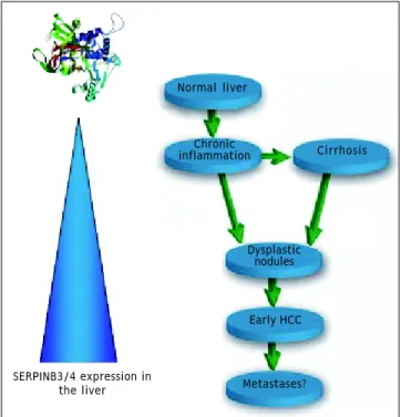
In the liver SERPINB3 and SERPINBB4 isoforms (known as squamous cell carcinoma antigen or SCCA), are undetectable in normal hepatocytes, but their expression progressively increases in chronic liver diseases24 as a cellular response to chronic
liv-er damage. Highliv-er levels are detectable in dysplastic Figure 1. Schematic representation of the main biological activities ascribed to SERPINB3. → indicates induction, i n -dicates inhibition. EMT: epithelial to mesenchymal transition.
Oxidative stress conditions chronic inflammation
TGF-β
Antiprotease activity
(papain-like cysteine proteinases)
EMT Proliferation
MYC Apoptosis
nodules25 and in hepatocellular carcinoma,26,27
sug-gesting that they may be also involved in relatively early events of hepatocarcinogenesis (Figure 2). Re-cent studies indicate that SERPINB3 is highly ex-pressed in hepatic stem/progenitor cells of both foetal and adult livers.28 This compartment is
com-posed by quiescent cells that proliferate under oxida-tive stress conditions. The occurrence of progenitor cell proliferation in humans has been described in the late stages of cirrhosis and tumors showing he-patic progenitor cell features have a worse progno-sis and a higher recurrence rate compared to tumors lacking these characteristics.29,30 In agreement with
these findings, high SERPINB3 levels have been de-tected in HCC of patients with early tumor recur-rence after surgical resection.31 In this subset, a
significant correlation of the serpin with over-ex-pression of TGF-β and of β-catenin was typically found. Transcriptome data-metanalysis further sup-ported these findings, showing accumulation of SERPINB3 in the S1 poor prognosis subclass.31
SERPINB3 has been detected recently also in hepa-toblastoma, the embryonal tumor of the liver, where a direct correlation was observed between its gene
expression, the up-regulation of Myc oncogene and tumor extension.22
Diagnostic and prognostic significance of SERPINB3/4 in serum
• HCC-diagnosis. On the basis of the oncogenic potential of SERPINB3 and of the reported find-ings of the presence of SERPINB3/4 isoforms (or SCCA) in the vast majority of HCCs specimens, in the last years ELISA assays have been devel-oped to assess the presence of SCCA as free pro-tein and/or as circulating immune complexes in serum.24,32 Free SCCA was not detected at
signif-icant levels in HCV infected patients with HCC, but this molecule was found coupled to IgMs (SCCA-IgM) to form circulating immunecomplex-es in the majority of patients with HCC, whereas in the healthy control population their levels were below the limit of detection.24 The
concen-tration of circulating SCCA-IgM, detected by a commercially available ELISA assay (SCCA-IC, Xeptagen), increased progressively at different stages of liver disease, from chronic hepatitis to cirrhosis and HCC, reflecting the extent of SCCA overexpression detected by immunohistochemis-try in liver specimens. SCCA-IgM did not overlap with AFP, offering the possibility to increase the sensitivity for HCC detection without loosing specificity. The occurrence of this biomarker-IgM immune complex seems to be supported by the re-cent immunoediting model that considers natural IgMs as one of the most important players of the innate immune system.33 This pathway likely
re-flects host immune protective mechanisms trying to apply selective pressure on newly developed transformed cells in order to control tumor growth. The neo-epitopes, present on the tumor cells surface, recognized by commonly circulating natural IgM, are able to enhance phagocytic clearance of transformed cell by macrophages and dendritic cells.34 The possible interfering
ef-fect of rheumatoid factor in SCCA-IgM reactivi-ty, found more frequently in HCV infected patients, has been excluded in artificially created samples, where the same results in terms of reac-tivity for SCCA-IgM were obtained, regardless of the presence of rheumatoid factor.35
• Fibrosis progression in chronic hepatitis. The behaviour of SCCA-IgM in serum over time has been also analysed in untreated patients with chronic hepatitis in relation to histological pro-gression of the fibrosis stage.36 After a median
Metastases?
Figure 2. Schematic diagram of the extent of SERPINB3 and SERPINB4 (SERPINB3/4) isoforms expression in the liver. Normal hepatocytes do not express this serpin and its ex-pression progressively increases in relation to the extent of liver damage. The highest levels are detectable in dysplastic nodules and early HCC. No data are available to date on HCC metastases.
SERPINB3/4 expression in the liver
Normal liver
Chronic
inflammation Cirrhosis
Dysplastic nodules
period of 6 years a significant increase of SCCA-IgM levels was observed in patients with histo-logical fibrosis score increase > 2, but not in those without histologic deterioration. These findings suggest that monitoring SCCA-IgM lev-els over time might become a useful approach to identify patients with chronic hepatitis at higher risk for cirrhosis development.
• Antiviral therapy. In HCV infected patients treated with pegylated interferon and ribavirin a significant decrease in median SCCA-IgM levels at the end of treatment, persisting up to a year of follow-up, has been described in patients with sustained virologic response, both in patients with chronic hepatitis37 and with cirrhosis.38 No
significant modifications were observed in non responder patients, indicating that SCCA-IgM monitoring in serum may be a reliable independ-ent prognostic marker of therapeutic effective-ness in anti-HCV positive patients undergoing antiviral therapy.
• Risk of HCC development in patients with cirrhosis. The behaviour of SCCA-IgM in rela-tion to HCC development has been evaluated in a cohort of HCV infected patients with cirrhosis prospectively followed up for a median period 4 years.39 The increase over time of SCCA-IgM,
as-sessed within at least one year before clinical di-agnosis of HCC, was remarkably higher in the group of patients who developed HCC than in pa-tients who did not develop HCC during the same period of follow-up, while AFP increase was not significantly different. This initial study indi-cates that the assessment of SCCA-IgM behav-iour over time might be useful to identify cirrhotic patients at higher risk of HCC develop-ment. These data have been confirmed in another retrospective study, addressed to evaluate wheth-er the levels of SCCA-IgM in swheth-erum could identify HCV positive cirrhotic patients at risk of HCC development.40 The SCCA-IgM value ≤ 200 AU/
mL accurately identified patients at low risk of liver cancer in the subsequent year, with a nega-tive predicnega-tive value of 97%. Considering an
an-nual HCC incidence ≤ 3%, patients with
SCCA-IgM ≤ 200 AU/mL had an HCC risk below
the accepted threshold of a cost-effective surveil-lance (1.5%).41 If the results of this pilot study
will be confirmed in larger studies, the authors propose that SCCA-IgM serum measurement might implement the current protocol of surveil-lance of cirrhotic patients,7 introducing a two
step (with different costs) surveillance,
consist-ing in an initial serological surveillance, based on the annual monitoring of this biomarker, and the conventional surveillance by semiannual ul-trasound when SCCA-IgM becomes > 200 AU/mL. This proposal could improve the cost/effective-ness of surveillance of HCV infected patients at risk of HCC with an acceptable number of missed early diagnoses.
• HCC prognosis. In a recent study SCCA-IgM proved efficient in the prediction of HCC progno-sis, identifying HCC patients with long overall and progression-free survival.42 Median survival
was indeed 48 months (C.I. 29-66) for patients
with low (≤ 130 AU/mL) SCCA-IgM and 26
months (C.I. 22-30) for those with high SCCA-IgM (> 130 AU/mL). At multivariate analysis tu-mour size and SCCA-IgM levels were identified as the only independent predictors of survival. In addition, SCCA-IgM levels correlated with overall response to treatment (including surgery, TACE, percutaneous ablation), with a median time to progression of 14 months in patients with low SCCA-IgM, vs. 6 months in those with high SCCA-IgM levels. Additional studies however are required to confirm these preliminary data and to better assess the behaviour of this oncomarker in relation to different methodologies of HCC treatment.
ACKNOWLEDGMENTS
I wish to thank my collaborators of the Molecular Hepatology Group (Alessandra Biasiolo, Santina Quar-ta, Mariagrazia Ruvoletto, Cristian Turato, Gian-marco Villano, Liliana Terrin, Natascia Tono, Andrea Martini, Davide Simionato), the colleagues of the Medical Clinic 5 and our Director Prof. Ange-lo Gatta, Giorgio Fassina and Andrea GalAnge-lotta (Xeptagen, Venice) for their important contribu-tions to the knowledge of SERPINB3 in the bio-logical and clinical fields of hepatology.
ABBREVIATIONS
• EMT: epithelial-to-mesenchymal transition. • HCC: hepatocellular carcinoma.
• SCCA: squamous cell carcinoma antigen. • TACE: transarterial chemoembolization.
REFERENCES
1. Sherman M. Epidemiology of hepatocellular carcinoma.
2. El-Serag HB, Kanwal F. Epidemiology of Hepatocellular Car-cinoma in the United States: Where Are We? Where Do We Go? Hepatology 2014. Doi: 10.1002/hep.27222 [Epub ahead of print].
3. El-Serag HB, Rudolph KL. Hepatocellular carcinoma: epide-miology and molecular carcinogenesis. Gastroenterology 2007; 132: 2557-76.
4. Zhang DY, Friedman SL. Fibrosis-dependent mechanisms of hepatocarcinogenesis. Hepatology 2012; 56: 769-75. 5. Fattovich G. Epidemiology of hepatocellular carcinoma. J
Hepatol 2003; 39(Suppl. 1): S50-S58.
6. Cabibbo G, Enea M, Attanasio M, Bruix J, Craxì A, Cammà C. A meta-analysis of survival rates of untreated patients in randomized clinical trials of hepatocellular carcinoma.
Hepatology 2010; 51: 1274-83.
7. Bruix J, Sherman M. Management of hepatocellular carci-noma. Hepatology 2005; 42: 1208-36.
8. Llovet JM, Bruix J. Molecular targeted therapies in hepa-tocellular carcinoma. Hepatology 2008; 48: 1312-27. 9. Villanueva A, Newell P, Chiang DY, Friedman SL, Llovet JM.
Genomics and signaling pathways in hepatocellular carci-noma. Semin Liver Dis 2007; 27: 55-76.
10. Hoshida Y, Nijman SM, Kobayashi M, Chan JA, Brunet JP, Chiang DY, Villanueva A, et al. Integrative transcriptome analysis reveals common molecular subclasses of human hepatocellular carcinoma. Cancer Res 2009; 69: 7385-92. 11. Kato H. Expression and function of squamous cell
carcino-ma antigen. Anticancer Res 1996; 199616(4B): 2149-53. 12. Suminami Y, Nagashima S, Vujanovic NL, Hirabayashi K,
Kato H, Whiteside TL. Inhibition of apoptosis in human tu-mor cells by tutu-mor-associated serpin, SCC antigen. Br J
Cancer 2000; 82: 981-9.
13. Vidalino L, Doria A, Quarta S, Zen M, Gatta A, Pontisso P. SERPINB3, apoptosis and autoimmunity. Autoimmun Rev 2009; 9: 108-12.
14. Katagiri C, Nakanishi J, Kadoya K, Hibino T. Serpin squa-mous cell carcinoma antigen inhibits UV-induced apoptosis via suppression of c-JUN NH2-terminal kinase. J Cell Biol 2006; 172: 983-90.
15. Murakami A, Suminami Y, Hirakawa H, Nawata S, Numa F, Kato H. Squamous cell carcinoma antigen suppresses ra-diation-induced cell death. Br J Cancer 2001; 84: 851-8. 16. Ciscato, Sciacovelli M, Villano G, Turato C, Bernardi P,
Ra-sola A, Pontisso P. SERPINB3 protects from oxidative da-mage by chemotherapeutics through inihibition of mitochondrial respiratory complex I. Oncotarget 2014; 5: 2418-27.
17. Quarta S, Vidalino L, Turato C, Ruvoletto M, Calabrese F, Valente M, Cannito S, et al. SERPINB3 induces epithelial-mesenchymal transition. J Pathol 2010; 221: 343-56. 18. Migliaccio E, Mele S, Salcini AE, Pelicci G, Lai KM,
Superti-Furga G, Pawson T, et al. Opposite effects of the p52shc/p46shc and p66shc splicing isoforms on the EGF receptor-MAP kinase-fos signalling pathway. EMBO J 1997; 16: 706-16.
19. Villano G, Turato C, Quarta S, Ruvoletto M, Ciscato F, Te-rrin L, Semeraro R, et al. Hepatic progenitor cells express SerpinB3. BMC Cell Biol 2014; 15: 5. Available at: http:// www.biomedcentral.com/1471-2121/15/5
20. Villano G, Quarta S, Ruvoletto MG, Turato C, Vidalino L, Biasiolo A, Tono N, et al. Role of squamous cell carcinoma antigen-1 on liver cells after partial hepatectomy in trans-genic mice. Int J Mol Med 2010; 25: 137-43.
21. Suminami Y, Nagashima S, Muratami A, Nawata S, Gondo T, Hirakawa H, Numa F, et al. Suppression of Squamous Cell Carcinoma (SCC)-related serpin, SCC Antigen, inhibits
tu-mor growth with increased intratutu-moral infiltration of ki-ller cells. Cancer Res 2001; 61: 176-80.
22. Turato C, Buendia MA, Fabre M, Redon MJ, Branchereau S, Quarta S, Ruvoletto M, et al. Over-expression of SERPINB3 in hepatoblastoma: A possible insight into the genesis of this tumour? Eur J Cancer 2012; 48: 1219-26.
23. Catanzaro JM, Sheshadri N, Pan JA, Sun Y, Shi C, Li J, Powers RS, et al. Oncogenic Ras induces inflammatory cytokine production by upregulating the squamous cell carcinoma antigens SerpinB3/B4. Nat Commun 2014; 5: 3729. Doi: 10.1038/ncomms4729.
24. Beneduce L, Castaldi F, Marino M, Quarta S, Ruvoletto M, Benvegnù L, Calabrese F, et al. Squamous cell carcinoma antigen-immunoglobulin M complexes as novel biomarkers for hepatocellular carcinoma. Cancer 2005; 103: 2558-65. 25. Guido M, Roskams T, Pontisso P, Fassan M, Thung SN,
Giaco-melli L, Sergio A, et al. Squamous cell carcinoma antigen in human liver carcinogenesis. J Clin Pathol 2008; 61: 445-7. 26. Pontisso P, Calabrese F, Benvegnù L, Lise M, Belluco C,
Ruvoletto MG, Marino M, et al. Overexpression of squa-mous cell carcinoma antigen variants in hepatocellular carcinoma. Br J Cancer 2004; 90: 833-7.
27. Trerotoli P, Fransvea E, Angelotti U, Antonaci G, Lupo L, Mazzocca A, Mangia A, et al. Tissue expression of Squa-mous Cellular Carcinoma Antigen (SCCA) is inversely corre-lated to tumor size in HCC. Mol Cancer 2009; 8: 29. Doi: 10.1186/1476-4598-8-29.
28. Villano G, Turato C, Quarta S, Ruvoletto M, Ciscato F, Te-rrin L, Semeraro R, et al. Hepatic progenitor cells express SerpinB3. BMC Cell Biol 2014; 15: 5. Available at: http:// www.biomedcentral.com/1471-2121/15/5.
29. Lee JS, Chu IS, Heo J, Calvisi DF, Sun Z, Roskams T, Durnez A, et al. Classification and prediction of survival in hepa-tocellular carcinoma by gene expression profiling.
Hepato-logy 2004; 40: 67-76.
30. Roskams T. Different types of liver progenitor cells and their niches. J Hepatol 2006; 45: 1-4.
31. Turato C, Vitale A, Fasolato S, Ruvoletto M, Terrin L, Quarta S, Ramirez Morales R, et al. SERPINB3 is associated with TGF-beta1 and cytoplasmic beta-catenin expression in hepatocellular carcinomas with poor prognosis. Br J
Cancer 2014; 110: 2708-15.
32. Giannelli G, Fransvea E, Trerotoli P, Beaugrand M, Marinos-ci F, Lupo L, Nkontchou G, et al. Clinical validation of com-bined serological biomarkers for improved hepatocellular carcinoma diagnosis in 961 patients. Clin Chim Acta 2007; 383: 147-52.
33. Dunn GP, Bruce AT, Ikeda H, Old LJ, Schreiber RD. Cancer immunoediting: from immunosurveillance to tumor escape.
Nat Immunol 2002; 3: 991-8.
34. Silverman GJ. Regulatory natural autoantibodies to apop-totic cells: pallbearers and protectors. Arthritis Rheum 2011; 63: 597-602.
35. Biasiolo A, Tono N, Zaninotto M, Merkel C, Fassina G, Ple-bani M, Gatta A, et al. Specificity of squamous cell carci-noma antigen (SCCA)-IgM detection in patients with HCV infection and rheumatoid factor seropositivity. J Med
Vi-rol 2013; 85: 1005-8.
36. Biasiolo A, Chemello L, Quarta S, Cavalletto L, Bortolotti F, Caberlotto C, Beneduce L, et al. Monitoring SCCA-IgM complexes in serum predicts liver disease progression in patients with chronic hepatitis. J Viral Hepat 2008; 15: 246-9.
an-tiviral therapy: a multicentric prospective study. J Viral
Hepat 2012; 19: 704-10.
38. Giannini EG, Basso M, Bazzica M, Contini P, Marenco S, Savarino V, Picciotto A. Successful antiviral therapy de-termines a significant decrease in squamous cell carcino-ma antigen-associated (SCCA) variants’ serum levels in anti-HCV positive cirrhotic patients. J Viral Hepat 2010; 17: 563-8.
39. Pontisso P, Quarta S, Caberlotto C, Beneduce L, Marino M, Bernardinello E, Beneduce L, et al. Progressive increase of SCCA-IgM immune complexes in cirrhotic patients is asso-ciated with development of hepatocellular carcinoma. Int J
Cancer 2006; 119: 735-40.
40. Buccione D, Fatti G, Gallotta A, Loggi E, Di Donato R, Testa L, Saitta C, et al. Serum Scca-IgM as a predictor of hepa-tocellular carcinoma in patients with cirrhosis. O J Gas 2012; 2: 56-61.
4 1 . Bruix J, Sherman M. Management of hepatocellular carcinoma: An update. Hepatology 2011; 53: 1 0 2 0 - 2 .