Prosopamnesia and Visuolimbic
Disconnection Syndrome: A Case Study
FRANCISCO LOPERA
Universidad de Antioquia Hospital San Vicente de Paul Medellin, ColombiaALFREDO ARDILA
Institute Neurologico de Colombia Bogota, Colombia
Abstract: After a head injury, a 58-year-old patient presented a severe deficit in face
recognition. Bilateral temporo-occipital hematomas were drained. Associated with this deficit in face recognition, the patient presented visual hypoemotionality (hyporeactivity to visual stimuli) and transient gustatory amnesia. Some topographic amnesia and mild anomia were also present. Five years later, difficulties in face recognition and visual hypoemotionality remained unchanged. It was observed that the major impairment was related to a lack of memory for faces. Defects in visual imagery were also present. Some distinctions between agnosia for faces (prosopagnosia) and amnesia for faces (prosopamnesia) are introduced. The brain mechanisms of prosopagnosia,prosopamnesia, and visual hypoemotionality are considered.
Keywords: Prosopamnesia, prosopagnosia, visuolimbic disconnection.
Bodamer (1947) published the first case of a brain-damaged patient with difficulties in face cognition. He proposed the name of prosopagnosia (prosopos means face). By 1974, the number of published cases had risen to 42 (Meadows, 1974). Until 1983, there were only approximately a dozen computed tomography (CT) studies and a comparable number of autopsy studies published (Damasio & Damasio, 1983; Nardelli et al., 1982). During the last 6 years this number has increased notably. This increase stimulated cognitive analyses of normal and abnormal face recognition (Ellis, 1986). Only one case of prosopagnosia in children has been reported (Young & Ellis, 1989), and only one case of developmental prosopagnosia has been reported (McConnachie, 1976).
Prosopagnosia is a distinct form of visual agnosia that affects a patient's ability to recognize familiar faces (Benton & Van Allen, 1972; Bruyer, 1983; Damasio & Damasio,
1983). The patient should be able to recognize people through other modalities, such as the voice. This impairment cannot be explained by a visual deficit, an intellectual impairment, a confusional state, or a psychotic disorder. On the basis of observations that some patients have difficulties in discriminating individuals within a given category (birds, fruits, cars),
Address correspondence to Alfredo Ardila, Institute Colombiano de Neuropsicologia, Apartado
some authors have proposed that prosopagnosia is not a specific-face agnosia, but a more general semantic impairment (Macrae & Trolle, 1956; De Renzi, Scotti, & Spinnler, 1969; Lhermitte, Chain, Scourolle, Ducarne, & Pillon, 1972).
Prosopagnosia is usually associated with other symptoms. In 90% of the cases, visual field deficits are present (Hecaen & Angeler^uss, 1962; Meadows, 1974). In a variable percentage of cases, color agnosia or achromatopsia (Green & Lessell, 1977; Kay & Levin, 1982), object agnosia (Levine, 1978), zooagnosia (Assal, Favre, & Anderes, 1984; Bornstein, Sroka, & Munitz, 1969), topographic memory deficits, constructional apraxia, spatial agnosia, somatosensory deficits, oculomotor defects, body-scheme impairments, dressing apraxia, and metamorphopsias (Hecaen & Angelergues, 1962; Damasio & Damasio, 1983, 1986) may also be present. It also has been reported that asymbolia (Romero-Lopez, Gonzalez-Elipe, Rallo-Gutierrez, & Garcia, 1982), visual imagery defects (Levine, 1978; Levine, Warach, & Farah, 1985), alexia without agraphia (Benson, Segarra, & Albert, 1974), palinopsia (Sergent & Villemure, 1989), and visual hypoemotionality (Bauer, 1982; Habib, 1986) can accompany prosopagnosia.
Clinical and anatomical correlations have been advanced. Hecaen and Angelergues (1962) proposed that damage to the basal posterior right hemisphere was sufficient to produce face agnosia. Cole and Perez-Cruet (1964) and Whiteley and Warrington (1977) supported this finding for localization. Tzavaras, Merienne, and Masure (1973) reported a left-handed patient who presented prosopagnosia after left temporal damage. The patient also presented aphasia and amnesia. Tzavaras et al. proposed that two different types of prosopagnosia could be distinguished. The first corresponded to a perceptual prosopagnosia with right hemisphere lesions; the second corresponded to an amnesic deficit associated with left hemisphere damage. Meadows (1974) proposed that prosopagnosia was related to bilateral damage. However, considering that 33 out of his 34 patients presented a left superior quadrantanopsia, he proposed that right damage was critical, whereas left damage was usually smaller and more variable. Damasio, Damasio, and Van Hoesen (1982) proposed that prosopagnosia requires a bilateral and relatively symmetrical lesion. The critical area would be the mesial basal area at the level of the occipital-temporal junction, including the lingual and fusiform gyri. However, De Renzi (1986), and Landis, Cummings, Christen, Bogen, and Imhof (1986), using CT scans, found cases of prosopagnosia in patients with exclusively right-sided damage.
Hypoemotionality is not usually mentioned in cases of prosopagnosia. It has been reported to be associated with face agnosia in at least two cases (Bauer, 1982; Habib, 1986). It is not clear if it is a frequent disorder occasionally overlooked or just an incidentally correlated symptom.
In this article, we analyze the case of a patient with a severe deficit in face recognition associated with hypoemotionality; the patient was observed for more than 5 years. We also introduce a distinction between prosopagnosia (inability to perform a perceptual analysis of faces) and prosopamnesia (inability to learn new faces and imagine familiar faces). This distinction is useful to help clarify the discussion about unilateral versus bilateral occipito-temporal involvement in face recognition impairments.
Case Report
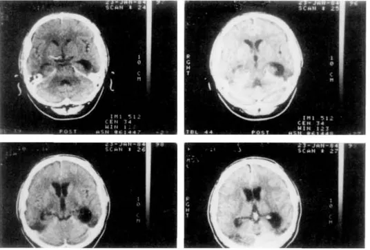
super-ficial coma for 2 weeks. Upon recovering consciousness, he complained about visual difficulties. He could not recognize his wife and children by their faces, but he could recognize them by their voices. He said that all faces looked the same. A campimetry (visual field exam) disclosed a right superior quadrantanopsia with normal visual acuity. A new CT scan (see Figure 1) disclosed a right basal occipito-temporal lesion and a deeper lesion involving the hippocampus in the left posterior temporal region; mild enlargement was observed in the temporal horn of the left lateral ventricle.
Neuropsychological Evaluation
The first neuropsychological evaluation was carried out in May, 1984. FE was found to be a cooperative, well-oriented, right-handed man. His language was fluent, grammatically correct, and prosodic. Paraphasias were not ordinarily observed in conversational language. However, some mild anomia was disclosed. Except for his left superior quadrantanopsia, the rest of the neurological exam was normal. However, he mentioned that, since the car accident, faces did not have any meaning forhim: "I see the mouth, nose, eyes, everything, but it doesn't have any meaning for me; even when I look at myself in the mirror, I just see the eyes, the mouth and so on, but nothing else; I can't even imagine how I was before." In addition, FE complained that visual stimuli did not have any emotional interest for him: "I see a landscape, the country, the flowers, but everything looks artificial, like plastic; I don't know what is beautiful and what is ugly." Furthermore, he mentioned that during the first months after the head trauma he had difficulties in recognizing foods; he observed special difficulties in
Figure 1. Computed tomography scan of the patient FE. (A right basal occipito-temporal lesion and
recognizing fruits. Tasting the fruit did not help him to recognize it. He could not imagine how fruits tasted. When eating a fruit, he reported feeling that it was the first time he had eaten this particular fruit (gustatory amnesia). Consequently, there was a defect not only visually but also gustatorily when discriminating fruits. FE could not distinguish between different types of flowers, despite being very interested in flowers before the accident. Neither could he recognize his own car unless he looked at the tag number, although he easily recognized it as a car. He mentioned that initially he had severe difficulties in spatial and topographic orientation.
When assessed, FE presented a total amnesia for the 6 months before the head trauma and some partial retrograde amnesia for several years before the accident. He did not remember any face, including his own. In addition, he reported (and his wife confirmed) that he became especially emotional in response to auditory information; music had become especially intense for him. An extensive neuropsychological evaluation was conducted to pinpoint (a) general neuropsychological defects, (b) agnosic deficits, (c) constructional impairments, and (d) characteristics of prosopagnosia.
Testing Results
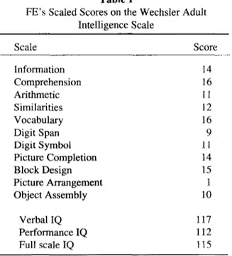
Wechsler Adult Intelligence Scale (Wechsler, 1955). FE obtained a full scale IQ of 115, a
verbal IQ of 117, and performance IQ of 112. He performed normal or above normal on all subtests except Picture Arrangement, on which he totally failed (see Table 1).
Figure Recognition. FE could recognize without mistakes the picture of objects. However,
he could correctly recognize only some (6 out of 18) superimposed Poppelreuter-type figures. In recognition of pictures of animals, mistakes were evident (15 of 15 pictures identified correctly). Recognition of fruits was impossible (0 correct out of 10), although FE
Table 1
FE's Scaled Scores on the Wechsler Adult Intelligence Scale
Scale Score
Information Comprehension Arithmetic Similarities Vocabulary Digit Span Digit Symbol Picture Completion Block Design Picture Arrangement Object Assembly
Verbal IQ Performance IQ Full scale IQ
14 16 11 12 16 9 11 14 15 1 10
could recognize that they were fruits. The same difficulty was observed for flowers (1 out of 12 pictures identified correctly).
Recognition of Family Photographs. One hundred and fifty photographs from a family
album were presented to FE. He was unable to recognize any of them, including his own photographs. However, he correctly described the different features of each face. Men and women were usually distinguished.
Matching Animal Faces. FE correctly matched 12 animal faces (6 front and 6 profile views).
However, he failed to identify exactly what each animal was (zooagnosia).
Recognition of Emotional Expressions. FE successfully recognized seven emotional
ex-pressions in photographs. However, he stipulated, "I do not see a smile, but I conclude it is a smile because the teeth are partially seen through the mouth."
Recognition of Costumes. FE easily recognized people wearing special costumes (the Pope,
a soccer player, a nun, etc.). In his everyday life, however, he does not recognize his own clothes and uses a special tag to identify the date he wears every shirt, coat, and necktie.
Color Recognition. FE obtained a score of 9.5 (out of 10) on the Ishihara test. He successfully
classified colors. After being presented with 24 different colors, he was asked to color the national flag (yellow, blue, and red); the task was correctly performed.
Rey-Osterrieth Complex Figure (Osterrieth, 1944). When copying the Rey-Osterrieth
Complex Figure, FE obtained a score of 25.5 (out of 36) according to the Taylor (1959) scoring system; this score corresponds to the 15th percentile in Colombian norms (Ardila, Rosselli, & Puente, in press). The delayed reproduction score 3 min later was 2 out of 36, corresponding to the 4th percentile. Consequently, a mild constructional apraxia (impair-ment in combinatory or organizing activity) was apparent. Poor performance on delayed reproduction implies severe nonverbal memory deficits.
Recognition of Car Models. FE failed to recognize 10 different brands and models of cars.
He mentioned that he could distinguish only gross characteristics; for instance, he could distinguish a car from a van because of the size.
Tactile Recognition. No astereognosia or agraphesthesia was disclosed. However, FE
mentioned that, because he worked in a textile factory, he had previously been able to recognize different materials by touch. After the head injury, he was no longer able to recognize different materials by their texture (ahylognosia, inability to differentiate the objects' material, i.e., density, weight, roughness, etc.; Botez, 1985; Delay, 1935).
Summary. During the first evaluation, performed in May of 1984, a severe deficit in face
recognition was observed. Difficulties in recognizing faces were associated with impairments in recognizing animals, cars, flowers, fruits, and, in general, individual members of a category. Six months of total retrograde amnesia and several years of partial retrograde amnesia were observed. Mild anterograde amnesia was disclosed, as was topographic amnesia. Gustatory amnesia and ahylognosia were reported in the anamnesis. Visual hypoemotionality was evident. Table 2 presents the summary of the first evaluation.
Follow-Up Test Results
Table 2
FE's Test Scores in the First Evaluation
Test
No. correct/ no. possible
Recognition of pictures of objects Superimposed Poppelreuter-type figures Schematized figures
Pictures of animals Recognition of fruits Recognition of flowers
Recognition of family photographs
Recognition of photographs of famous people Classification of photographs and cartoons Matching photographs and cartoons Recognition of cartoons of famous people Classification of human and animal faces Matching animal faces
Matching photograph compositions Recognition of emotional expressions Recognition of races
Recognition of costumes Recognition of colors
Three-dimensional block constructions Rey-Osterrieth Complex Figure
Copy
Delayed reproduction Recognition of car models Right-left orientation Logical memory
Corsi's block-tapping test
20/20 8/18 2/5 0/15 0/10 1/12 0/150 0/15 7/7 4/4 0/7 10/10 12/12 10/10 7/7 9/9 6/6 9.5/10 29/29 25.5/36 2/36 0/10 20/20 7/10 4/
quadrantanopsia. He strongly complained of the lack of emotion perceived in visual stimuli and a total failure in recognizing faces. This second evaluation was directed toward analysis of perceptual and amnesic impairments for faces. The following tests were given.
Test of Facial Recognition, Form FL (Benton, Van Allen, Hamsher, & Levin, 1975). He
obtained a score of 40 out of 54, which fell within normal limits.
Denman Memory for Faces (Denman, 1987). Onfour consecutive presentations, FEobtained
the following scores (out of 16): 7,6,8, and 8. Performance at this level is considered severely abnormal.
Lopera's Memory for Faces Test (Lopera, 1989). This tests consists of two parts. Initially,
photographs (with right and left hemifaces). FE's score was 35 (out of 63; chance level is 31.5). Hence, learning effaces was virtually impossible.
Rey-Osterrieth Complex Figure. When copying the Rey-Osterrieth Complex Figure, FE
obtained a score of 34.5 out of 36, which corresponds to the 80th percentile. The delayed reproduction score 3 min later was 7 out of 36, corresponding to the 17th percentile.
Visual Memory. FE had difficulty verbally describing the route required to go from one place
to another in a city that was well known to him (his hometown); he omitted important places to the right and left. He mentioned that he did not frequently dream and that, when he did dream, he never recognized the peoples' faces. He made gross mistakes when verbally describing fruits that were well known to him; for instance, he did not know the color that is inside a coconut. He drew a squirrel and a rabbit virtually identically, without using any distinguishing features.
Summary. In this follow-up evaluation, FE showed a very severe impairment in learning
faces, although he was able to match faces. Visual hypoemotionality remained. Construc-tional deficits were no longer observed, but nonverbal memory defects were virtually unchanged. Some deficits in visual imagery were disclosed. Table 3 summarizes the results.
Discussion
After more than 5 years, some deficits disclosed during the first evaluation had dramatically improved. Other deficits remained virtually unchanged: recognition of faces, nonverbal memory, and visual hypoemotionality. It was evident that FE could use perceptual cues to match faces, according to the results on the Test of Facial Recognition. However, he totally failed on all memory-for-faces tests (Denman Memory Test for Faces, Lopera's Memory for Faces Test). His performance was just at the chance level. Visual perceptual ability for face discrimination (i.e., the ability to use distinguishing cues) seemed to be much better preserved than visual memory for faces. This observation points to a more amnesic than perceptual defect. FE presents a very severe retrograde and anterograde amnesia for faces.
FE was unable to verbally describe fruits and places that were well-known to him, and he failed to use distinguishing features when drawing different animals. Visual imagery was severely impaired. Imagery obviously depends on memory. Levine et al. (1985) proposed the existence of a double system in visual imagery: the what system and the where system (e.g.,
Table 3
FE's Test Scores in the Follow-Up Evaluation
Test Score
Benton's Test of Facial Recognition 40/54 Denman Memory for Faces Test (average) 7.2 Lopera's Memory for Faces Test
Learning 14 trials Previously presented photographs 35/63 Rey—Osterrieth Complex Figure
what is the object, and where is the object?). They found that 14 of 28 patients with
prosopagnosia without visual disorientation had difficulties imagining objects, faces, and places. In these cases, visual imagery (what) was impaired, but spatial imagery (where) was preserved.
Tiberghien and Clerc (1986), analyzing the effects of context and familiarity in faces recognition, suggested that prosopagnosia usually corresponds only partially to a real visual agnosia. They proposed replacing the term prosopagnosia with prosopamnesia. We suggest, however, that prosopagnosia and prosopamnesia correspond to two different clinical syndromes. The former is an apperceptive disorder, according to Lissauer's (1890) and Warrington's (1985) perception models; the latter corresponds more strictly to a specific memory deficit for faces. FE presents a very severe amnesia for faces, but he is able to distinguish facial perceptual cues. More than a prosopagnosia, he presents a prosopamnesia. Interpretation of prosopagnosia has been contentious (Benton, 1990). Bay (1953) proposed that prosopagnosia was a special type of simultanagnosia. Warrington and James (1967) interpreted prosopagnosia as a deficit in visuoperceptual classification; they proposed that visual percepts are grouped according to certain homogenous features and that the prosopagnosic fails to distinguish individuals belonging to a specific visual-perceptual category. Benton (1980) pointed out that many patients with severe visuoperceptual deficits do not present defects in face recognition. He mentioned that, although prosopagnosia is not frequently observed, difficulties in discriminating unfamiliar faces are very frequently found. Benton proposed that different neuropsychological processes underlie the ability to discriminate unfamiliar faces and the ability to recognize familiar faces. Discrimination of unfamiliar faces is basically a visuoperceptual task, but recognition of familiar faces is also a memory and affective process; this implies the participation of more extended cerebral areas, and hence a lower frequency. FE fails in familiar face recognition and performs normally in unfamiliar face recognition. Hecaen and Angelergues (1962) identified a possible memory factor, given the observation that prosopagnosic patients frequently complain of an inability to imagine faces, as is evident in our patient.
However, to interpret prosopagnosia just as an amnesic deficit can be problematic. Usually, amnesic patients do not present with prosopagnosia. Frequently, a patient with a severe hippocampal amnesia has difficulty in learning new faces but continues to recognize previously known faces. Damasio et al. (1982) mentioned that face recognition implies the recall of multiple memories; this leads to a feeling of familiarity. A face should release multiple verbal and nonverbal memories; not all of these memories are required for face recognition, although they do facilitate face recognition. FE's low scores in other nonverbal memory tasks (e.g., delayed reproduction of the Rey-Osterrieth Complex Figure) is noteworthy.
Face recognition is closely related to emotional memory. Bauer (1982) described a case of visual hypoemotionality associated with bilateral occipito-temporal damage. Some reported cases of prosopagnosia point to some hypoemotionality for faces. This affective content may be related to the ability to assign individuality to each face. In this case, FE's auditory affect increased as his visual affect decreased.
Acknowledgment
We are very grateful to Raymond Bruyer, Nancy Echeverry, Juan Fernando Perez, Irene Gonzalez, and Adolfo Cumplido for their support and very valuable suggestions in the analysis of this case.
References
Ardila, A., Rosselli, M., & Puente, A. (in press). A practical guide to the neuropsychological
evalu-ation. New York: Plenum Press.
Assal G., Favre, C., & Anderes, J. (1984). Non-reconnaissance d'animaux familiers chez un paysan: Zoo-agnosie ou prosopagnosie pour les animaux [Nonrecognition of animal faces in a peasant: Zooagnosia or prosopagnosia for animals]. Revue de Neurologic, 140, 580-584.
Bauer, R. M. (1982). Visual hypoemotionality as a symptom of visual-limbic disconnection in man.
Archives of Neurology, 39, 702-708.
Bay, E. (1953). Disturbances of visual perception and their examination. Brain, 76, 515-551. Benson, D. F., Segarra, J., & Albert, M. L. (1974). Visual agnosia—prosopagnosia. Archives of
Neurology, 30, 307-310.
Benton, A. L. (1980). The neuropsychology of facial recognition. American Psychologist, 35,176-186. Benton, A. L. (1990). Facial recognition 1990. Cortex, 26, 491-500.
Benton, A. L., & Van Allen, M. W. (1972). Prosopagnosia and facial discrimination. Journal of
Neurological Sciences, 15, 167-172.
Benton, A. L., Van Allen, M. W., Hamsher, K. S., & Levin, H. S. (1975). Test of facial recognition,
Form SL. Iowa City: University of Iowa Hospitals.
Bodamer, J. (1947). Prosopagnosie [Prosopagnosia]. Archives Psychiatric undNervenkr, 179, 6—54. Botez, M. I. (1985). Parietal lobe syndromes. In J. A. M. Frederiks (Ed.), Handbook of clinical
neu-rology: Vol. 45. Clinical neuropsychology (pp. 63-85). Amsterdam: Elsevier.
Bornstein, B., Sroka, H., & Munitz, H. (1969). Prosopagnosia with animal face agnosia. Cortex, 5,164— 169.
Bruyer, R. (1983)Le visages et I' expression faciale: Approache neuropsychologique [Faces and facial expressions: A neuropsychological approach]. Brussels, Belgium: Pierre Mardage.
Cole, M., & Perez-Cruet, J. (1964). Prosopagnosia. Neuropsychologia, 2, 237-246.
Damasio, A. R., & Damasio, H. (1983). Localization of lesions in achromatopsia and prosopagnosia. In A. Kertesz (Ed.), Localization in neuropsychology (pp. 419-428). San Diego, CA: Academic Press.
Damasio, A. R., & Damasio, H. (1986). The anatomical substrate of prosopagnosia. In R. Bruyer (Ed.),
The neuropsychology of face perception andfacialexpression(pp. 31-38).Hillsdale,NJ:Erlbaum.
Damasio, A. R., Damasio, H., & Van Hoesen, G.W. (1982). Prosopagnosia: Anatomical basis and behavioral mechanisms. Neurology, 32, 331-341.
Delay, J. (1935). Les astereognosies: Pathologie du toucher [Astereagnosia: Pathology of touch]. Paris: Masson et Cie.
Denman, S. B. (1987). Denman Neuropsychology Memory Scale. Charleston, SC: Author.
De Renzi, E. (1986). Prosopagnosia in two patients with CT scan: Evidence of the damage confined to the right hemisphere. Neuropsychologia, 24, 385-389.
De Renzi, E., Scotti, G., & Spinnler, H. (1969). Perceptual and associative disorders of visual recognition. Neurology, 19, 634—642.
Ellis, H. D. (1986). Processes underlying face recognition. In R. Bruyer (Ed.), The neuropsychology
of face perception and facial expression (pp. 1-27). Hillsdale, NJ: Erlbaum.
Green, G. J., & Lessell, S. (1977). Acquired cerebral dyschormatopsia. Archives of Ophthalmology, 95, 121-128.
Habib, M. (1986). Visual hypoemotionality and prosopagnosia associated with right temporal lobe isolation. Neuropsychologia, 24, 577-582.
92-100.
Kay, M. C., & Levin, H. S. (1982). Prosopagnosia. American Journal of Ophthalmology, 94, 75-80. Landis, T., Cummings, J. L., Christen, L., Bogen, J. E., & Imhof, H. G. (1986). Are unilateral right posterior cerebral lesions sufficient to cause prosopagnosia? Clinical and radiological findings in six additional patients. Cortex, 22, 243-252.
Levine, D. N. (1978). Prosopagnosia and visual object agnosia: A behavioral study. Brain and
Lan-guage, 5, 341-365.
Levine, D. N., Warach, J., & Farah, M. (1985). Two visual systems in mental imagery: Dissociation of "what" and "where" in imagery disorders due to bilateral posterior cerebral lesions. Neurology,
35, 1010-1018.
Lhermitte, J., Chain, F., Scourolle, R., Ducarne, B., & Pillon, B. (1972) Etude anatomo-clinique d'un cas de prosopagnosie [Clinical/anotomical study of acase of prosopagnosia]. Revue Neurologique,
126, 329-346.
Lissauer, H. (1890). Ein Fal von Seelenblindheit nebst einem Beitrag zur Theorie derselbem [A case of psychic blindness as a contribution to the theory]. Archives Psychiatric undNervenkr, 21,222-270.
Lopera, F. (1989). Apprentissage et reconnaissance de visages. Unpublished manuscript, Universite Catholique de Louvain, Brussels, Belgium.
Macrae, D., & Trolle, E. (1956). The defect of function in visual agnosia. Brain, 79, 94-110. McConnachie, H. (1976). Developmental prosopagnosia: A single case study. Cortex, 12, 76-82. Meadows, J. C. (1974). The anatomical basis of prosopagnosia. Journal of Neurology, Neurosurgery
and Psychiatry, 37, 489-501.
Nardelli, E., Buonanno, F., Coccia, G., Fiaschi, A., Terzian, H., & Rizzuto, N. (1982). Prosopagnosia: Report of four cases. European Neurology, 21, 289-297.
Osterrieth, P. A. (1944). Le test de copie d'une figure complexe [The test of copying a complex figure].
Archives de Psychologie, 30, 206-256.
Romero-Lopez, J., Gonzalez-Elipe, J., Rallo-Gutierrez, B., & Garcia, J. (1982). Prosopagnosia: Hallazgos clinicos and neurorradiologicos (TAG) de un nuevo caso [Prosopagnosia: Clinical and neuroradiological findings of a new case]. Revista Clinica Espanola, 167, 331-333.
Sergent, J., & Villemure, J. G. (1989). Prosopagnosia in a right hemispherectomized patient. Brain,! 12, 975-995.
Taylor, E. M. (1959). The appraisal of children with cerebral deficits. Cambridge, MA: Harvard University Press.
Tiberghien, G., & Clerc, I. (1986). The cognitive locus of prosopagnosia. In R. Bruyer (Ed.), The
neuropsychology of face perception and facial expression (pp. 39-63). Hillsdale, NJ: Erlbaum.
Tzavaras, A., Merienne, L., & Masure, M. C. (1973). Prosopagnosie, amnesia, et troubles du language par lesion temporale gauche chez un sujet gaucher [Prosopagnosia, amnesia, and language defects resulting from a left temporal lesion in a left-handed subject]. L'Encephale, 62, 382-394. Young, A., & Ellis, H. D. (1989). Childhood prosopagnosia. Brain and Cognition, 9, 16-47. Warrington, E. K. (1985). Agnosia: The impairment of object recognition. In J. A. M. Frederiks (Ed.),
Handbook of clinical neurology, Vol. 45: Clinical neuropsychology (pp. 333-349). Amsterdam:
Elsevier.
Warrington, E. K., & James, M. (1967). An experimental investigation of facial recognition in patients with unilateral cerebral lesions. Cortex, 3, 317-326.
Wechsler, D. (1955). Wechsler Intelligence Scale: Manual. New York: Psychological Corporation. Whiteley, A. M., & Warrington, E. (1977). Prosopagnosia: A clinical, psychological and anatomical