Practical Considerations of Real Life of
Hepatocellular Carcinoma in a Tertiary Center of Brazil
Sandra R. Almeida-Carvalho,* Maria L. Gomes-Ferraz,* Carla A. Loureiro-Matos,* Antônio E. Benedito-Silva,* Roberto J. Carvalho-Filho,* Rogério Renato-Perez,† Adriano Miziara-Gonzalez,† Alcides A. Salzedas-Netto,‡ Denis Szejnfeld,§ Giuseppe D’Ippolito,§ Valéria Pereira-Lanzoni,|| Ivonete S. Souza-Silva*
* Department of Gastroenterology, Hepatology Unit. † Department of Surgery, Liver Transplant Unit. ‡ Department of Pediatric Surgery. § Department of Diagnostic Radiology.|| Department of Diagnostic Pathology. Federal University of Sao Paulo (Unifesp), Sao Paulo, SP, Brazil.
March-April, Vol. 16 No. 2, 2017: 255-262
INTRODUCTION
Hepatocellular carcinoma (HCC) is the sixth most common tumor in the world and its incidence and risk factors vary widely.1 This tumor is three-fold more
com-mon in men than in women and affects individuals at a mean age of 64 years.2 Liver cirrhosis is the main risk
fac-tor for the development of HCC.3,4 The most frequent
eti-ologies are hepatitis C infection in the United States,3,5
Europe and Japan, and hepatitis B infection in Asia and Af-rica.6 Mortality from HCC is high, with an estimated
600,000 deaths per year worldwide.6
In Brazil, the incidence of HCC ranges from 3.3% to 6% per 100,000 persons and mortality rates range from 3.6% to 6% per 100,000 persons per year.1 Data from a
Brazilian national survey indicate a predominance of the disease in men older than 50 years with cirrhosis and HCV infection. However, these findings differ between the different regions of the country, especially between the North/Northeast and South/Southeast.7 Moreover, there
is a paucity of data from these regions regarding diag-nosis, type of study population (from private or public services), and treatment. Imaging equipment is critical for the diagnosis of HCC and is rare in Brazilian public hospitals. In addition, liver transplantation (LT) is only available in large Brazilian cities. Therefore, more stud-ies on HCC are needed to elaborate diagnostic and treat-ment strategies for this population. The aim of this study was to evaluate the clinical profile, epidemiological char-acteristics, laboratory parameters, treatment and survival The Official Journal of the Mexican Association of Hepatology,
the Latin-American Association for Study of the Liver and the Canadian Association for the Study of the Liver
Manuscript received: Manuscript received:Manuscript received:
Manuscript received:Manuscript received: December 18, 2015. Manuscript accepted:Manuscript accepted:Manuscript accepted:Manuscript accepted:Manuscript accepted: July 23, 2016.
DOI:10.5604/16652681.1231584.
A B S T R A C T A B S T R A C T A B S T R A C T A B S T R A C T A B S T R A C T
Background. Background.Background. Background.
Background. Hepatocellular carcinoma (HCC) is the most common malignancy that develops in cirrhotic livers. Its clinical and epi-demiological characteristics and mortality rates vary according to geographical region. The objective of this study was to evaluate the clinical profile, epidemiological characteristics, laboratory parameters, treatment and survival of patients with HCC. Material andMaterial andMaterial andMaterial andMaterial and methods.
methods.methods. methods.
methods. Patients with HCC seen between 2000 and 2012 were studied. The Kaplan-Meier method was used for survival analysis according to variables in question. Results.Results.Results.Results.Results. The study included 247 patients with a mean age of 60 ± 10 years. There was a pre-dominance of males (74%). The main etiologies of HCC were HCV infection (55%), excessive alcohol consumption (12%), and HBV infection (8%). Liver cirrhosis was present in 92% of cases. The mean tumor number and diameter were 2 and 5 cm, respectively. Patients meeting the Milan criteria corresponded to 43% of the sample. Liver transplantation was performed in 22.4% of patients of the Milan subset and in 10% of the whole sample. The overall mean survival was 60 months, with a 1-, 3- and 5-year survival proba-bility of 74%, 40% and 29%, respectively. Lower survival was observed among patients with alcoholic etiology. Survival was higher among patients submitted to liver transplantation (P < 0.001), TACE (P < 0.001), or any kind of treatment (P < 0.001). However, no difference was found for surgical resection (P = 0.1) or sorafenib (P = 0.1). Conclusion.Conclusion.Conclusion.Conclusion.Conclusion. Patients with HCC were mainly older men diagnosed at an advanced stage. Treatment was associated with better overall survival, but few patients survived to be treated.
Key words. Key words.Key words. Key words.
of patients with HCC seen at a specialized public center in Brazil.
MATERIAL AND METHODS
We studied patients with HCC seen between 2000 and 2012 at a specialized center of the Federal University of São Paulo, São Paulo, Brazil. The Ethics Committee of the Federal University of São Paulo approved the study.
At the time of diagnosis, the patients were investigated for clinical, demographic, laboratory, and radiological fea-tures. Differences in survival were analyzed according to etiology and to the different therapeutic modalities used. For survival analysis, follow-up was censored on Decem-ber 30, 2012. Data regarding death were obtained from the hospital records in more than 90% of cases. In some cases, this information was obtained by contacting the family.
Clinical and laboratory assessment
Clinical evaluation consisted of a detailed clinical his-tory and physical examination. The laborahis-tory tests includ-ed serum measurement of albumin (g/dL), alpha-fetoprotein (AFP, IU/mL), aspartate aminotrans-ferase (AST, U/L), alanine aminotransaminotrans-ferase (ALT, U/L), alkaline phosphatase (U/L), gamma-glutamyltransferase (GGT, U/L), total bilirubin (mg/dL), international nor-malized ratio (INR), platelets (n/μL), and creatinine (male < 1.2; female < 0.9 mg/dL).
Viral markers such as HBsAg, HBc, HBeAg, anti-HBe and anti-HCV were assayed using commercial ELI-SA kits. Detection of HBV DNA and HCV RNA was performed by RT-PCR with a detection limit of 20 IU/ mL (Cobas TaqMan HBV, Roche Diagnostics) and 50 IU/ mL, respectively.
Liver cirrhosis was confirmed by histological analysis and/or based on clinical-laboratory parameters such as as-cites, hepatic encephalopathy, portal hypertension (splenomegaly, esophageal varices, thrombocytopenia), and ultrasonographic signs suggestive of liver cirrhosis. Excessive alcohol consumption as a cause of cirrhosis was defined as ethanol consumption > 40 g/day in women and > 60 g/day in men for more than 5 years. Patients with liv-er cirrhosis wliv-ere evaluated using the Child-Turcotte-Pugh and MELD scores.
Diagnosis of hepatocellular carcinoma
The diagnosis of HCC was based on triple-phase imag-ing by computed tomography or magnetic resonance of the liver that showed a hypervascular tumor in the arterial
phase (wash in) and washout in the portal or equilibrium phase. Tumors not defined by the imaging exams were submitted to image-guided biopsy. The tumor diameter and number, tumor macrovascular invasion, and extrahe-patic metastases were analyzed by chest/abdominal tom-ography and bone scanning.
Treatment of hepatocellular carcinoma
When indicated, treatment consisted of LT, surgical re-section, transarterial chemoembolization (TACE), radiof-requency thermal ablation (RFA), percutaneous ethanol
Table 1. Demographic, clinical, laboratory and tumor
character-istics of patients with hepatocellular carcinoma (n = 247).
Characteristics
Demographic/clinical
Age (years)† 60 ± 10
Male/female 74%/26%
Etiology
Hepatitis C 55%
Hepatitis C/alcohol 14%
Alcohol 12%
Hepatitis B 8%
Other 11%
Cirrhosis 92%
Child-Pugh A/B/C 57%/36%/7%
Laboratory parameters†
MELD 11 ± 4
AST (U/L) 93 ± 71
ALT (U/L) 71 ± 56
Alkaline phosphatase (U/L) 161 ± 132
GGT (U/L) 191 ± 186
Bilirubin (mg/dL) 1.8 ± 1.9
Platelets (10³/μL) 123 ± 74
Creatinine (mg/dL) 1.0 ± 0.7
Albumin (g/dL) 3.6 ± 0.64
Alpha-fetoprotein (IU/mL) 4,475 ± 13,754
INR 1.24 ± 0.23
Tumor characteristics
Number of nodules† 2 ± 1
Nodule size† (cm) 5 ± 3
Extrahepatic metastases, n (%) 24 (10) Tumor vascular invasion, n (%) 31 (13)
Tumor treatment, n (%)
Liver transplant 25 (10)
Surgical resection 13 (5)
Transarterial chemoembolization 122 (49) Radiofrequency ablation 3 (1) Percutaneous ethanol injection 5 (2)
Sorafenin 35 (14)
Symptomatic therapy 68 (28)
† Expressed as mean ± standard deviation. MELD: model for end-stage liver
Laboratory analysis
The mean ± SD, median and range of serum AFP lev-els were 4,475 ± 13,754, 57 and 1-102,436 IU/mL, respec-tively. The mean serum levels of AST, ALT, alkaline phosphatase, GGT, platelets, albumin, total bilirubin, cre-atinine and INR were 93 U/L, 71 U/L, 161 U/L, 191 U/L, 123,000/μL, 3.6 g/dL, 1.8 mg/dL, 1.0 mg/dL and 1.24, re-spectively. The mean MELD score was 11 (Table 1).
Tumor characteristics
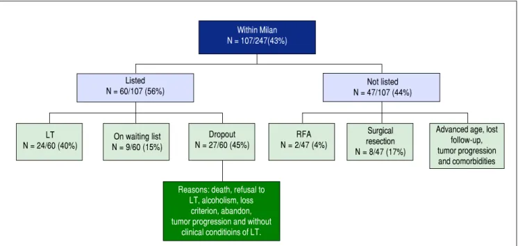
The mean number of nodules was 2 ± 1 (range: 1-7 nod-ules) and the mean diameter was 5 ± 3 cm (range: 0.9-21 cm). Extrahepatic metastases and tumor macrovascular inva-sion were observed in 10% and 13% of cases, respectively. Extrahepatic metastases were detected mainly in regional lymph nodes, lungs, and bone. The number of patients meeting the Milan criteria was 107 (43%) (Table 1).
Tumor treatment
• Group within Milan criteria (Figure 1). Among pa-tients meeting the Milan criteria (n=107), 56% (60/ 107) were on the liver transplant list. Of these, 40% (24/60) underwent LT, 15% were waiting for LT, and 45% (27/60) left the list. Among the patients not listed (47/107), 4% (2/47) were submitted to RFA, 8% (4/47) injection, and administration of sorafenib. The patients
were classified according to the Milan criteria,8 and each
therapeutic modality was performed according to the Bar-celona Clinic Liver Cancer (BCLC) staging system9 or
experience of the service.
Statistical analysis
The results are expressed as the mean ± standard devia-tion. Survival was defined as the interval between the date of HCC diagnosis and either the date of liver-related death or the last follow-up on December 30, 2012. The Ka-plan-Meier method was used for survival analysis. A level of significance of P < 0.05 was adopted. Statistical analysis was performed using the SPSS 16 program (Chicago, IL, USA).
RESULTS
There were 247 HCC patients in the study. The mean age of these patients was 60 ± 10 years and there was a pre-dominance of men (74%). The main etiologies were in-fection with HCV (55%), followed by inin-fection with HCV associated with excessive alcohol consumption (14%), ex-cessive alcohol consumption (12%), infection with HBV (8%), and other causes (11%). Liver cirrhosis was present in 92% of the patients who were classified as Child-Tur-cotte-Pugh A (57%), B (36%), and C (7%) (Table 1).
Figure 1. Figure 1. Figure 1.
Figure 1. Figure 1. Algorithm of HCC patients within the Milan criteria according to treatment offered and outcomes (n = 247). LT: liver transplantation. RFA: radiofrequency thermal ablation.
Within Milan N = 107/247(43%)
Listed
N = 60/107 (56%) N = 47/107 (44%)Not listed
LT N = 24/60 (40%)
On waiting list N = 9/60 (15%)
Dropout N = 27/60 (45%)
RFA N = 2/47 (4%)
Surgical resection N = 8/47 (17%)
Advanced age, lost follow-up, tumor progression and comorbidities
Reasons: death, refusal to LT, alcoholism, loss
criterion, abandon, tumor progression and without
to percutaneous ethanol injection, and 17% (8/47) to surgical resection. The remaining patients were not listed because of advanced age, severe comorbidities and tumor progression, or were lost to follow-up. • Group outside Milan criteria (Figure 2). TACE,
sorafenib, RFA and surgical resection were offered to
patients outside the Milan criteria (140/247). Downstag-ing to within Milan by TACE was achieved in only one patient. The procedure was well-tolerated. Sorafenib was offered to 14% (35/247) of the patients with ad-vanced-stage HCC at a dose of 800 mg/day. Sorafenib combined with TACE was offered to 71% (25/35) of the
Figure 2. Figure 2. Figure 2. Figure 2.
Figure 2. Algorithm of HCC patients outside the Milan criteria according to treatment offered and outcomes (n = 247). TACE: transarterial chemoemboliza-tion. RFA: radiofrequency thermal ablachemoemboliza-tion. * One patient was submitted to liver transplantation by downstaging.
Outside Milan N = 140/247(57%)
Surgical resection N = 5 (4%)
TACE* N = 64 (46%)
Sorafenib N = 10 (7%)
TACE + Sorafenib N = 25 (18%)
RFA N = 1 (10.7%)
A. A. A. A.
A. Alcohol x HCV
Survival probability Survival probability Survival probability
Survival probability Survival probability
1.0 0.8 0.6 0.4 0.2 0.0
1.0 0.8 0.6 0.4 0.2 0.0
1.0 0.8 0.6 0.4 0.2 0.0 1.0
0.8 0.6 0.4 0.2 0.0
1.0 0.8 0.6 0.4 0.2 0.0
0 50 100 150
Time (months)
0 20 40 60
Time (months) 0 40Time (months)80 120 160
0 40 80 120 160
Time (months)
0 40 80 120 160
Time (months)
P < 0.004 P = 0.1
P < 0.03 P = 0.09
P = 0.1 B.
B. B. B.
B. Alcohol x HBV C. C. C. C. C. HCV x Alcohol + HCV
D. D. D.
D. D. HCV x HBV E .E .E .E .E .
HCV-censored HCV + Alcohol-censored
HBV-censored
HCV-censored Alcohol
Alchohol-censored HCV
HCV-censored HBV HBV-censored HCV + Alcohol HCV + Acohol-censored Alcohol-censored
HCV-censored
Figure 3. Figure 3. Figure 3. Figure 3.
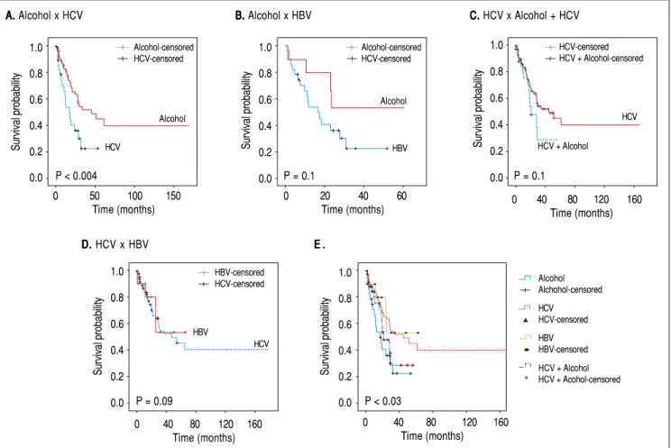
Figure 3. Survival probability according to etiology comparing (A) excessive alcohol consumption with HCV infection, (B) excessive alcohol consumption with HBV infection, (C) HCV infection with and without excessive alcohol consumption, (D) HCV infection with HBV infection, and (E) all etiologies.
Alcohol-censored HCV-censored
Alcohol
HBV Alcohol
HCV
HCV
HCV + Alcohol
patients. The mean daily dose of the drug was 630 mg. Side effects were observed in 37% (13/35) of the patients who received the drug. Diarrhea, headache and hand-foot skin reaction were the main adverse events.
Survival analysis
At the time the data were censored, 89 (46.4%) patients had died. The overall mean survival was 60 months, with a 1-, 3- and 5-year survival probability of 74%, 40% and 29%, respectively.
A significant difference in survival was observed be-tween etiologies. Survival was lower among patients with alcoholic etiology or alcoholic etiology and HCV infec-tion. Figure 3 shows the differences in survival according to the different etiologies.
There was also a significant difference in survival be-tween treatments. Individual analysis of survival according to the different treatments showed higher survival among patients submitted to LT (P < 0.001), TACE (P < 0.001), or any kind of treatment (P < 0.001). However, no differ-ence was found for surgical resection (P = 0.1) or soraf-enib (P = 0.1) (Figure 4).
Resection Liver transplantation TACE Sorafenib No treatment Censored A AA A
A BBBBB
Survival probability Survival probability
Survival probability Survival probability
Survival probability Survival probability Survival probability
1.0 0.9 0.8 0.7 0.6 0.5 0.4 0.3 0.2
0 30 60 90 120 150 Time (months)
0 20 40 60 80 100120140 160 Time (months) Function Censored 1.0 0.9 0.8 0.7 0.6 0.5 0.4 0.3 0.2 0.1 0.0 No Non-censored Yes Censored 1.0 0.9 0.8 0.7 0.6 0.5 0.4 0.3 0.2 0.1 0.0
0 27 54 81 108 135 162 189 Time (months) C CC C C 1.0 0.8 0.6 0.4 0.2 0.0 D D D D D
0 30 60 90 120 150 180 210 Time (months) P = 0.1
Treatments resection TACE E EE EE 1.0 0.8 0.6 0.4 0.2 0.0
0 30 60 90 120 150 180 210 Time (months) P < 0.001
1.0 0.8 0.6 0.4 0.2 0.0 P = 0.1 F
FF FF
0 30 60 90 120 150 180 210 Time (months) Sorafenib No Non-censored Yes Censored G G G G G 1.0 0.8 0.6 0.4 0.2 0.0
0 30 60 90 120 150 180 210 Time (months) P < 0.001
Figure 4. Figure 4. Figure 4.
Figure 4. Figure 4. Survival probability (A) in all patients of the entire cohort, (B) according to the different treatments, (C) according to liver transplantation (LT), (D) according to surgical resection, (E) according to transarterial chemoembolization (TACE), (F) according to systemic therapy (sorafenib), and (G) according to any treatment. No Non-censored Yes Censored Any treatment No Non-censored Yes Censored No Non-censored Yes Censored Resection LT Survival
P < 0.001
DISCUSSION
We found a predominance of men in the study and the mean age was 60. These findings were similar to those reported in a Brazilian national survey on the prev-alence of HCC.7 However, some differences were
ob-served when compared to other countries. For example, in Asia, the tumor incidence peaks around 60 years and HBV infection predominates.6,10 These findings agree
with our study regarding age but not in relation to etiolo-gy because we found a predominance of HCV. Another discrepancy is seen in Japan where HCV predominates, while age ranges from 70 to 75 years.10,11 These findings
show that epidemiological differences in HCC between regions cannot always be explained by different etiologies. With respect to etiology, we observed lower survival among patients with a history of alcohol abuse and survival was also lower when alcohol abuse was associated with HCV infection. This fact has also been reported in the study of Bucci et al. in which patients with alcoholic etiol-ogy had significantly lower survival than those with HCV infection alone.12
Liver cirrhosis is the main risk factor for HCC3,8,9,13
and was present in 92% of the cases, in agreement with other studies. Another important finding was that most pa-tients with HCC were classified as Child A (57%). HCC with compensated cirrhosis is commonly found in liver cancer patients.14-16
Abnormal AFP serum values were detected in most pa-tients (63%), but 37% had AFP values within the normal range. This was expected because 30-40% of tumors have normal AFP values.6 Moreover, the utility of AFP for the
diagnosis of HCC is known to be limited.17-19
With respect to tumor characteristics, the number of patients outside the Milan criteria was high. The number of patients with advanced HCC differs between studies. In an Indian study, 44% of the patients with HCC had ad-vanced disease,20 while in a larger study conducted by
Ku-mar, et al. (2008), there were 82% advanced cases of HCC.21
In the present study, in contrast to the significant number of patients with advanced disease, the frequency of distant metastases (10%) and macrovascular tumor inva-sion (13%) was low. Addario, et al. (2011) observed distant metastases and tumor macrovascular invasion in 10% of cases.22 In a large American study, Yang, et al. (2014) found
distant metastases in 16% of cases.23 Paul, et al. (2009)
ob-served distant metastases in 13% of cases and macrovascu-lar tumor invasion in 40% .20
Treatment of HCC such as LT, RFA and surgical re-section were offered to a small proportion of patients. LT was performed in 22.4% of patients within the Milan cri-teria and in 10% of the whole sample. These findings are consistent with other studies. In the study of Kitisin, et al.
(2011), 13% of patients with HCC underwent LT.15 This
percentage was only 7% in the study of Yang, et al. (2014).23
These numbers reflect not only the presence of advanced disease, but also the low availability of donor organs. As expected, the survival rate of patients submitted to LT was higher than the survival of patients receiving no transplant. Surgical resection had no impact on patient survival. In fact, surgical resection was offered to only 13 patients who had advanced disease, which likely explains the findings. In the study of Carrilho, et al. (2010), the rate of surgical re-section was 7% .7 Higher rates of 11% and 12% have been
reported in other studies.15,23 The use of TACE for some
intermediate tumors as downstaging treatment to within Milan criteria may explain in part the small number of surgical resections found here.
Downstaging of patients with HCC outside the Milan criteria using TACE has been associated with high rates of successful downstaging to within Milan criteria but also with post-LT recurrence and survival.24 However, in our
study few patients showed a decrease in tumor size to within Milan criteria for subsequent LT. TACE has also been proposed as a bridging treatment to LT because it may improve survival and reduce the recurrence of HCC after LT.25,26 In our study, 27% of the patients who
re-ceived TACE were on the waiting list for LT. This proce-dure resulted in a very low rate of dropout and was well tolerated. In addition, better survival was observed among patients who received TACE compared to those who did not. Although RFA has gained widespread use over recent years as an effective procedure, especially for small HCC not amenable to surgical resection, this procedure is not available at our center.
In the present study, 14% of the patients received soraf-enib either as monotherapy or combined with TACE. The drug was well tolerated by most patients. Data indicate that sorafenib might have a beneficial therapeutic effect on advanced-stage HCC27 by inhibiting tumor cell
prolifera-tion and tumor angiogenesis. In addiprolifera-tion, sorafenib has been shown to increase the rate of apoptosis in a wide range of tumor models, increasing survival.28 However, no
association between survival and the use of sorafenib was observed in our study.
The indication of sorafenib is still controversial. There-fore, the number of patients receiving the drug varies widely. Sanyal, et al. (2010) reported that 6% of patients with HCC used sorafenib.29 This percentage was only 3%
in the study of Kitisin, et al. (2011).15 These differences
suggest that the populations studied differ in terms of the indications and contraindications to drug use.
organs and advanced disease restrict the access to LT. These findings and those of similar Brazilian studies high-light the need to intensify the screening of risk patients for HCC in order to identify this cancer early, which should permit to increase the success rates of conventional treat-ments.
ABBREVIATIONS
• AFP: alpha-fetoprotein • ALT: alanine aminotransferase • Anti-HBc: hepatitis B core antibody • Anti-HBe: hepatitis B e antibody • Anti-HCV: hepatitis C virus antibody • AP: alkaline phosphatase
• AST: aspartate aminotransferase • BCLC: Barcelona clinic liver câncer. • CI: confidence interval.
• CTP: Child-Turcotte-Pugh. • GGT: gamma-glutamyltransferase. • HBeAg: hepatitis B e antigen. • HBsAg: hepatitis B surface antigen. • HBV: hepatitis B vírus.
• HBV DNA: hepatitis B virus DNA. • HCC: hepatocellular carcinoma. • HCV: hepatitis C vírus.
• HCV RNA: hepatitis C virus RNA. • INR: international normalized ratio. • LT: liver transplantation.
• MELD: model for end-stage liver disease. • PEI: percutaneous ethanol injection. • RF: radiofrequency ablation.
• RT-PCR: reverse transcription polymerase chain re-action.
• TACE: transarterial chemoembolization. • TMI: tumor macrovascular invasion.
COMPETING INTERESTS
The authors declare that they have no competing inter-ests.
REFERENCES
1. IARC (International Agency for Research on Cancer), GLO-BOCAN 2012. (<http://globocan.iarc.fr/>; 2012 [accessed 05.09.14])
2. CDC - Centers for Disease Control and Prevention. Hepatocel-lular Carcinoma - United States 2001-2006 2010; 59: 517-20. 3. Colombo M, Franchis R, Ninno ED, Sangiovanni A, Fazio CD,
Tommasini M, Donato MF, et al. Hepatocellular carcinoma in Italian patients with cirrhosis. N Engl J Med 1991; 325:
675-80.
4. Okuda H. Hepatocellular carcinoma development in cirrhosis.
Best Pract Res Clin Gastroenterol 2007; 21: 161-73.
5. Davila JA, Morgan RO, Shaib Y, McGlynn KA, EL-Serag HB. Hepatitis C infection and the increasing incidence of hepato-cellular carcinoma: A Population-Based Study. Gastroenter-ology 2004; 127: 1372-80.
6. Ferenci P, Fried M, Labrecque D, Bruix J, Sherman M, Omata M, Heathcote J. et al. Hepatocellular Carcinoma (HCC): A Global Perspective. J Clin Gastroenterol 2010; 44: 239-45. 7. Carrilho FJ, Kikuchi L, Branco F, Gonçalves CS, Mattos AA,
Brazilian HCC Study Group. Clinical and epidemiological as-pects of hepatocellular carcinoma in Brazil. Clinics 2010; 65: 1285-90.
8. Mazzaferro V, Regalia E, Doci R, Andreola S, Pulvirenti A, Bozzetti F, Montalto F. et al. Liver transplantation for the treatment of small hepatocellular carcinomas in patients with cirrhosis. N Engl J Med 1996; 334: 693-9.
9. Forner A, Josep ML, Bruix J. Hepatocellular carcinoma. Lan-cet 2012; 379: 1245-55.
10. Bosch FX, Ribes J, Cléries R, Díaz M. Epidemiology of Hepa-tocellular Carcinoma. Clin Liver Dis 2005; 9: 191-211. 11. Llovet JM, Burroughs A, Bruix J. Hepatocellular carcinoma.
Lancet 2003; 362: 1907-17.
12. Bucci L, Garuti F, Camelli V, Lenzi B, Farinati F, Giannini EG, Ciccarese F, et al. Comparison between alcohol- and hepati-tis C virus-related hepatocellular carcinoma: clinical presen-tation, treatment and outcome. Aliment Pharmacol Ther
2016; 43: 385-99.
13. Somboon K, Siramolpiwat S, Vilaichone RK. Epidemiology and Survival of Hepatocellular Carcinoma in the Central Region of Thailand. Asian Pac J Cancer Prev 2014; 15: 3567-70.
14. Cheung TK, Lai CL, Wong BCY, Fung J, Yuen MF. Clinical features, biochemical parameters, and virological profiles of patients with hepatocellular carcinoma in Hong Kong. Ali-ment Pharmacol Ther 2006; 24: 573-83.
15. Kitisin K, Packiam V, Steel J, Humar A, Gamblin TC, Geller DA, Marsh JW, et al. Presentation and outcomes of hepato-cellular carcinoma patients at a western centre. HPB (Ox-ford) 2011; 13: 712-22.
16. Kneuertz PJ, Demirjian A, Firoozmand ADA, Villalobos CC, Bhagat N, Herman J, Cameron A, et al. Diffuse Infiltrative Hepatocellular Carcinoma: assessment of presentation, treatment, and outcomes. Ann Surg Oncol 2012; 19: 2897-907.
17. Sherman M. Hepatocellular carcinoma: epidemiology, surveil-lance, and diagnosis. Semin Liver Dis 2010; 30: 3-16. 18. Trevisani F, D’Intino PE, Morselli-Labate AM, Mazzella G,
Ac-cogli E, Caraceni P, De Notariis S, et al. Serum alpha-fetopro-tein for diagnosis of hepatocellular carcinoma in patients with chronic liver disease: influence of HBsAg and anti-HCV status. J Hepatol 2001; 34: 570-5.
19. Marrero JA, Feng Z, Wang Y, Nguyen MH, Befeler AS, Roberts LR, Reddy KR, et al. AlfaFetoprotein, DesCarboxy-prothrombin, and Lectin-Bound. AlfaFetoprotein in Early Hepatocellular Carcinoma. Gastroenterology 2009; 137: 110-18.
20. Paul SB, Chalamalasetty SB, Vishnubhatla S, Madan K, Gamanagatti SR, Batra Y, Gupta SD, et al. Clinical Profile, Eti-ology and Therapeutic Outcome in 324 Hepatocellular Carci-noma Patients at a Tertiary Care Center in India. Oncology
2009; 77: 162-71.
metas-tasis of hepatocellular carcinoma. Digestive and Liver Dis-ease 2011; 43: 319-24.
23. Yang D, Hanna DL, Usher J, LoCoco J, Chaudhari P, Lenz HJ, Setiawan VW, et al. Impact of sex on the survival of pa-tients with hepatocellular carcinoma: A Surveillance, Epide-miology, and End Results analysis. Cancer 2014; 120:
3707-16.
24. Parikh ND, Waljee AK, Singal AG. Downstaging hepatocellu-lar carcinoma: A systematic review and pooled analysis.
Liver Transpl 2015; 21: 1142-52.
25. Graziadei IW, Sandmueller H, Waldenberger P, Koenigsrainer A, Nachbaur K, Jaschke W, Margreiter R, et al. Chemoembol-ization followed by liver transplantation for hepatocellular carcinoma impedes tumor progression while on the waiting list and leads to excellent outcome. Liver Transpl 2003; 9: 557-63.
26. Tsochatzis E, Garcovich M, Marelli L, Papastergiou V, Fatourou E, Rodriguez-Peralvarez ML, Germani G, et al. Transarterial embolization as neoadjuvant therapy pretrans-plantation in patients with hepatocelular carcinoma. Liver Int
2013; 33: 944-9.
27. Llovet JM, Ricci S, Mazzaferro V, Hilgard P, Gane E, Blanc JF, Oliveira AG, et al. Sorafenib in advanced hepatocellular carcinoma. N Engl J Med 2008; 359: 378-90.
28. Chang YS, Adnane J, Trail PA, Levy J, Henderson A, Xue D, Bortolon E, et al. Sorafenib (BAY 43-9006) inhibits tumor growth and vascularization and induces tumor apoptosis and hypoxia in RCC xenograft models. Cancer chemother pharmacol 2007; 59: 561-74.
29. Sanyal A, Poklepovic A, Moyneur E, Barghout V. Population-based risk factors and resource utilization for HCC: US per-spective. Curr Med Res Opin 2010; 26: 2183-91.
Correspondence and reprint request: Ivonete S. Souza-Silva, M.D.
Department of Gastroenterology, Federal University of São Paulo. Street: Loefgren - number: 1726, São Paulo, SP, Brazil