A Duplicated Gallbladder in
a Patient Presenting with Acute Cholangitis.
A Case Study and a Literature Review
Antonios Vezakis,* Eirini Pantiora,* Dimitrios Giannoulopoulos,* Sofia Fontara,** Elissaios Kontis,* Andreas Polydorou,* Georgios Fragulidis*
* 2nd Dept. of Surgery, “Aretaieio” Hospital, National and Kapodistrian University of Athens, School of Medicine, Greece. ** 1st Dept. of Radiology, “Aretaieio” Hospital, National and Kapodistrian University of Athens, School of Medicine, Greece.
January-February, Vol. 18 No. 1, 2019: 240-245
The Official Journal of the Mexican Association of Hepatology, the Latin-American Association for Study of the Liver and
the Canadian Association for the Study of the Liver
Manuscript received: Manuscript received: Manuscript received: Manuscript received:
Manuscript received: September 25, 2017. Manuscript accepted:Manuscript accepted:Manuscript accepted: December 28, 2017.Manuscript accepted:Manuscript accepted:
DOI:10.5604/01.3001.0012.7932
A B S T R A C T A B S T R A C T A B S T R A C T A B S T R A C T A B S T R A C T
Gallbladder duplication can present a clinical challenge primarily due to difficulties with diagnosis and identification. Recognition of this anomaly and its various types is important since it can complicate a gallbladder disease or a simple hepatobiliary surgical proce-dure. The case report of a 63-year-old woman who presented with cholangitis and underwent a successful laparoscopic management of symptomatic gallbladder duplication is described, emphasizing several important considerations. Using ERCP, MRCP and 3D re-constructions the two cystic ducts with one common bile duct were identified. A review of the literature in referral of this variant, its anatomical classifications and significance to clinical and surgical practice is included. In conclusion, gallbladder anomalies should be anticipated in the presence of a cystic lesion reported around the gallbladder when evaluating radiologic studies. In case of surgery, preoperative diagnosis is essential to prevent possible biliary injuries or reoperation if accessory gallbladder has been overlooked dur-ing initial surgery. Laparoscopic cholecystectomy remains feasible for intervention can be safely done and awareness is necessary to avoid complications or multiple procedures.
Key words. Key words.Key words. Key words.
Key words. Gallbladder duplication. Congenital malformations. Gallbladder disease. Cholangitis. Laparoscopic surgery.
INTRODUCTION
Congenital malformations and anatomical variations of the gallbladder can present a clinical challenge due to dif-ficulties with diagnosis and identification. They can be classified mainly as duplicated gallbladder (split-primor-dium) or accessory gallbladder, based on the number of associated cystic ducts and how the corresponding cystic duct connects to the common bile duct. The prevalence is relatively equal between the two genders, however due to higher occurrence of gallbladder disease in women the re-ported cases of duplication are higher in females than in males, 1.7:1.1,2
A duplicated gallbladder is a rarely reported diagnosis and it is also not often considered in symptomatic patients with gallbladder disease. Clinical symptoms are not dif-ferent or more specific than in single gallbladder patholo-gy and in many cases duplicated gallbladders are found
intraoperative or at the time of a reoperation. With an inci-dence of 1 in 3800-5000 these rare congenital anatomical variants of the hepatobiliary system are considered one of the most important predisposing factors for iatrogenic bile duct injuries during cholecystectomy.
Thus, clinical consideration of a duplicated gallbladder in a symptomatic patient plays a crucial role in planning surgery and to prevent possible surgical complications and repeated laparotomies.3,4 This is a case of a patient with a
gallbladder disease presented with cholangitis who was found to have a duplicated gallbladder after having been worked up.
CASE REPORT
pre-Figure 1. Figure 1.Figure 1.
Figure 1.Figure 1. ERCP showing, (A)(A)(A)(A) the gallbladder (white arrow) and a second pear shaped cystic formation (black arrow) and (B)(A) (B)(B)(B)(B) a normal cystic duct (white arrow).
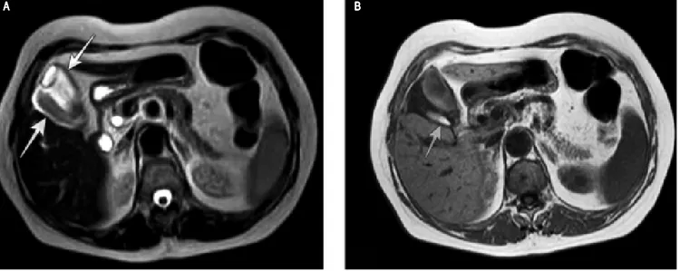
Figure 2. A. Figure 2. A.Figure 2. A.
Figure 2. A.Figure 2. A. Axial single-shot fast SE T2-weighted MR image and, B.B.B.B.B. Axial T1-weighted MR image shows two pear-shaped structures in the gallbladder fossa (arrows). The posterior structure (arrow) has low T2 signal intensity and high T1 signal intensity due to dependent material within the gallbladder lumen. There is wall thickening and pericholecystic fluid indicating inflammation.
sented with symptoms of acute cholangitis. Initially, she was evaluated with an abdominal ultrasound (U/S) where both chololethiasis and choledocholithiasis were found. Patient underwent ERCP (Endoscopic Retrograde Cholangio-Pancreatography) achieving common bile stone clearance. However, ERCP imaging showed an ab-normality suspicious of a duplicated gallbladder (Figure 1). A focused ultrasound examination revealed two adjacent, though, separate and distinct cystic structures within the
gallbladder fossa. There were ultrasound features consist-ent with the presence of two separate cystic ducts. How-ever it was not clear whether they eventually fused before inserting into the common duct. She was further investi-gated with an MRI (magnetic resonance imaging) and MRCP (Magnetic Resonance Cholangio-Pancreatogra-phy). The MRI revealed two pear-shaped structures in the gallbladder fossa with wall thickening and pericholecystic fluid indicating inflammation (Figure 2). The MRCP
A A A A
A BBBBB
A AA
Figure 3. (A and B). Figure 3. (A and B). Figure 3. (A and B). Figure 3. (A and B).
Figure 3. (A and B). Three-dimensional MR cholangiopancreatogram shows two separate cystic ducts (arrows).
Figure 4. Figure 4. Figure 4. Figure 4.
Figure 4. Three-dimensional MR cholangiopancreatogram shows a sepa-rated cystic duct (arrow) that joins together with the main cystic duct (arrow-head) to become a shared, single “common” cystic duct just before the later joins the common bile duct; indicative of Y- shaped subtype I duplicated gallbladder.
showed separated cystic ducts that joined together in a main cystic duct (single “common” cystic duct), just be-fore the later joined the common bile duct (Figures 3 and 4). A diagnosis of duplicated gallbladder was consequently made and the patient was scheduled for laparoscopic cholecystectomy. Intraoperative a duplication of the gallbladder was confirmed as a Y-shaped type (vesica fellea duplex) with two cystic ducts joining into a common cystic duct clearly identified before entering the bile duct and a single cystic artery. Both gallbladders lying in the gallblad-der fossa was successfully removed laparoscopically (Figure 5). Pathology report confirmed the presence of two gallbladders with features of chronic cholecystitis and adenomyomatosis. The patient had an uneventful recovery and she was discharged home on the 2nd postoperative day.
DISCUSSION
Congenital malformations of the gallbladder have been categorized into morphological and positional abnormali-ties including malformation, deformation, multiple gall-bladder, ectopias, intrahepatic position and heterotopic mucosa. Gallbladder duplication is a morphological ab-normality.5 It is considered to result from the incorrect
differentiation or excessive division of embryonic organs during the 5th and 6th gestational week, when the caudal bud of the hepatic diverticulum splits into separate buds or outpunching’s. The later the single primordium
bifur-A AA
cates, the less complete is the resulting duplication of the gallbladder. As a result, a true duplication of gallbladder takes place earlier in the gestation and involves the exist-ence of an accessory gallbladder and two distinct cystic ducts. The accurate incidence of duplicated gallbladders is difficult to calculate because only symptomatic cases or incidental surgical, radiological and cadaveric findings are registered.2 The first reported human case was noted in a
sacrificial victim of Emperor Augustus in 31 BC. Sherren, reported the first documented case of double accessory gallbladder in a living human in 1911.6
Several authors have classified the anatomical variations of duplicated gallbladders. Classification is based on their relation to the cystic duct and mainly is classified into two major groups, as a duplicated (split-primordium) or as an accessory gallbladder, according to the presence or ab-sence of a common cystic duct respectively. Accessory gallbladders are characterized by separate cystic ducts en-tering the biliary tree and arise from two or more separate cystic primordia forming. The most widely accepted clas-sification for double gallbladder is the Boyden’s classifica-tion.5 Boyden was first to describe the duplicate
gallbladder and its variable anatomy in 1926. Based on their relation to the cystic duct, he described “vesica fellea divisa”, (bilobed gallbladder which is drained by a sole cystic duct), and “vesica fellea duplex” (true gallbladder dupli-cation). The latter is sub classified into “Y-shaped type” (two cystic ducts uniting before entering the common bile duct), and “H-shaped or ductular type” (two cystic ducts enter separately into the common bile duct). In 1936, Gross also described congenital abnormalities of gallblad-der and classified them into six types labeled A-F. He also stated that a bilobed gallbladder is an anomaly, but not a true duplication.7 In 1977, Harlaftis, et al. further modified
the classification by describing two main types based on morphology and embryogenesis.8 In type 1 (split
primordi-um), the duplicate gallbladders have a single cystic duct that enters the common bile duct and is further subdivid-ed into septatsubdivid-ed, V shaped, or Y shaped. In type 2 (dual pri-mordium), the accessory gallbladders have two or more cystic ducts drain independently into the biliary tree. The organ more proximal to the liver is regarded as the acces-sory one. This type is subdivided into ductular gallbladder
(H type) and trabecular gallbladder. In the ductular type
Figure 5. (A and B). Figure 5. (A and B).Figure 5. (A and B).
Figure 5. (A and B).Figure 5. (A and B). Intraoperative view of a duplicated gallbladder (a and b), and the main cystic duct (arrow). C.C.C.C.C. Specimen of Y-shaped duplicated gall-bladders within a common serosal coat each with a cystic duct (arrows). Clips can be seen on either side of the V-shaped ducts.
Table 1. Cases of duplicated gallbladder associated with other entities.
First author, year reported Gender of patient/age Concurrent entities
Galambos, 1953 (2 cases) Duodenojejunal diverticulum
Roeder, 1971 Male/36 yrs (Triple GB) - papillary adenocarcinoma
Granot, 1983 Female/4 yrs Biliary cirrhosis
Udelsman, 1985 Female/60 yrs Anteriorly displaced rt. hepatic artery
Bailie, 2003 Female/7 yrs Heterotopic gastric mucosa
Sasaki, 2005 Male/69 yrs Double gallbladder of the duodenal type
Lefemine, 2009 Male/55 yrs Traumatic neuroma
Kawanishi, 2010 Male/75 yrs Well differentiated tubular adenocarcinoma
Kachare, 2013 Female/55 yrs Ectopic thyroid
Girish 2013 Male/3-day-old Duodenal atresia
Menon, 2013 Male/4 yrs Duodenal duplication cyst
Gupta, 2016 Male/2-day-old Gastro-intestinal atresia
Gupta, 2016 Male/12-day-old Duodenal atresia
A AA
the accessory cystic duct connects to the common bile duct. In the trabecular type the accessory cystic duct con-nects to the left or right hepatic duct. Finally, Harlaftis
type III includes any anatomical variation that cannot be classified in either of the two-aforementioned categories. The most common variant found in the literature (48.6% of cases), is the H or ductular type 2 variant, where there are two separate gallbladders and the cystic and accessory cystic ducts enter the common bile duct separately.
While gallbladder duplication is typically associated with gallstones and cholecystitis, there are sporadic re-ports in the literature of other anomalies and diseases, which have been presented with this entity.2 Concurrent
entities that have been reported alongside a duplicated gallbladder are presented in table 1. In addition to double gallbladders there have been cases of triple gallbladders reported in the literature.9-11
There are no specific symptoms or signs associated with duplicate gallbladders. Higher frequency of patholo-gy (malignant or not) in a duplicated gallbladder compar-ing to a scompar-ingle gallbladder has not been confirmed. Patients present atypical symptoms for biliary disease or the common pathologies that can occur as a normal, single gallbladder, including acute cholecystitis, cholelithiasis, empyema, torsion, cholecystocolic fistula, lump in the ab-domen.12 Since the most common imaging modality for
biliary disease is operator dependent, (i.e. ultrasound), this disease entity can be misdiagnosed. As a result, many cases will proceed to cholecystectomy without the pres-ence of a double gallbladder being considered. Many clin-ical studies, however, have demonstrated that congenital anomalies of the gallbladder are associated with an in-creased risk of complications postoperative and repeated surgery.13 If symptoms appear post cholecystectomy, the
clinician has to include a possibly duplicated gallbladder in his diagnostic algorithm, in case a missed second gall-bladder has remained inside the abdomen.
Therefore, defining ductal anatomy is important to ap-preciate these anatomic variations of the biliary system and imaging plays a fundamental role in the clinical evalua-tion for gallstone disease. However, none of the imaging modalities is sensitive enough, in view of the fact that suc-cessful imaging diagnosis is noted in slightly more than half of the cases. Abdominal ultrasound (US) is often the first-line imaging modality used in the assessment of a patient with gallbladder disease; however does not always allow a precise diagnosis of gallbladder malformations. Even though it can identify a duplicate gallbladder in the presence of two cystic structures occupying the gallblad-der fossa, US is not accurate enough to depict properly the anatomy of the cystic duct(s) and to exclude a wide range of alternative diagnoses.12 The most common
enti-ties that may imitate a duplicate gallbladder on US
exam-ination are a choledochal cyst, gallbladder diverticulum and a phrygian cap. In a review study of 17 cases reports, abdominal ultrasound confirmed duplicate gallbladder in only 3 cases.14
ERCP can provide a detailed imaging of ductal anato-my, however it is invasive and carries a risk of serious complications and is not commonly used as a diagnostic tool. In addition, it may not be indicated in every case of cholelithiasis or cholecystitis. Apparently in our patient ERCP management was imperative as the patient present-ed with cholangitis. In our case, ERCP showpresent-ed the gall-bladder and a second pear shaped cystic formation filled with contrast, through one common cystic duct. Accord-ingly, a high index suspicion was maintained for the diag-nosis of a duplicate gallbladder. In such cases, while ERCP is reserved for therapeutic indications, an inciden-tal diagnosis of gallbladder malformations can be made.
As a result, MRCP is becoming the initial imaging tool for the biliary tract imaging in case of suspected gallblad-der duplication during the preoperative managing.1
How-ever, although MRCP and 3D reconstructions may be able to evaluate ductal anatomy without the use of radiation, CT cholangiography should be used where there are any contraindications to MRCP or where it is unable to iden-tify adequately the biliary anatomy.12 In a study of potential
living liver donors, CT cholangiography allowed the visu-alization of at least the second level branches of the intra-hepatic biliary tree in all patients (up to the fourth level in some patients), whereas MRCP only reliably displayed the intrahepatic biliary anatomy up to the bifurcation of the common hepatic duct.15
Surgery should be the treatment of choice only in symptomatic patients. Surgery is not indicated when du-plicated gallbladders are discovered incidentally and prophylactic cholecystectomy in an asymptomatic pa-tient with gallbladder duplication is not recommended. Yet, it is recommended to remove both gallbladders in symptomatic patients at one stage to prevent subsequent disease in the remnant gallbladder and repeated surgical procedures. There has been an emphasis on the need for open cholecystectomy to identify and manage the differ-ent types of gallbladder duplication in the literature.13 On
the other hand, with the advent of newer imaging modal-ities and expertise, these rare anomalies can be diagnosed preoperatively and can be successfully treated by laparos-copy with minimal morbidity. Laparoscopic cholecystec-tomy is the mainstay of treatment, and has been successfully utilized, as it can be found in literature with the first of these procedures reported by Garcia, et al. in 1993.16,17 A recent review identified 3 out of 13
laparo-scopically managed cases that require conversion to an open cholecystectomy.3 An important issue performing a
proce-dure should be converted to an open cholecystectomy. This is a judgment call dependent on the experience of the surgeon and the degree of difficulty and suspicion of anatomical variations face during surgery after a fine landmark dissection. In an uncertain situation intraoper-ative cholangiography and inspection of the gallbladder specimen is indicated to properly evaluate the anatomy and the necessity for further actions.18,19
CONCLUSION
In conclusion, a duplicated gallbladder should be an additional consideration when typical gallbladder disease symptoms are present under certain circumstances while uncharacteristic imaging is found, and further diagnostic imaging is important. Clinicians have to consider these entities and anatomic variations when evaluating patients with biliary disease, in order to avoid unnecessary biliary injury in case of surgical treatment. Total laparoscopic re-moval can be performed safely; however these cases prob-ably do better in the hand of an experienced laparoscopic surgeon or a hepatobiliary surgeon.
ABBREVIATIONS
• ERCP: endoscopic retrograde cholangio-pancreatog-raphy.
• MRI: magnetic resonance imaging.
• MRCP: magnetic resonance cholangio-pancreatogra-phy.
CONFLICT OF INTEREST
The authors declares that there is no conflict of interest regarding the publication of this article.
REFERENCES
1. Botsford A, McKay K, Hartery A, Hapgood C. MRCP imaging of duplicate gallbladder: a case report and review of the lit-erature. Surg Radiol Anat 2015; 30: 425-9. doi: 10.1007/ s00276-015-1456-1. Epub 2015 Mar 11.
2. Paraskevas GK, Raikos A, Ioannidis O, Papaziogas B. Dupli-cated gallbladder: surgical application and review of the liter-ature. Ital J Anat Embryol 2011; 116: 61-6.
3. Yu W, Yuan H, Cheng S, Xing Y, Yan W. A double gallblad-der with a common bile duct stone treated by laparoscopy accompanied by choledochoscopy via the cystic duct: A case report. Exp Ther Med 2016; 12: 3521-6. doi: 10.3892/ etm.2016.3834. Epub 2016 Oct 25.
4. Borghi F, Giraudo G, Geretto P, Ghezzo L. Perforation of Missed Double Gallbladder after Primary Laparoscopic Cholecystectomy: Endoscopic and Laparoscopic Manage-ment. J Laparoendosc Adv Surg Tech A 2008; 18: 429-31. doi: 10.1089/lap.2007.0088.
5. Boyden EA. The accessory gallbladder. An embryological and comparative of aberrant biliary vesicles occurring in man and the domestic mammals. Am J Anat 1926; 32: 177-231.
6. Sherren J. A double gallbladder removed by operation. Ann Surg 1911; 54: 204-5.
7. Gross RE. Congenital anomalies of the gallbladder. Arch Surg
1932; 32: 131-62.
8. Harlaftis N, Gray SW, Skandalakis JE. Multiple gallbladders.
Surg Gynecol Obstet 1977; 145: 928-34.
9. Alicioglu B. An incidental case of triple gallbladder. World J Gastroentrol 2007: 13; 2004-6.
10. Barnes S, Nagar H, Levine C, Santo M, Sold A, Mercer D, Kessler A. Triple gallbladder: preoperative sonographic diag-nosis. J Ultrasound Med 2004; 23: 1399-402.
11. Schroeder C, Draper KR. Laparoscopic cholecystectomy for triple gallbladder. Surg Endosc 2003; 17: 1322. Epub 2003 Jun 13.
12. Mann CD, Briggs CD, Neal CP, Rajesh A, Berry DP. Defining ductal anatomy using CT cholangiography in a patient with gallbladder duplication. Br J Radiol 2009; 82: e175-7. doi: 10.1259/bjr/57068706.
13. Walbolt TD. Lalezarzadeh F. Laparoscopic management of a duplicated gallbladder: a case study and anatomic history.
Surg Laparosc Endosc Percutan Tech 2011; 2: e156-8. doi: 10.1097/SLE.0b013e31821d47ce.
14. Al Rawahi A, Al Azri Y, Al Jabri S, Alfadli A, Al Aghbari S. Successful laparoscopic management of duplicate gallblad-der: A case report and review of literature. Int J Surg Case Rep 2016; 21:142-6. doi: 10.1016/j.ijscr.2016.03.002. Epub 2016 Mar 6.
15. Schroeder T, Malago M, Debatin JF, Goyen M, Nadalin S, Ruehm SG. All-in-one imaging protocols for the evaluation of potential living liver donors: comparison of magnetic reso-nance imaging and multidetector computed tomography. Liver Transplantation 2005; 11: 776-87.
16. Cueto GJ, Weber A, Serrano BF, Tanur TB. Double gallblad-der treated successfully by laparoscopy. J Laparoendosc Surg 1993; 3: 153-5.
17. Desolneux G, Mucci S, Lebigot J, Arnaud JP, Hamy A. Dupli-cation of the gallbladder. A case report. Gastroenterol Res Pract 2009; 2009: 483473. doi: 10.1155/2009/483473. Epub 2009 Nov 30.
18. Guajardo-Salinas GE, Martinez-Ugarte ML, Abourjaily G. The use of intraoperative cholangiogram during laparoscopic double cholecystectomy. J Surg Case Rep 2010; 2010(7): 5. doi: 10.1093/jscr/2010.7.5.
19. Otaibi W, Quach G, Burke B. Double Cystic Duct in a Septat-ed Gallbladder. J Investig Med High Impact Case Rep 2015; 3: 2324709615579105. doi: 10.1177/2324709615579105. eCollection 2015 Apr-Jun.
Correspondence and reprint request: Georgios P. Fragulidis, M.D., Ph.D.
2nd Department of Surgery, Aretaieio Hospital, National and Kapodistrian University of Athens Medical School, 76, Vas. Sophias Ave., 11528, Athens, Attica, Greece
Cel. : +30 6972 910955,
Tel.: +30 210 9690177, Fax: +30 210 9690184 E-mail: gfragulidis@aretaieio.uoa.gr