CorSalud 2018 Ene-Mar;10(1):13-20
RNPS 2235-145 © 2009-2018 Cardiocentro Ernesto Che Guevara, Villa Clara, Cuba. Todos los derechos reservados. 13
Sociedad Cubana de Cardiología
_________________________Artículo Original
Duración del QRS como predictor de baja fracción de eyección en el
infarto miocárdico con elevación del ST
Dra. Ailed E. Rodríguez Jiménez
1, Dr. Hugo Cruz Inerarity
1, Lic. Blanca Valdés Arias
2, Dr.
Guillermo Quintana Cañizares
1y Dr. Enrique Toledo Rodríguez
11 Servicio de Cardiología y 2 Departamento de Psicología. Hospital General Universitario Camilo Cienfuegos. Sancti
Spíritus, Cuba.
Full English text of this article is also available
INFORMACIÓN DEL ARTÍCULO
Recibido: 16 de febrero de 2017 Aceptado: 18 de abril de 2017
Conflictos de intereses
Los autores declaran que no existen conflictos de intereses
Abreviaturas
dQRS: duración del QRS
FEVI: fracción de eyección del ventrículo izquierdo
SCACEST: síndrome coronario agudo con elevación del segmento ST
Versiones On-Line: Español - Inglés
AE Rodríguez Jiménez
Bayamo 151, e/ Frank País y Silvestre Alonso.Sancti Spíritus, Cuba. Correo
electrónico: [email protected]
RESUMEN
Introducción: La duración del QRS es un elemento pronóstico y se ha asociado a una disminución de la fracción de eyección del ventrículo izquierdo en pacientes con síndrome coronario agudo.
Objetivo: Evaluar la implicación pronóstica de la duración del QRS en la reduc-ción de la fracreduc-ción de eyecreduc-ción del ventrículo izquierdo al egreso.
Método: Se realizó un estudio analítico transversal con 347 pacientes con infarto agudo de miocárdico con elevación del segmento ST, ingresados en el Hospital Universitario Camilo Cienfuegos del 1 de enero de 2013 al 31 de diciembre de 2015. Las variables estudiadas fueron: edad, sexo, factores de riesgo cardiovascular clá-sicos, tensión arterial, estrategia de reperfusión, clase de Killip-Kimbal, filtrado glo-merular, duración del QRS y la fracción de eyección del ventrículo izquierdo. Las variables cualitativas se analizaron con el método estadístico Chi cuadrado, las cuantitativas con la t de Student y la regresión lineal. Se construyó la curva ROC para la capacidad de discriminación y se realizó un análisis multivariado para determinar la independencia de variables.
Resultados:La duración del QRS tuvo una correlación negativa con la fracción de eyección (r=-0,267; p<0,001) y una adecuada capacidad de discriminación como predictor de una fracción de eyección inferior a 35% (c=0,643).
Conclusiones: La duración del QRS superior a 90 milisegundos se asoció de ma-nera independiente a una fracción de eyección menor de 35% al egreso.
Palabras clave: Duración del QRS, Fracción de eyección del ventrículo izquierdo, Infarto de miocardio con elevación del ST
QRS duration as a predictor of low ejection fraction in the
ST-segment elevation myocardial infarction
ABSTRACT
Introduction: The QRS duration is a prognostic element and it has been asso-ciated with a decrease in the ejection fraction of the left ventricular in patients with acute coronary syndrome.
Objective: To assess the prognostic implications of the QRS duration in the de-pression of the left ventricular ejection fraction at discharge.
Cienfuegos from January 1st, 2013 to December 31st, 2015. The variables studied were: age, sex, classical cardiovascular risk factors, blood pressure, reperfusion strategy, Killip-Kimball class, glomerular filtration rate, QRS duration and left ven-tricular ejection fraction. The qualitative variables were analyzed with the Chi-square statistical method and the quantitative with the t of Student and linear re-gression. The ROC curve was constructed for the discrimination capacity and a multivariate analysis was performed to determine the independence of variables.
Results: The QRS duration was negatively correlated with the ejection fraction r=-0.267; p<0.001) and an adequate discrimination ability as a predictor ejection frac-tion less than 35% (c=0.643).
Conclusions: The QRS duration greater than 90 milliseconds was independently associated with an ejection fraction lower than 35% at discharge.
Key words: QRS duration, Left ventricular ejection fraction, ST-segment elevation myocardial infarction
INTRODUCCIÓN
El electrocardiograma, por su amplia disponibilidad, su bajo costo y su simplicidad, es una herramienta imprescindible para el diagnóstico y la estratifica-ción pronóstica del síndrome coronario agudo con elevación del segmento ST (SCACEST). La anchura del QRS es uno de los elementos que se obtienen en el electrocardiograma y ha sido un pilar fundamen-tal para el empleo de la terapia de resincronización
cardíaca1. Se plantea además, que en pacientes con
insuficiencia cardíaca, la mayor duración del QRS
(dQRS) empeora el pronóstico2,3.
La asociación del QRS y la fracción de eyección del ventrículo izquierdo (FEVI) ha sido ampliamente
explorada en la insuficiencia cardíaca4,5, no tanto así
en la cardiopatía isquémica. La isquemia induce un daño celular que altera las propiedades eléctricas
del músculo cardíaco6. Fluctuaciones en las
concen-traciones y las corrientes iónicas, así como los cam-bios locales en las propiedades de las uniones
inter-celulares en hendidura (gab junctions) pudieran
resultar en un enlentecimiento de la conducción y una dispersión de la refractariedad del potencial de
acción en las fibras isquémicas7, lo cual pudiera
prolongar la dQRS.
El objetivo de este estudio ha sido evaluar la im-plicación pronóstica de la dQRS en la reducción de la FEVI al egreso.
MÉTODO
Se realizó un estudio analítico transversal que inclu-yó a todos los pacientes ingresados con el diagnósti-co de SCACEST tipo I (aterosclerótidiagnósti-co) en la Unidad
de Cuidados Intensivos Coronarios (UCIC) del Hos-pital General Universitario Camilo Cienfuegos de la provincia Sancti Spíritus, Cuba, en el período com-prendido del 1 de enero de 2013 al 31 de diciembre de 2015.
Se conformó una muestra no intencionada con todos los casos que cumplieron los criterios de in-clusión y exin-clusión (n=347).
Criterios de inclusión
Pacientes atendidos en la UCIC con el referido diag-nóstico.
Criterios de exclusión Pacientes con:
Diagnóstico previo de miocardiopatía dilatada y
FEVI basal previa inferior a 35%.
Bloqueo de rama o ritmo de marcapaso que
en-sanchara el QRS basal.
Fibrilación auricular previa al diagnóstico del
SCACEST, ya que impide o interfiere la medición de los parámetros electrocardiográficos.
Pacientes fallecidos antes de la realización del
ecocardiograma pre-alta.
Variables
Las variables estudiadas fueron: edad, sexo, factores de riesgo coronario (hipertensión arterial y diabetes mellitus con diagnóstico previo al evento coronario, hábito de fumar, hipercolesterolemia, obesidad), tensión arterial y frecuencia cardíaca al ingreso, estrategia de reperfusión, clase de Killip-Kimbal, filtrado glomerular, duración del QRS y fracción de eyección del ventrículo izquierdo.
Rodríguez Jiménez AE, et al.
CorSalud 2018 Ene-Mar;10(1):13-20 15
valores de referencia del hospital; la obesidad se tuvo en cuenta en aquellos pacientes con un índice
de masa corporal superior a 30 kg/m2, y el
antece-dente de cardiopatía isquémica cuando existía diag-nóstico previo al evento coronario.
Se estudiaron además la clase de Killip-Kimbal, y el uso y éxito de la trombólisis (signos clínicos o eléctricos de reperfusión: alivio del dolor, mejoría hemodinámica, regresión del supradesnivel del ST mayor del 50% o aparición de arritmias de
reperfu-sión)8. Resulta válido señalar la imposibilidad de
utilizar el criterio de enzimático por no tener tropo-ninas disponibles.
Otras variables estudiadas fueron el filtrado glo-merular (FG), estimado mediante la fórmula de
Co-ckcroft-Gault9 y la FEVI, por el método de Simpson
biplano, obtenida del ecocardiograma realizado an-tes del egreso.
La medición del QRS se realizó de forma
ma-nual10,11 en las 12 derivaciones del
electrocardiogra-ma estandarizado, donde 10 milímetros (mm) equi-valen a 1 milivoltio, y la velocidad del papel fue de 25 mm/segundo. El QRS se midió desde la deflexión inicial de la onda hasta su terminación en el punto J, y se tomó el QRS de mayor duración.
Procesamiento de la información
La información se obtuvo a partir de las historias clínicas ubicadas en el Departamento de Archivo y Estadística de la entidad hospitalaria.
Los datos fueron procesados con el software S
ta-tistical Package for the Social Sciences (SPSS), ver-sión 17.0, instalado en una microcomputadora con sistema operativo Windows 8, y se analizaron de la siguiente manera: las variables cualitativas se expre-saron en frecuencias absolutas y relativas, las cuan-titativas se expresaron en su media y desviación estándar.
Para comprobar la fortaleza de asociación entre variables cualitativas fue seleccionada la prueba no paramétrica Chi Cuadrado; ante situaciones en que más del 20% de las frecuencias esperadas presenta-ron valores menores de cinco se utilizó la prueba exacta de Fisher. Para comparar las medias de
va-riables cuantitativas se empleó el estadígrafo t de
Student para muestras independientes. Para la rela-ción entre la dQRS y la FEVI se empleó la regresión lineal y se calculó su capacidad de discriminación mediante la construcción de la curva operador-receptor (ROC) y el área bajo la curva (índice «c»). Teniendo en cuenta los resultados de la curva ROC, se determinó un punto de corte para dicotomizar las
variables continuas e incluirlas en el análisis univa-riado.
Como contribución relativa a la determinación de la dQRS como factor independiente de predicción de baja FEVI al egreso, se realizó un análisis multi-variado con un modelo de regresión logística binaria donde resultó ser la FEVI menor de 35% la variable dependiente (dicotómica). En el análisis multivaria-do se consideró identificar como factores propensos a la predicción, aquellos aspectos contenidos en las variables para los que el estadígrafo Wald mostró una probabilidad menor a 5% (p<0,05), al ser anali-zado el exponencial de los coeficientes del modelo
exponencial de β (Exp β) como estimador de la
razón de productos cruzados u odds ratio (OR).
El carácter agregado de la recogida de la infor-mación contribuyó al mantenimiento de la privaci-dad de los sujetos implicados en el estudio y los resultados solo han sido utilizados con fines científi-cos.
RESULTADOS
Se estudiaron un total de 347 pacientes, 211 hombres
(60,8%) y 136 mujeres (39,2%) (Tabla 1). La media
de edad en los pacientes con FEVI mayor de 35% fue de 75,88 años, superior a la de los pacientes con FEVI menor de 35% (68,14). El factor de riesgo más frecuente en la muestra fue la HTA con 252 pacien-tes (76,2%), también se encontraron diabepacien-tes melli-tus (25,9%), hábito de fumar (56,5%),
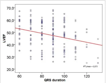
Figura 1: Regresión lineal entre la FEVI y la duración del
lemia (37,8%) y obesidad (21,9%). La estrategia de reperfusión empleada fue la trombólisis con estrep-toquinasa recombinante cubana, utilizada en el 53% de los pacientes, con un éxito de 11,2%. La disfun-ción ventricular fue frecuente, con una clase de Ki-llip-Kimbal superior a I en 46,4% de los pacientes. La media de la tensión arterial sistólica fue inferior en
los pacientes con FEVI menor de 35% (79,08±35,82 vs. 124,51±35,03 mmHg), al igual que el filtrado
glo-merular (47,85±12,72 vs. 70,09±30,05 ml/min/m2). La
letalidad en la muestra estudiada fue de 10,9%.
La figura 1 muestra, mediante un análisis de
re-gresión lineal, la correlación inversa entre la dQRS y la FEVI, con una significación estadística muy
signi-Tabla 1. Características generales de la población. Hospital Camilo Cienfuegos Sancti Spíritus.
Enero 2013-Diciembre 2015.
Variables FEVI > 35% (n=298) FEVI < 35% (n=49) Total
Características demográficas
Edad 68,14 ± 11,17 75,88 ± 7,34 69,23 ± 11,04
Sexo masculino 185 (53,31) 26 (7,49) 211 (60,8)
Factores de riesgo
Hipertensión arterial 224 (64,55) 28 (8,07) 252 (72,6)
Diabetes mellitus 60 (17,29) 30 (8,65) 90 (25,9)
Hipercolesterolemia 114 (32,85) 17 (4,90) 131 (37,8)
Obesidad 71 (20,46) 5 (1,44) 76 (21,9)
Hábito de fumar 176 (50,72) 20 (5,76) 196 (56,5)
Cardiopatía isquémica crónica 139 (40,06) 17 (4,90) 156 (45)
Estrategia de reperfusión
Estreptoquinasa 157 (45,24) 27 (7,78) 184 (53)
Reperfusión exitosa 38 (10,95) 1 (0,29) 39 (11,2)
Tiempo de isquemia (minutos) 225,67 ± 143,78 280,0 ± 168,48 233,64 ± 148,41 Killip-Kimbal
Clase I 158 (45,5) 28 (8,07) 186 (53,6)
Clase II 48 (13,83) 3 (0,86) 51 (14,7)
Clase III 68 (19,60) 15 (4,32) 83 (23,9)
Clase IV 24 (6,92) 3 (0,86) 27 (7,8)
Variables clínicas y de laboratorio
Frecuencia cardíaca 83,30 ± 27,89 86,16 ± 24,31 83,7 ± 27,4
Tensión arterial sistólica (mmHg) 124,51 ± 35,03 79,08 ± 35,82 118,09 ± 38,5 Filtrado glomerular (ml/min/m2) 70,09 ± 30,05 47,85 ± 12,72 66,94 ± 29,28
Variables electrocardiográficas
Duración del QRS (mseg.) 84,33 ± 15,87 93,39 ± 17,17 85,6 ± 16,34
Estado al egreso
Vivo 281 (80,98) 28 (8,07) 309 (89,1)
Rodríguez Jiménez AE, et al.
CorSalud 2018 Ene-Mar;10(1):13-20 17
ficativa (p<0,001). El área bajo la curva ROC es el mejor indicador global de la precisión de una prue-ba diagnóstica o pronóstica, y hace factible expresar su desempeño mediante un número simple. La
figura 2 muestra la curva ROC para la relación de
la dQRS con la FEVI menor de 35%, con un índice
bajo la curva c=0,643 y p=0,001.
El punto de corte hallado para incluir la dQRS en el análisis multivariado con mejor sensibilidad y
especificidad fue de 90 mseg. (Figura 3).En el
aná-lisis univariado (Tabla 2) resultaron como factores
de riesgo con significación estadística la edad mayor de 75 años (p<0,001), la diabetes mellitus (p<0,001), la reperfusión no exitosa (p=0,010), la clase Killip II, III, IV (p<0,001), la tensión arterial sistólica menor de 100 mmHg (p<0,001), el filtrado glomerular menor de
60 ml/min/m2 (p<0,001), y la dQRS>90 mseg. (p=
0,004).
En el análisis multivariado (Tabla 3), la dQRS>90
mseg. resultó ser un predictor independiente de FEVI menor de 35% (p=0,002), asociado a la edad
Figura 2: Curva ROC de dQRS para la determinación
pronóstica de FEVI menor de 35%. Figura 3: Punto de corte de la duración del QRS para predecir una FEVI menor de 35%.
Tabla 2. Variables predictoras de FEVI<35% al egreso: Análisis univariado.
Variables FEVI > 35% (n=298) FEVI < 35% (n=49) OR Nº % Nº % Inferior Superior IC 95% p
Edad mayor de 75 años 76 21,90 32 9,22 5,50 2,89 10,46 <0,001
Hipertensión arterial 224 64,55 28 8,07 0,44 0,24 0,82 0,011
Diabetes mellitus 60 17,29 30 8,65 6,26 3,30 11,89 <0,001
Obesidad 71 20,46 5 1,44 0,36 0,14 0,95 0,022
Hábito de fumar 176 50,72 20 5,76 0,48 0,26 0,88 0,017
Reperfusión no exitosa 260 74,93 48 13,83 7,02 0,94 52,32 0,010
KillipKimbal II,III, IV 116 33,43 45 12,97 17,65 6,18 50,38 <0,001
TAS < 100 mmHg 55 15,85 36 10,37 12,24 6,09 24,60 <0,001
FG < 60 ml/min/m2 149 42,94 44 12,68 8,80 3,40 22,81 <0,001
QRS > 90 mseg. 117 33,72 30 8,65 2,44 1,31 4,54 0,004
mayor de 75 años, la diabetes mellitus, la clase de Killip-Kimbal mayor de I (II, III y IV), y la tensión arterial sistólica menor de 100 mmHg.
DISCUSIÓN
La dQRS ha sido asociada a alteraciones en la
es-tructura y la función del ventrículo izquierdo12. La
mayor dQRS posterior a un SCACEST ha sido rela-cionada con la afectación del sistema de conducción por la extensión de la escara necrótica, lo cual
po-dría predisponer a arritmias ventriculares6,13,14. La
FEVI, tras un SCACEST se ha convertido en un pre-dictor de muerte súbita independiente con elevada
capacidad predictiva15, lo cual es tomado en cuenta
en nuestro estudio donde se evalúa la capacidad predictiva de la dQRS sobre la FEVI al egreso.
Zou et al.16 en un estudio reciente llevado a cabo en pacientes con miocardiopatía dilatada, demostra-ron que la normalización de la FEVI con tratamiento medicamentoso estaba en relación con la disminu-ción de la dQRS. Nuestros resultados muestran una correlación negativa de la dQRS con la FEVI al egre-so, así como una adecuada capacidad de discrimi-nación de la dQRS para una FEVI inferior a 35%.
En un modelo de predicción de riesgo basado en variables ecocardiográficas, la FEVI resultó ser un predictor independiente en el análisis multivariado
(índice de riesgo [hazard ratio, HR] 1,45; intervalo
de confianza [IC] 95%: 1,02-2,08; p=0,040), y el pro-nóstico resultó ser inversamente proporcional a la
FEVI cuando esta fue inferior al 40%17.
Nuestros resultados coinciden con los de Shah et
al.7, quienes en un estudio llevado a cabo con 536
pacientes con síndrome coronario agudo sin eleva-ción del ST demostraron que un QRS>90 mseg. re-sultó ser un predictor independiente de enfermedad de tres vasos coronarios y de grave reducción de la
FEVI. Por otra parte, Winter et al.18 en un estudio
realizado con 132 pacientes con enfermedad arterial coronaria, y una FEVI basal inferior a 30%, no encon-traron asociación entre la dQRS y la FEVI. Resulta válido señalar que este estudio no se realizó en pa-cientes con síndrome coronario agudo e incluyó una FEVI basal inferior al 30%, lo cual difiere con el dise-ño de nuestro estudio (y, por tanto, con nuestros resultados), ya que los pacientes con FEVI basal inferior al 35% conocida fueron excluidos.
En el análisis multivariado, la dQRS superior a 90 mseg. se asoció como predictor independiente a la FEVI menor del 35% al egreso; y al ser ajustado por la edad, también se asociaron la diabetes mellitus, la clase de Killip-Kimbal y la tensión arterial sistólica (p<0,05), además del filtrado glomerular menor de
60 ml/min/m2 y la reperfusión no exitosa (p>0,05).
Jiménez-Candil et al.19 en un estudio llevado a
cabo en pacientes con síndrome coronario agudo sin elevación del ST encontraron que las variables independientes asociadas a mayor mortalidad intra-hospitalaria, fueron el aumento de las tropininas (HR=8,1; IC 95%: 1,04-62,8; p=0,045); la FEVI menor de 40% (HR=12,6; IC 95%: 4,7-34; p<0,001) y la edad mayor de 65 años (HR=2,1; IC 95%: 1,2-3,4; p=0,03). Y en el seguimiento de pacientes tras el alta hospitala-ria, la dQRS superior a 90 mseg. resultó ser un pre-dictor independiente de mortalidad cardiovascular (HR=2,4; IC 95%: 1,2-4,8; p=0,009). Por su parte,
Nwa-Tabla 3. Variables predictoras de FEVI<35% al egreso: Análisis multivariado.
Variables B estándar Error Wald Significa-ción Exp(B) Inferior Superior IC 95%
Edad mayor de 75 años 0,95 0,44 4,59 0,032 2,58 1,08 6,13
Diabetes mellitus 0,87 0,44 4,03 0,045 2,40 1,02 5,62
KillipKimbal II,III, IV 2,87 1,04 7,64 0,006 17,64 2,30 135,02
TAS < 100 mmHg 1,92 0,40 22,82 <0,001 6,85 3,11 15,08
FG < 60 ml/min 1,34 0,98 1,85 0,174 0,26 0,04 1,80
QRS > 90 mseg. 1,27 0,42 9,20 0,002 3,57 1,57 8,14
Reperfusión no exitosa 0,55 1,27 0,19 0,664 0,58 0,05 6,96
Constante 4,62 1,20 14,93 <0,001 0,01
Rodríguez Jiménez AE, et al.
CorSalud 2018 Ene-Mar;10(1):13-20 19
kile et al.20 también encontraron una asociación de
la dQRS y la mortalidad por arritmias ventriculares. La dQRS>10 mseg. se asoció con una incidencia de 21,8% de taquicardia ventricular y de 3,2% de fibrila-ción ventricular comparado con un 10,3% y un 0,9%, respectivamente, en los pacientes con dQRS<110 mseg.
La dQRS ha sido utilizada para evaluar la disin-cronía mecánica en el empleo de la terapia de
resin-cronización cardíaca21. Su utilidad también fue
de-mostrada por Joseph et al.4 como predictor de
muer-te por causa cardiovascular, de parada cardíaca recuperada y de hospitalizaciones por insuficiencia cardíaca. Esto último pudiera estar relacionado con los resultados encontrados en nuestro estudio don-de se evidon-dencia una correlación negativa entre la dQRS y la FEVI. Otro estudio llevado a cabo en pa-cientes con SCACEST demostró un incremento de la mortalidad a los 30 días, en pacientes con dQRS>100 mseg., resultado que fue ajustado por la FEVI, la
función renal, la hipotensión y la taquicardia22.
CONCLUSIONES
El electrocardiograma continúa siendo una herra-mienta útil en la estratificación de riesgo del sín-drome coronario agudo con elevación del segmento ST. La dQRS superior a 90 mseg. se asoció de mane-ra independiente a una FEVI menor de 35% tmane-ras el egreso. La dQRS y la FEVI presentaron una correla-ción negativa con significacorrela-ción estadística. La mayor dQRS presentó una adecuada capacidad de discri-minación como predictor de una baja FEVI al egre-so.
BIBLIOGRAFÍA
1. Brignole M, Auricchio A, Barón-Esquivias G,
Bor-dachar P, Boriani G, Breithardt OA, et al. Guía de
práctica clínica de la ESC 2013 sobre estimulación cardiaca y terapia de resincronización cardiaca. Rev Esp Cardiol. 2014;67(1):58.e1-e60.
2. Baldasseroni S, Gentile A, Gorini M, Marchionni
N, Marini M, Masotti G, et al. Intraventricular
conduction defects in patients with congestive heart failure: Left but not right bundle branch block is an independent predictor of prognosis. A report from the Italian Network on Congestive Heart Failure (IN-CHF database). Ital Heart J. 2003;4(9):607-13.
3. Khan NK, Goode KM, Cleland JG, Rigby AS,
Free-mantle N, Eastaugh J, et al. Prevalence of ECG
abnormalities in an international survey of pa-tients with suspected or confirmed heart failure at death or discharge. Eur J Heart Fail. 2007;9(5):491-501.
4. Joseph J, Claggett BC, Anand IS, Fleg JL, Huynh T,
Desai AS, et al. QRS duration is a predictor of
ad-verse outcomes in heart failure with preserved ejection fraction. JACC: Heart Fail. 2016;4(6):477-86.
5. Park HS, Kim H, Park JH, Han S, Yoo BS, Shin MS,
et al. QRS prolongation in the prediction of clini-cal cardiac events in patients with acute heart failure: Analysis of data from the Korean Acute Heart Failure Registry. Cardiology. 2013;125(2):96-103.
6. Zipes DP, Jalife J, Eds. Cardiac Electrophysiology:
From Cell to Bedside. 6th Ed. Philadelphia: Else-vier-Saunders; 2014.
7. Shah M, Maludum O, Bhalla V, De Venecia TA,
Patil S, Curet K, et al. QRS duration and left
ven-tricular ejection fraction (LVEF) in non-ST seg-ment elevation myocardial infarction (NSTEMI). Int J Cardiol. 2016;221:524-8.
8. Steg G, James SK, Atar D, Badano LP, Blomstrom
Lundqvist C, Borger MA, et al. Guía de práctica
clínica de la ESC para el manejo del infarto agudo de miocardio en pacientes con elevación del segmento ST. Rev Esp Cardiol. 2013;66(1):53.e1-e46.
9. Cabrerizo García JL, Zalba Etayo B, Pérez Calvo
JI. Valor pronóstico del filtrado glomerular en el síndrome coronario agudo: ¿Índice de Cockcroft o ecuación MDRD? Med Clin (Barc). 2010;134(14): 624-9.
10.Gadaleta FL, Llois SC, Sinisi VA, Quiles J, Avanzas
P, Kaski JC. Prolongación del intervalo QT corre-gido: Nuevo predictor de riesgo cardiovascular en el síndrome coronario agudo sin elevación del ST. Rev Esp Cardiol. 2008;61(6):572-8.
11.Chávez González E, Alonso Herrera A, Carmona
Puerta R, Pérez Cabrera D, Ramos Ramírez RR,
Gómez Paima W, et al. Dispersión del QRS como
índice de disincronía en el bloqueo de rama iz-quierda y de sincronía tras la terapia de resin-cronización cardíaca, una variable de respuesta exitosa. CorSalud [Internet]. 2015 [citado 14 Ene 2017];7(2):106-16. Disponible en:
http://www.corsalud.sld.cu/sumario/2015/v7n2a1 5/dispersionqrs.html
Schaffer A, Ambrose JA. A prolonged QRS dura-tion on surface electrocardiogram is a specific in-dicator of left ventricular dysfunction. J Am Coll Cardiol. 1998;32(2):476-82.
13.Tjandrawidjaja MC, Fu Y, Westerhout CM,
Wag-ner GS, Granger CB, Armstrong PW, et al.
Useful-ness of the QRS score as a strong prognostic marker in patients discharged after undergoing primary percutaneous coronary intervention for ST-segment elevation myocardial infarction. Am J Cardiol. 2010;106(5):630-4.
14.Horwich T, Lee SJ, Saxon L. Usefulness of QRS
prolongation in predicting risk of inducible mon-omorphic ventricular tachycardia in patients re-ferred for electrophysiologic studies. Am J Cardi-ol. 2003;92(7):804-9.
15.Shiga T, Hagiwara N, Ogawa H, Takagi A,
Nagashi-ma M, YaNagashi-mauchi T, et al. Sudden cardiac death
and left ventricular ejection fraction during long-term follow-up after acute myocardial infarction in the primary percutaneous coronary interven-tion era: Results from the HIJAMI-II registry. Heart. 2009;95(3):216-20.
16.Zou CH, Zhang J, Zhang YH, Wei BQ, Wu XF,
Zhou Q, et al. Frequency and predictors of
nor-malization of left ventricular ejection fraction in recent-onset nonischemic cardiomyopathy. Am J Cardiol. 2014;113(10):1705-10.
17.Bedetti G, Gargani L, Sicari R, Gianfaldoni ML,
Molinaro S, Picano E. Comparison of prognostic
value of echographic [corrected] risk score with the Thrombolysis in Myocardial Infarction (TIMI) and Global Registry in Acute Coronary Events (GRACE) risk scores in acute coronary syn-drome. Am J Cardiol. 2010;106(12):1709-16.
18.De Winter O, Van de Veire N, Van Heuverswijn F,
Van Pottelberge G, Gillebert TC, De Sutter J. Rela-tionship between QRS duration, left ventricular volumes and prevalence of nonviability in pa-tients with coronary artery disease and severe left ventricular dysfunction. Eur J Heart Fail. 2006;8(3):275-7.
19.Jiménez-Candil J, Cruz González I, Martín F,
Pa-bón P, León V, Hernández J, et al. Relationship
between QRS duration and prognosis in non-ST-segment elevation acute coronary syndrome. Int J Cardiol. 2008;126(2):196-203.
20.Nwakile C, Purushottam B, Bhalla V, Ukpong D,
Shah M, Yun J, et al. Significance of QRS duration
in non-ST elevation myocardial infarction. Int J Cardiol. 2015;187:146-7.
21.Poole JE, Singh JP, Birgersdotter-Green U. QRS
duration or QRS morphology: What really matters in cardiac resynchronization therapy? J Am Coll Cardiol. 2016;67(9):1104-17.
22.Nwakile C, Purushottam B, Yun J, Bhalla V,
CorSalud 2018 Jan-Mar;10(1):13-20
RNPS 2235-145 © 2009-2018 Cardiocentro Ernesto Che Guevara, Villa Clara, Cuba. All rights reserved. 13
Cuban Society of Cardiology
_________________________Original Article
QRS duration as a predictor of low ejection fraction in the ST-segment
elevation myocardial infarction
Ailed E. Rodríguez Jiménez
1, MD; Hugo Cruz Inerarity
1, MD; Blanca Valdés Arias
2, BS;
Guillermo Quintana Cañizares
1, MD, and Enrique Toledo Rodríguez
1, MD
1Department of Cardiology and 2Department of Psychology. Hospital General Universitario Camilo Cienfuegos. Sancti
Spíritus, Cuba.
Este artículo también está disponible en español
ARTICLE INFORMATION
Received: February 16, 2017 Accepted: April 18, 2017
Competing interests
The authors declare no competing interests
Acronyms
LVEF: left ventricular ejection fraction
QRSd: QRS duration
STEACS: ST-segment elevation acute coronary syndrome
On-LineEnglish & Spanish versions
AE Rodríguez Jiménez
Bayamo 151, e/ Frank País y Silvestre Alonso.Sancti Spíritus, Cuba. E-mail address:
ABSTRACT
Introduction: The QRS duration is a prognostic element and it has been asso-ciated with a decrease in the left ventricular ejection fraction in patients with acute coronary syndrome.
Objective: To assess the prognostic implications of the QRS duration in the de-pression of the left ventricular ejection fraction at discharge.
Method:A cross-sectional study was conducted with 347 patients with ST-segment elevation myocardial infarction, admitted at the Hospital Universitario Camilo Cienfuegos from January 1st, 2013 to December 31st, 2015. The variables studied were: age, sex, classical cardiovascular risk factors, blood pressure, reperfusion strategy, Killip-Kimball class, glomerular filtration rate, QRS duration and left ventricular ejection fraction. The qualitative variables were analyzed with the Chi-square statistical method and the quantitative with the t of Student and linear regression. The ROC curve was constructed for the discrimination capacity and a multivariate analysis was performed to determine the independence of variables.
Results: The QRS duration was negatively correlated with the ejection fraction r= -0.267; p<0.001) and an adequate discrimination ability as a predictor ejection fraction less than 35% (c=0.643).
Conclusions: The QRS duration greater than 90 milliseconds was independently associated with an ejection fraction lower than 35% at discharge.
Key words:QRS duration, Left ventricular ejection fraction, ST-segment elevation myocardial infarction
Duración del QRS como predictor de baja fracción de eyección en
el infarto miocárdico con elevación del ST
RESUMEN
Introducción: La duración del QRS es un elemento pronóstico y se ha asociado a una disminución de la fracción de eyección del ventrículo izquierdo en pacientes con síndrome coronario agudo.
Objetivo: Evaluar la implicación pronóstica de la duración del QRS en la reduc-ción de la fracreduc-ción de eyecreduc-ción del ventrículo izquierdo al egreso.
Las variables estudiadas fueron: edad, sexo, factores de riesgo cardiovascular clá-sicos, tensión arterial, estrategia de reperfusión, clase de Killip-Kimbal, filtrado glo-merular, duración del QRS y la fracción de eyección del ventrículo izquierdo. Las variables cualitativas se analizaron con el método estadístico Chi cuadrado, las cuantitativas con la t de Student y la regresión lineal. Se construyó la curva ROC para la capacidad de discriminación y se realizó un análisis multivariado para determinar la independencia de variables.
Resultados: La duración del QRS tuvo una correlación negativa con la fracción de eyección (r=-0,267; p<0,001) y una adecuada capacidad de discriminación como predictor de una fracción de eyección inferior a 35% (c=0,643).
Conclusiones: La duración del QRS superior a 90 milisegundos se asoció de ma-nera independiente a una fracción de eyección menor de 35% al egreso.
Palabras clave: Duración del QRS, Fracción de eyección del ventrículo izquierdo, Infarto de miocardio con elevación del ST
INTRODUCTION
The electrocardiogram, due to its wide availability, low cost and simplicity, is an essential tool for the diagnosis and prognostic stratification of ST-segment elevation cute coronary syndrome (STEACS). The QRS width is one of the elements obtained in the electrocardiogram and has been a fundamental pillar
for the use of cardiac resynchronization therapy1. It
is also stated that in patients with heart failure, a
longer QRS duration (QRSd) worsens prognosis2,3.
The association between QRS and left ventricular ejection fraction (LVEF) has been widely explored in
heart failure4,5, but not so much in ischemic heart
disease. Ischemia induces cell damage which alters
the electrical properties of the heart muscle6.
Fluc-tuations in concentrations and ionic currents, as well as local changes in the properties of gap junctions may cause conduction delay and dispersion of the refractoriness of the action potential in the ischemic
fibers7, which could prolong the QRSd.
This study aimed at evaluating the prognostic implication of the QRSd in the reduction of LVEF at discharge.
METHOD
A cross-sectional analytical study was conducted, including all the patients admitted with a diagnosis of type I STACS (atherosclerotic) in the Coronary Intensive Care Unit (ICU) at the Hospital Universitar-io Camilo Cienfuegos of Sancti Spíritus province,
Cuba, from January 1st, 2013 to December 31st, 2015.
An unintentional sample was formed with all the cases that met the inclusion and exclusion criteria
(n=347).
Inclusion criteria
Patients treated at the ICU with the aforementioned diagnosis.
Exclusion criteria Patients with:
Previous diagnosis of dilated cardiomyopathy
and previous baseline LVEF less than 35%.
Branch block or pacing rhythm that may broaden
the baseline QRS.
Atrial fibrillation prior to the diagnosis of STEACS,
since it prevents or interferes with the measure-ment of electrocardiographic parameters.
Patients who died before performing the
pre-discharge echocardiogram.
Variables
The variables studied were: age, sex, coronary risk factors (high blood pressure and diabetes mellitus with diagnosis prior to coronary event, smoking, hypercholesterolemia, obesity), blood pressure and heart rate at admission, reperfusion strategy, Killip class-Kimbal, glomerular filtration rate, QRS duration and left ventricular ejection fraction.
Serum cholesterol levels greater than 6.71 mmol/L were considered hypercholesterolemia, according to hospital reference values; Obesity was taken into account in those patients with a body
mass index greater than 30 kg/m2, and history of
ischemic heart disease when there was a diagnosis prior to the coronary event.
Rodríguez Jiménez AE, et al.
CorSalud 2018 Jan-Mar;10(1):13-20 15
improvement, regression of ST segment elevation greater than 50% or appearance of reperfusion
ar-rhythmias)8. It is valid to point out the impossibility
of using the enzymatic criterion due to lack of avail-able troponins.
Other variables studied were glomerular filtration rate (GFR), estimated by the Cockcroft-Gault
formu-la9 and LVEF, by the Simpson biplane method,
ob-tained from the echocardiogram performed before discharge.
The QRS was manually measured10,11 in a
stand-ard 12-lead electrocstand-ardiogram, where 10 millimeters (mm) equals 1 millivolt, and the paper speed was 25 mm/second. The QRS was measured from the initial deflection of the wave to its termination at point J, and the QRS of longer duration was recorded.
Information processing
The information was obtained from the medical rec-ords located in the Archives and Statistics Depart-ment of the hospital.
The data were processed with the Statistical Pack-age for the Social Sciences (SPSS) software, version 17.0, installed in a microcomputer with Microsoft Windows 8 operating system, and analyzed as fol-lows: qualitative variables were expressed in abso-lute and relative frequencies; quantitative data were expressed in their mean and standard deviation.
To check the strength of association between qualitative variables, the non-parametric Chi square test was selected; in situations where more than 20% of the expected frequencies presented values lower than five, Fisher's exact test was used. To compare the means of quantitative variables, the Student's t-statistic was used for independent samples. For the relationship between QRSd and LVEF, linear regres-sion was used and its discrimination capacity was calculated by constructing the Receiver Operator Curve (ROC) and the area under the curve (index «c»). Considering the results of the ROC curve, a cut-off point was determined to dichotomize the contin-uous variables and include them in the univariate analysis.
LVEF at discharge, a multivariate analysis was performed with a binary logistic regression model where LVEF of less than 35% was the dependent variable (dichotomous). In the multivariate analysis we considered to identify as factors prone to predic-tion, those aspects contained in the variables for which the Wald statistic showed a probability lower than 5% (p<0.05), when we analyzed the exponential
of the coefficients of the exponential model of β (Exp
β) as an estimator for the ratio of cross products or
odds ratio (OR).
The aggregate nature of the information collection contributed to keep the privacy of the subjects in-volved in the study and the results have only been used for scientific purposes.
RESULTS
A total 347 patients were studied, 211 men (60.8%)
and 136 women (39.2%) (Table 1). The mean age in
patients with LVEF greater than 35% was 75.88 years, higher than in patients with LVEF less than 35% (68,14). The most frequent risk factor in the sample was HBP with 252 patients (76.2%), diabetes mellitus (25.9%), smoking habit (56.5%), hypercholesterole-mia (37.8%) and obesity (21.9%) were also found. The reperfusion strategy used was thrombolysis with Cuban recombinant streptokinase, used in 53% of patients, with a success of 11.2%. Ventricular dys-function was frequent, with a Killip-Kimbal class superior to I in 46.4% of patients. The mean systolic blood pressure was lower in patients with LVEF less than 35% (79.08 ± 35.82 vs. 124.51 ± 35.03 mmHg), as was the glomerular filtration rate (47.85 ± 12. 72 vs. 70.09 ± 30.05 ml/min/m2). The lethality in the sample was 10,9%.
Figure 1 shows, by means of a linear regression
analysis, the inverse correlation between the QRSd and the LVEF, with a very significant statistical
signif-Figure 1: Linear regression between LVEF and QRS
icance (p <0.001). The area under the ROC curve is the best overall indicator of the accuracy of a diag-nostic or progdiag-nostic test, and makes it possible to
express its performance by a simple number.
Fig-ure 2 shows the ROC curve for the ratio of the
QRSd with LVEF less than 35%, with an index under the curve c=0.643 and p=0,001.
The cut-off point found to include the QRSd in the multivariate analysis with better sensitivity and
specificity was 90 msec. (Figure 3). In the
univari-ate analysis (Table 2) were found as risk factors
with statistical significance age older than 75 years (p<0.001), diabetes mellitus (p<0.001), unsuccessful reperfusion (p=0.010), Killip class II, III, IV (p<0.001),
Table 1. General characteristics of the population. Hospital "Camilo Cienfuegos" Sancti Spíritus.
January 2013-December 2015.
Variables LVEF > 35% (n=298) LVEF < 35% (n=49) Total
Population characteristics
Age 68.14 ± 11.17 75.88 ± 7.34 69.23 ± 11.04
Male sex 185 (53.31) 26 (7.49) 211 (60.8)
Risk factors
High blood pressure 224 (64.55) 28 (8.07) 252 (72.6)
Diabetes mellitus 60 (17.29) 30 (8.65) 90 (25.9)
Hypercholesterolemia 114 (32.85) 17 (4.90) 131 (37.8)
Obesity 71 (20.46) 5 (1.44) 76 (21.9)
Smoking habit 176 (50.72) 20 (5.76) 196 (56.5)
Chronic ischemic heart disease 139 (40.06) 17 (4.90) 156 (45)
Reperfusion strategy
Streptokinase 157 (45.24) 27 (7.78) 184 (53)
Successful reperfusion 38 (10.95) 1 (0.29) 39 (11.2)
ischemic time (minutes) 225.67 ± 143.78 280.0 ± 168.48 233.64 ± 148.41
Killip-Kimbal
Class I 158 (45.5) 28 (8.07) 186 (53.6)
Class II 48 (13.83) 3 (0.86) 51 (14.7)
Class III 68 (19.60) 15 (4.32) 83 (23.9)
Class IV 24 (6.92) 3 (0.86) 27 (7.8)
Clinical and laboratory variables
Heart rate 83.30 ± 27.89 86.16 ± 24.31 83.7 ± 27.4
Systolic blood pressure (mmHg) 124.51 ± 35.03 79.08 ± 35.82 118.09 ± 38.5 Glomerular filtration (ml/min/m2) 70.09 ± 30.05 47.85 ± 12.72 66.94 ± 29.28
Electrocardiographic variables
QRS duration (mseg.) 84.33 ± 15.87 93.39 ± 17.17 85.6 ± 16.34
State at discharge
Alive 281 (80.98) 28 (8.07) 309 (89.1)
Rodríguez Jiménez AE, et al.
CorSalud 2018 Jan-Mar;10(1):13-20 17
systolic blood pressure less than 100 mmHg (p< 0.001), glomerular filtration rate less than 60 ml/min/ m2 (p<0.001), and QRSd>90 msec. (p=0.004).
In the multivariate analysis (Table 3), QRSd > 90
msec. was found to be an independent predictor of LVEF less than 35% (p=0.002), associated with age
older than 75 years, diabetes mellitus, Killip-Kimbal class higher than I (II, III, and IV), and stress systolic blood pressure less than 100 mmHg.
DISCUSSION
The QRSd has been associated with alterations in
the structure and function of the left ventricle12. A
greater QRSd after STEACS has been related to in-volvement of the conduction system due to the ex-tension of the necrotic eschar, which could
predis-pose to ventricular arrhythmias6,13,14. LVEF, after
STEACS, has become a predictor of sudden
inde-pendent death with high predictive capacity15, which
Figure 2: ROC curve of QRSd for the prognostic
determination of LVEF less than 35%. Figure 3: Cut-off point of QRS duration to predict LVEF less than 35%.
Table 2. Predictive variables of LVEF <35% at discharge: Univariate analysis.
Variables LVEF > 35% (n=298) LVEF < 35% (n=49) OR Nº % Nº % Inferior Superior CI 95% p
Age older than 75 years 76 21.90 32 9.22 5.50 2.89 10.46 <0.001
High blood pressure 224 64.55 28 8.07 0.44 0.24 0.82 0.011
Diabetes mellitus 60 17.29 30 8.65 6.26 3.30 11.89 <0.001
Obesity 71 20.46 5 1.44 0.36 0.14 0.95 0.022
Smoking habit 176 50.72 20 5.76 0.48 0.26 0.88 0.017
Unsuccessful reperfusion 260 74.93 48 13.83 7.02 0.94 52.32 0.010
KillipKimbal II, III, IV 116 33.43 45 12.97 17.65 6.18 50.38 <0.001
SBP < 100 mmHg 55 15.85 36 10.37 12.24 6.09 24.60 <0.001
GFR < 60 ml/min/m2 149 42.94 44 12.68 8.80 3.40 22.81 <0.001
QRS > 90 mseg. 117 33.72 30 8.65 2.44 1.31 4.54 0.004
is taken into account in our study where the predic-tive capacity of the QRSd on LVEF at discharge is evaluated.
A recent study by Zou et al.16 carried out in
pa-tients with dilated cardiomyopathy, showed that the normalization of LVEF with drug treatment was re-lated to a decrease in QRSd. Our results show a neg-ative correlation of the QRSd with the LVEF at dis-charge, as well as an adequate discrimination capac-ity of the QRSd for a LVEF less than 35%.
In a risk prediction model based on echocardio-graphic variables, LVEF was an independent predic-tor in the multivariate analysis (hazard ratio, [HR] 1.45, confidence interval [CI] 95%: 1.02-2.08; p=0.040), and the prognosis was inversely proportional to the
LVEF when was less than 40%17.
Our results coincide with those of Shah et al.7,
who in a study carried out with 536 patients with acute coronary syndrome without ST elevation showed that a QRS>90 msec. turned out to be an independent predictor for three-vessel coronary disease and of serious reduction in LVEF. On the
other hand, Winter et al.18 in a study conducted with
132 patients with coronary artery disease, and a baseline LVEF less than 30%, found no association between QRSd and LVEF. It is worth noting that this study was not performed in patients with acute cor-onary syndrome and included a baseline LVEF of less than 30%, which differs from our study design (and, therefore, with our results), since patients with known baseline LVEF less than 35% were excluded.
In the multivariate analysis, the QRSD exceeds 90 msec. LVEF less than 35% at discharge was
associat-ed as an independent prassociat-edictor; and when adjustassociat-ed for age, diabetes mellitus, Killip-Kimbal class and systolic blood pressure (p<0.05) were also associat-ed, besides glomerular filtration rate of less than 60
ml/min/m2 and unsuccessful reperfusion (p>0.05).
ST elevation, found that the independent varia-bles associated with higher in-hospital mortality were the increase in tropinins (HR=8.1, 95% CI: 1.04-62.8, p=0.045); LVEF less than 40% (HR=12.6, 95% CI: 4.7-34, p<0.001) and age over 65 years (HR=2.1, 95% CI: 1.2-3,4, p=0.03). And in the follow-up of patients after hospital discharge, the QRSd greater than 90 msec. turned out to be an independent predictor for cardiovascular mortality (HR=2.4, 95% CI: 1.2-4.8,
p=0.009). On the other hand, Nwakile et al.20 also
found an association of QRSd and mortality due to ventricular arrhythmias. The QRSd>10 msec. was associated with an incidence of 21.8% of ventricular tachycardia and 3.2% of ventricular fibrillation com-pared with 10.3% and 0.9%, respectively, in patients with QRSd<110 msec.
The QRSd has been used to evaluate mechanical dyssynchrony in the use of cardiac
resynchroniza-tion therapy21. Its usefulness was also demonstrated
by Joseph et al.4 as a predictor of death due to
car-diovascular causes, recovered cardiac arrest and hospitalizations due to heart failure. The latter could be related to the results found in our study where there is a negative correlation between the QRSd and LVEF. Another study carried out in patients with STEACS demonstrated an increase in mortality at 30 days, in patients with QRSd>100 msec, a result that was adjusted by LVEF, renal function, hypotension
Table 3. Predictive variables for LVEF <35% at discharge: Multivariate analysis.
Variables B Standard error Wald Signifi-cance Exp(B) Inferior Superior CI 95%
Age older than 75 years 0.95 0.44 4.59 0.032 2.58 1.08 6.13
Diabetes mellitus 0.87 0.44 4.03 0.045 2.40 1.02 5.62
KillipKimbal II, III, IV 2.87 1.04 7.64 0.006 17.64 2.30 135.02
SBP < 100 mmHg 1.92 0.40 22.82 <0.001 6.85 3.11 15.08
GFR < 60 ml/min 1.34 0.98 1.85 0.174 0.26 0.04 1.80
QRS > 90 mseg. 1.27 0.42 9.20 0.002 3.57 1.57 8.14
Unsuccessful reperfusion 0.55 1.27 0.19 0.664 0.58 0.05 6.96
Constant 4.62 1.20 14.93 <0.001 0.01
Rodríguez Jiménez AE, et al.
CorSalud 2018 Jan-Mar;10(1):13-20 19
and tachycardia22.
CONCLUSIONS
The electrocardiogram continues to be a useful tool for risk stratification of ST-segment elevation acute coronary syndrome. The QRSd greater than 90 msec. was independently associated with an ejec-tion fracejec-tion lower than 35% at discharge. QRSd and LVEF presented a negative correlation with statisti-cal significance. The highest QRSd presented an ade-quate discrimination capacity as a predictor of low LVEF at discharge.
REFERENCES
1. Brignole M, Auricchio A, Barón-Esquivias G,
Bor-dachar P, Boriani G, Breithardt OA, et al. Guía de
práctica clínica de la ESC 2013 sobre estimulación cardiaca y terapia de resincronización cardiaca. Rev Esp Cardiol. 2014;67(1):58.e1-e60.
2. Baldasseroni S, Gentile A, Gorini M, Marchionni
N, Marini M, Masotti G, et al. Intraventricular
conduction defects in patients with congestive heart failure: Left but not right bundle branch block is an independent predictor of prognosis. A report from the Italian Network on Congestive Heart Failure (IN-CHF database). Ital Heart J. 2003;4(9):607-13.
3. Khan NK, Goode KM, Cleland JG, Rigby AS,
Free-mantle N, Eastaugh J, et al. Prevalence of ECG
abnormalities in an international survey of pa-tients with suspected or confirmed heart failure at death or discharge. Eur J Heart Fail. 2007;9(5):491-501.
4. Joseph J, Claggett BC, Anand IS, Fleg JL, Huynh T,
Desai AS, et al. QRS duration is a predictor of
ad-verse outcomes in heart failure with preserved ejection fraction. JACC: Heart Fail. 2016;4(6):477-86.
5. Park HS, Kim H, Park JH, Han S, Yoo BS, Shin MS,
et al. QRS prolongation in the prediction of clini-cal cardiac events in patients with acute heart failure: Analysis of data from the Korean Acute Heart Failure Registry. Cardiology. 2013;125(2):96-103.
6. Zipes DP, Jalife J, Eds. Cardiac Electrophysiology:
From Cell to Bedside. 6th Ed. Philadelphia: Else-vier-Saunders; 2014.
7. Shah M, Maludum O, Bhalla V, De Venecia TA,
Patil S, Curet K, et al. QRS duration and left
ven-tricular ejection fraction (LVEF) in non-ST seg-ment elevation myocardial infarction (NSTEMI). Int J Cardiol. 2016;221:524-8.
8. Steg G, James SK, Atar D, Badano LP, Blomstrom
Lundqvist C, Borger MA, et al. Guía de práctica
clínica de la ESC para el manejo del infarto agudo de miocardio en pacientes con elevación del segmento ST. Rev Esp Cardiol. 2013;66(1):53.e1-e46.
9. Cabrerizo García JL, Zalba Etayo B, Pérez Calvo
JI. Valor pronóstico del filtrado glomerular en el síndrome coronario agudo: ¿Índice de Cockcroft o ecuación MDRD? Med Clin (Barc). 2010;134(14): 624-9.
10.Gadaleta FL, Llois SC, Sinisi VA, Quiles J, Avanzas
P, Kaski JC. Prolongación del intervalo QT corre-gido: Nuevo predictor de riesgo cardiovascular en el síndrome coronario agudo sin elevación del ST. Rev Esp Cardiol. 2008;61(6):572-8.
11.Chávez González E, Alonso Herrera A, Carmona
Puerta R, Pérez Cabrera D, Ramos Ramírez RR,
Gómez Paima W, et al. Dispersión del QRS como
índice de disincronía en el bloqueo de rama iz-quierda y de sincronía tras la terapia de resin-cronización cardíaca, una variable de respuesta exitosa. CorSalud [Internet]. 2015 [citado 14 Ene 2017];7(2):106-16. Disponible en:
http://www.corsalud.sld.cu/sumario/2015/v7n2a1 5/dispersionqrs.html
12.Murkofsky RL, Dangas G, Diamond JA, Mehta D,
Schaffer A, Ambrose JA. A prolonged QRS dura-tion on surface electrocardiogram is a specific in-dicator of left ventricular dysfunction. J Am Coll Cardiol. 1998;32(2):476-82.
13.Tjandrawidjaja MC, Fu Y, Westerhout CM,
Wag-ner GS, Granger CB, Armstrong PW, et al.
Useful-ness of the QRS score as a strong prognostic marker in patients discharged after undergoing primary percutaneous coronary intervention for ST-segment elevation myocardial infarction. Am J Cardiol. 2010;106(5):630-4.
14.Horwich T, Lee SJ, Saxon L. Usefulness of QRS
prolongation in predicting risk of inducible mon-omorphic ventricular tachycardia in patients re-ferred for electrophysiologic studies. Am J Cardi-ol. 2003;92(7):804-9.
15.Shiga T, Hagiwara N, Ogawa H, Takagi A,
Nagashi-ma M, YaNagashi-mauchi T, et al. Sudden cardiac death
interven-tion era: Results from the HIJAMI-II registry. Heart. 2009;95(3):216-20.
16.Zou CH, Zhang J, Zhang YH, Wei BQ, Wu XF,
Zhou Q, et al. Frequency and predictors of
nor-malization of left ventricular ejection fraction in recent-onset nonischemic cardiomyopathy. Am J Cardiol. 2014;113(10):1705-10.
17.Bedetti G, Gargani L, Sicari R, Gianfaldoni ML,
Molinaro S, Picano E. Comparison of prognostic value of echographic [corrected] risk score with the Thrombolysis in Myocardial Infarction (TIMI) and Global Registry in Acute Coronary Events (GRACE) risk scores in acute coronary syn-drome. Am J Cardiol. 2010;106(12):1709-16.
18.De Winter O, Van de Veire N, Van Heuverswijn F,
Van Pottelberge G, Gillebert TC, De Sutter J. Rela-tionship between QRS duration, left ventricular volumes and prevalence of nonviability in pa-tients with coronary artery disease and severe
left ventricular dysfunction. Eur J Heart Fail. 2006;8(3):275-7.
19.Jiménez-Candil J, Cruz González I, Martín F,
Pa-bón P, León V, Hernández J, et al. Relationship
between QRS duration and prognosis in non-ST-segment elevation acute coronary syndrome. Int J Cardiol. 2008;126(2):196-203.
20.Nwakile C, Purushottam B, Bhalla V, Ukpong D,
Shah M, Yun J, et al. Significance of QRS duration
in non-ST elevation myocardial infarction. Int J Cardiol. 2015;187:146-7.
21.Poole JE, Singh JP, Birgersdotter-Green U. QRS
duration or QRS morphology: What really matters in cardiac resynchronization therapy? J Am Coll Cardiol. 2016;67(9):1104-17.
22.Nwakile C, Purushottam B, Yun J, Bhalla V,