Onset of heart failure
determines the hepatic cell death pattern
Kerstin Herzer,*,† Guntje Kneiseler,* Lars Peter Bechmann,* Felix Post,‡ Martin Schlattjan,* Jan-Peter Sowa,*
Till Neumann,§ Günter Marggraf,|| Raimund Erbel,§ Guido Gerken,* Ali Canbay,* Christoph Jochum*
* Department for Gastroenterology and Hepatology, University Hospital Essen, Hufelandstr, Germany. † Department of Medicine, Evangelisches Krankenhaus Muehlheim. Hufelandstr, Germany. ‡ Department of Medicine, Johannes Gutenberg University, Mainz, Hufelandstr, Germany.
§ Department of Cardiology, West-German Heart Center Essen, University Hospital Essen, Hufelandstr, Germany.
|| Department of Thoracic and Cardiovascular Surgery, West-German Heart Center Essen, University Hospital Essen, Hufelandstr, Germany.
ABSTRACT
Background and rationale. Acute and chronic heart failure (HF) may affect the liver, but the underlying mechanisms that lead to progressive liver damage are poorly understood. The hepatic cytokeratin-18 (CK18) epitopes M65 and M30 have been reported to distinguish between overall (necrotic) and apoptotic cell death, respectively. We aimed to evaluate the predominant hepatic cell death pattern in acute vs.
chronic heart failure and examined if these assays predict the course of the disease. Main results. In a prospective study comprising 21 patients with acute HF (AHF) and 18 patients with chronic HF (CHF) serum levels of M65 and M30 were assessed. Compared with CHF, M65 levels were significantly increased in pa-tients with AHF (CHF: 1,283 ± 591.6U/l vs. AHF: 20,912 ± 15,132U/l, p < 0.001). In addition, M30 levels were significantly increased in AHF (CHF: 642.2 ± 177.4U/l vs. AHF: 3,844 ± 5,293U/l, p < 0.05), but the M30/M65 ratio was significantly higher in CHF (CHF: 0.54 ± 0.15 vs. AHF: 0.20 ± 0.19, p < 0.001), indicating a greater contribution of apoptotic cell death in CHF. AHF patients with higher M30 values had a worse prognosis.
Conclusions. The ratio of CK18 M30/M65 is a potential marker to discriminate AHF from CHF induced LF and M30 might be a prognostic marker for survival in AHF induced liver injury.
Key words. Heart failure. Liver injury. Cell death. Cytokeratin 18. M30. M65.
Correspondence and reprint request: Ali Canbay, M.D., Professor of Medicine. Dept of Gastroenterology and Hepatology. University Hospital Essen. Hufelandstr 55. 45122 Essen Tel.: +49 (201) 723-84713
Fax: +49 (201) 723-5719
Manuscript received: January 20, 2011. Manuscript accepted: February 12, 2011.
INTRODUCTION
Acute liver failure (ALF) is defined as an abrupt onset of jaundice and coagulopathy in the absence of pre-existing liver disease. Various etiologies (toxic, viral, secondary, etc.) have been identified to induce ALF, including heart failure (HF).1 Liver
abnorma-lities are common in heart disease and typically seen in patients with a cardiac index of below 1.5 l/min/ m2.2 Chronic heart disease causes liver injury that
may further progress to cardiac cirrhosis and car-diogenic ischemic hepatitis.3 Interestingly, although
hepatic symptoms in this patient cohort are genera-lly mild and right-ventricular heart failure related
symptoms predominate, serum liver function tests (LFT’s) are of good prognostic value.4
In contrast, acute cardiac congestion secondary to myocardial infarction or major surgical interventions frequently causes a dramatic increase in LFT’s.5
Such liver injury often poses a diagnostic and thera-peutic dilemma, since it might progress to ALF and it is generally difficult to distinguish from other causes of liver injury. In patients without obvious symptoms of heart failure, the exclusion of other known causes of ALF might consume valuable time necessary for clinicians to restore cardiac function.
175
Cell death in acute heart failure. , 2011; 10 (2): 174-179
compared with healthy individuals and helped dis-tinguish cardiogenic from other (i.e. HBV) causes in this cohort.6 Here, we report that peripheral cell
death markers help to discriminate between acute and chronic heart failure induced liver injury and propose that M30 might be a prognostic marker for survival in patients with heart failure.
EXPERIMENTAL PROCEDURES
Patients
The study was carried out according to the Decla-ration of Helsinki and the guidelines of the Interna-tional Conference for Harmonization for Good Clinical Practice. We included 21 consecutive patients with ALF due to acute HF (AHF), which was diag-nosed by right heart catheterisation, transthoracic echocardiography and laboratory data. We further-more included 18 consecutive patients who were refe-rred to the II. Department of Medicine, University Hospital Mainz, with the established clinical diagno-sis of chronic HF (CHF) and consecutive liver injury (LI). Since heart failure induced LI is a subacute se-condary condition, we applied the term liver injury to the patients rather than ALF in the acute setting. All patients had presented without apparent pre-existing liver disease and were otherwise excluded.
Laboratory data
Sera from CHF patients were collected during routine check-up presentation in the outpatient cli-nic. Sera from AHF patients were collected upon ad-mission and throughout hospitalization. All sera were stored within 2h at -20 °C until testing. Indivi-dual values of clinical and standard laboratory data, markers of overall cell death and apoptosis (M65 ELISA and M30-Apoptosense, respectively, both Pe-viva; Bromma, Sweden) were determined.
Standard procedure for patients with acute liver injury
Patients, who were admitted with the diagnosis of LI underwent the following diagnostic procedures: an ultrasound Doppler, in particular of the liver, to exclude an acute Budd-Chiari syndrome, blood flow measurements were performed using sonographic equipment with colour Doppler capability (Siemens Sonoline, Erlangen, Germany) and a 3.5MHz probe to determine tracing in the portal vein, the hepatic artery and the hepatic vein. Moreover, special
labo-ratory investigation for all other possible diagnoses that are associated with ALF were performed, e.g. viral hepatitis (A and B and E), autoimmune hepati-tis, M. Wilson, drug-related toxicity (in particular acetaminophen). Laboratory routine workup was performed daily and included at least total bilirubin, AST, ALT and INR.
Statistics
Differences between laboratory values were eva-luated by one-way ANOVA, repeated-measure ANO-VA, or paired Student’s t-test and t-test for independent-samples. For categorical variables, fre-quencies and percentages were estimated. χ2 or
Fisher’s exact tests were used for categorical fac-tors. ROC calculations were undertaken where applicable. Screening, optimal and diagnostic cutoff values were calculated. All values are given as means ± standard errors of means. Analyses were performed with SPSS 17.0.1, version 2008 (SPSS, Chicago, IL, USA).
RESULTS
Patients and clinical outcome
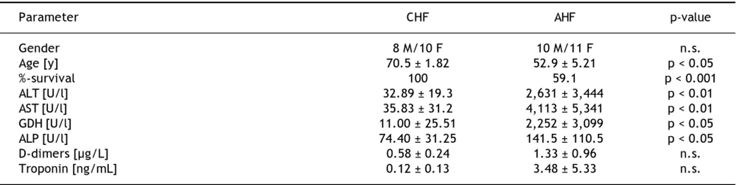
We included 21 cases of acute LI (10 males, 11 fe-males) aged 52.9 ± 5.21. The diagnosis of AHF and/ or cardiogenic shock at admission was associated with a mortality rate of 40.9 %. Leading cause of death was the underlying heart failure. We found significantly elevated serum concentrations of ALT, AST, ALP and GDH in the AHF-patients compared to CHF patients (Table 1). The serum markers tro-ponin and D-dimers were also increased in AHF, but this difference did not reach significance. CHF pa-tients were older and had a lower mortality rate.
Cell death markers
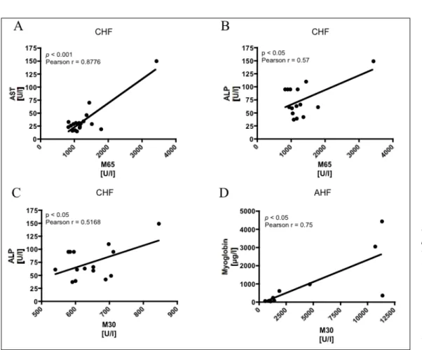
correlate with LFTs and myoglobin
Cell death markers differ between CHF-induced and acute AHF-induced liver injury
As previously published, cell are death markers were elevated in both CHF- and AHF-patients, indi-cating that a large number of cells are undergoing apoptotic and necrotic cell death.6 Levels of M65 and
M30, however, were significantly higher in AHF (M65: chronic 1,283 ± 591.6U/l vs. acute 20,912 ± 15,132U/l, p < 0.001; M30: chronic 642.2 ± 177.4U/l
vs. acute 3,844 ± 5,293U/l, p < 0.05; Figure 2A and B).
Apoptosis is the predominant cell death mecha-nism in CHF-induced liver injury
Patients with CHF exhibited a significantly higher M30/M65-ratio than AHF patients (M30/ M65: chronic 0.54 ± 0.15 vs. acute 0.20 ± 0.19, p < 0.001; Figure 2C), suggesting that a greater proportion of cells are undergoing apoptotic cell dea-th in CHF. In contrast, AHF-patients were polarized towards necrotic cell death.
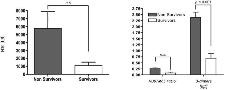
Cell death markers may predict survival in AHF
In order to evaluate the potential of the analy-sed markers to predict the outcome after AHF, we grouped the patients into survivors (n = 9) and non-survivors (n = 13). We found a trend for in-creased M30-levels (survivors: 1,115 ± 408 non-survivors: 5,734 ± 2,106; p = 0.06; Figure 3A) and the M30/M65 ratio (survivors: 0.08 ± 0.04, non-survivors: 0.25 ± 0.07; p = 0.08; Figure 3B) in non-survivors vs. survivors. D-dimers, which have previously been shown to correlate with survival in HF differed significantly between the groups (sur-vivors: 0.69 ± 0.20, non-sur(sur-vivors: 2.38 ± 0.22; p < 0.001; Figure 3B).7
DISCUSSION
177
Cell death in acute heart failure. , 2011; 10 (2): 174-179
acute LI. Patients with LI, secondary to AHF had significantly higher serum CK18 levels (cleaved and uncleaved) compared to patients with CHF. Howe-ver, the M30/M65 ratio, representing the ratio bet-ween apoptosis and necrosis was higher in CHF patients. This is of clinical relevance and helps dis-tinguish the cause of liver injury in cardiologic pa-tients. Furthermore, we found that M30 ELISA might be utilised to distinguish patients with good
vs. bad prognosis. However, a larger cohort of pa-tients would be required to validate these results.
Recent studies reported an increase in caspase cleaved CK18 in the serum of patients admitted for acute myocardial infarction (AMI).8 The M30 assay
was utilised in a study of 39 patients with AMI,
and was shown to be superior to troponin T and creatine kinase in the diagnosis of AMI.9 An
abun-dance of CK18 antibodies is seen in patients with coronary heart disease.10 However, the origin of
the CK18 remains unclear; in one study, weak stai-ning for CK18 in cardiomyocytes virtually excluded the cardiomyocyte as the source of caspase cleaved CK18, despite the identification of cleaved CK18 fragments within myocardial lysosomes.11 The
authors concluded that the source of the cleaved CK18 was not the myocardium, but rather endo-thelial cells or extracardial tissues. Indeed, macro-phage uptake of serum cleaved CK18 was suggested to account for the abundance of M30 positive mate-rial within the myocardium. No studies, however, Figure 2. Elevated cell death in chronic heart failure (CHF) and acute heart failure (AHF). M65 (A) and M30 (B) were signifi-cantly higher in AHF than in CHF. In contrast the ratio of M30/M65 was higher in patients with CHF than AHF patients (C).
A B C
Figure 3. Potential prognostic value of cell death for outcome after acute congestive heart failure. Patients were grouped as survivors (n = 9) or non-survivors (n = 13). While no significant difference for M30 (A) as well as the M30/M65 ratio (B) were ob-served, d-dimers differed significantly between the groups (B).
M30/M65 ratio D-dimers
[µg/l]
Table 1. Demographic and basic clinical data of CHF and AHF patients.
Parameter CHF AHF p-value
Gender 8 M/10 F 10 M/11 F n.s.
Age [y] 70.5 ± 1.82 52.9 ± 5.21 p < 0.05
%-survival 100 59.1 p < 0.001
ALT [U/l] 32.89 ± 19.3 2,631 ± 3,444 p < 0.01
AST [U/l] 35.83 ± 31.2 4,113 ± 5,341 p < 0.01
GDH [U/l] 11.00 ± 25.51 2,252 ± 3,099 p < 0.05
ALP [U/l] 74.40 ± 31.25 141.5 ± 110.5 p < 0.05
D-dimers [µg/L] 0.58 ± 0.24 1.33 ± 0.96 n.s.
Troponin [ng/mL] 0.12 ± 0.13 3.48 ± 5.33 n.s.
examined for the extra-cardiac, or hepatic contri-bution to CK18 levels. Since we and others have shown that liver injury is associated with the sys-temic release of cleaved and uncleaved CK18, and that M30 levels correlate with serum LFT’s rather than markers for myocardial damage, this study suggests the hepatocyte is the primary source of CK18 epitopes in AMI.12,13
While systemic hypotension or shock may indu-ce LI in a patient with cardiac dysfunction, addi-tional mechanisms contribute to LI. For example, HF resulting in increased central venous pressure and hepatic venous congestion induce sinusoidal dilation, endothelial injury, replacement of hepa-tocytes with erythrocytes, and ultimately centrilo-bular tissue destruction.5 Consistent with this,
Lebray et al. observed increased liver stiffness, unrelated to hepatic fibrosis in a patient with HCV who underwent heart transplant.14 Millonig
et al. further reported that the clamping of the in-ferior vena cava in pigs led to a significant increa-se in liver stiffness, while treatment of congestive cardiac failure in patients led to the amelioration of liver stiffness, as assessed by transient elasto-graphy.15 Interestingly, changes in liver stiffness
did not correlate with serum LFT’s. In our cohort of CHF patients, serum LFT’s were also within normal limits, but M30/M65 ratio indicated an apoptotic cell death pattern. Thus, these assays might be a more sensitive screening tool for hepa-tocyte damage in hepatovenous congestion compa-red to standard LFT’s. Although it may be difficult to ascertain the source of CK18, the pat-tern of cell death markers could be a useful pre-dictive marker of hepatic outcomes.16
In contrast, patients with AHF exhibit high serum levels of cell death markers and highly eleva-ted LFT’s. This common clinical constellation is clearly dominated by the primary cardiac event and cardiologic/ cardiothoracic surgical treatment is
the curative management of choice. However, we identified the serum M30-epitope and the ratio of M30/M65 as possible prognostic markers for the sur-vival of AHF patients. These results are consistent with previous studies that demonstrated serum LFTs to be of good prognostic value in heart disea-se.4 Further validation of serum cell-death markers
in AHF and CHF patients will be important to help identify HF-patients at risk of severe liver injury, particularly in the pre-operative setting.
FINANCIAL SUPPORT
This work was supported by the Deutsche Fors-chungsgemeinschaft (DFG, grant 267/8-1), the Wil-helm Laupitz Foundation and the IFORES program of the University of Essen (L.P.B.)
CONFLICT OF INTEREST
None declared.
ABBREVIATIONS
• ALF: Acute liver failure. • LI: Liver injury.
• HF: Heart failure.
• AHF: Acute heart failure. • CHF: Chronic heart failure. • CK18: Cytokeratin-18.
• ELISA: Enzyme-linked immunosorbent assay. • AST: Aspartate aminotransferase.
• ALT: Alanine aminotransferase. • ALP: Alkaline phosphatase. • GDH: Glutamate dehydrogenase.
REFERENCES
179
Cell death in acute heart failure. , 2011; 10 (2): 174-179
2. Kubo SH, Walter BA, John DH, Clark M, Cody RJ. Liver function abnormalities in chronic heart failure. Influence of systemic hemodynamics. Arch Intern Med 1987: 147: 1227-30.
3. Moller S, Dumcke CW, Krag A. The heart and the liver. Ex-pert Rev Gastroenterol Hepatol 2009: 3: 51-64.
4. Batin P, Wickens M, McEntegart D, Fullwood L, Cowley AJ. The importance of abnormalities of liver function tests in predicting mortality in chronic heart failure. Eur Heart J
1995: 16: 1613-8.
5. Seeto RK, Fenn B, Rockey DC. Ischemic hepatitis: clinical pre-sentation and pathogenesis. Am J Med 2000: 109: 109-13. 6. Bechmann LP, Jochum C, Kocabayoglu P, et al.
Cytokera-tin 18-based modification of the MELD score improves pre-diction of spontaneous survival after acute liver injury. J Hepatol 2010: 53: 639-47.
7. Marcucci R, Gori AM, Giannotti F, et al. Markers of hyper-coagulability and inflammation predict mortality in patients with heart failure. J Thromb Haemost 2006: 4: 1017-22. 8. Senturk T, Aydinlar A, Yilmaz Y, Oral AY, Ozdabakoglu O,
Ulukaya E. Serial changes in circulating M30 antigen, a bio-marker of apoptosis, in patients with acute coronary syn-dromes: relationship with the severity of coronary artery disease. Coron Artery Dis 2009: 20: 494-8.
9. Adlbrecht C, Hoetzenecker K, Posch M, et al. Elevated le-vels of interleukin-1beta-converting enzyme and
caspase-cleaved cytokeratin-18 in acute myocardial infarction.
Eur J Clin Invest 2007: 37: 372-80.
10. Mattey DL, Dawes PT, Nixon NB, Goh L, Banks MJ, Kitas GD. Increased levels of antibodies to cytokeratin 18 in pa-tients with rheumatoid arthritis and ischaemic heart di-sease. Ann Rheum Dis 2004: 63: 420-5.
11. Soleiman A, Lukschal A, Hacker S, et al. Myocardial lipofus-cin-laden lysosomes contain the apoptosis marker caspa-se-cleaved cytokeratin-18. Eur J Clin Invest 2008: 38: 708-12.
12. Dechene A, Sowa JP, Gieseler RK, et al. Acute liver failure is associated with elevated liver stiffness and hepatic ste-llate cell activation. Hepatology 2010: 52: 1008-16. 13. Hetz H, Hoetzenecker K, Hacker S, et al. Caspase-cleaved
cytokeratin 18 and 20 S proteasome in liver degeneration.
J Clin Lab Anal 2007: 21: 277-81.
14. Lebray P, Varnous S, Charlotte F, Varaut A, Poynard T, Ratziu V. Liver stiffness is an unreliable marker of liver fi-brosis in patients with cardiac insufficiency. Hepatology
2008: 48: 2089
15. Millonig G, Friedrich S, Adolf S, et al. Liver stiffness is di-rectly influenced by central venous pressure. J Hepatol
2010:52: 206-10.
![ofsarcomapatients[ ].Amongthenovelanti-tumoragentsused,apoptosisligand2/TNF-related bothinhematologicalmalignanciesandepithelial-derivedcancers.Inthepresentstudy,wehavetestedLUV-TRAILinseveralhumansarcomatumorcelllineswithdifferentsensitivitytosoluble des](data:image/gif;base64,R0lGODlhAQABAIAAAP///wAAACH5BAEAAAAALAAAAAABAAEAAAICRAEAOw==)