Frequency and Characteristics of Occult
Hepatitis B Infection Among Hepatocellular
Carcinoma Patients in Japan
Jun Muto,*,**, Masaya Sugiyama,**, Ken Shirabe,* Motokazu Mukaide,** Ikue Kirikae-Muto,** Toru Ikegami,* Tomoharu Yoshizumi,* Yo-ichi Yamashita,* Yoshihiko Maehara,* Masashi Mizokami**
* Department of Surgery and Science, Graduate School of Medical Sciences, Kyushu University, Fukuoka, Japan. ** The Research Center for Hepatitis and Immunology, National Center for Global Health and Medicine, Ichikawa, Chiba, Japan.
These authors contributed equally to this work. July-August, Vol. 17 No. 4, 2018: 596-603
INTRODUCTION
Hepatocellular carcinoma (HCC) is the sixth most common cancer and the third most common cause of can-cer-related deaths worldwide.1 Chronic hepatitis B (CHB) infection with detectable circulating hepatitis B surface antigen (HBsAg) is a common cause of HCC. Ap-proximately 60% of the annual total of 530,000 HCC cases worldwide are related to CHB.2 In addition to CHB, a past history of hepatitis B virus (HBV) infection among patients with chronic hepatitis C (CHC), and positivity for hepatitis B core antigen (HBcAg) have also been re-ported as risk factors for HCC carcinogenesis.3,4 HBV
in-fects hepatocytes. For further investigations, specimens from the liver have to be used for the studies.
Occult HBV infection (OBI) is defined as a condition with undetectable HBsAg, but detectable HBV DNA in the serum or liver tissues. The incidence of OBI among CHC patients is known to be high, and about a third of CHC patients in the Mediterranean region were reported to be positive for OBI.5,6 OBI is of interest as a cause of de novo hepatitis B and reactivation of hepatitis B during chemotherapy and has also been reported as a cause of hepatitis B after blood transfusion.6 A previous study found that the disease-free survival period and overall survival period of OBI patients were shorter among HCC patients
The Official Journal of the Mexican Association of Hepatology, the Latin-American Association for Study of the Liver and
the Canadian Association for the Study of the Liver
Manuscript received: Manuscript received: Manuscript received: Manuscript received:
Manuscript received: July 15, 2017. Manuscript accepted:Manuscript accepted:Manuscript accepted:Manuscript accepted:Manuscript accepted: October 20, 2017.
DOI: 10.5604/01.3001.0012.0927
A B S T R A C T A B S T R A C T A B S T R A C T A B S T R A C T A B S T R A C T
Introduction and aim. Introduction and aim.Introduction and aim. Introduction and aim.
Introduction and aim. Occult hepatitis B virus (HBV) infection (OBI) represents a state without detectable hepatitis B surface antigen, but positive for HBV DNA. The correlation between OBI and hepatocellular carcinoma (HCC) carcinogenesis is controver-sial. We studied the frequency and characteristics of OBI among HCC patients and metastatic liver cancer patients. Material andMaterial andMaterial andMaterial andMaterial and methods.
methods.methods. methods.
methods. DNA was obtained from tumor and non-tumor tissues from 75 HCC patients (15 chronic hepatitis B (CHB), 39 chronic hepatitis C (CHC), 21 cryptogenic) and 15 metastatic liver cancer patients who underwent liver resection. HBV DNA and covalently-closed circular (ccc) DNA were detected using real-time polymerase chain reaction (PCR), and four HBV DNA regions were detect-ed by nestdetect-ed PCR. Clinicopathological factors were compardetect-ed between patients with and without OBI. Results.Results.Results.Results.Results. HBV DNA was detected in 14 (93.3%) CHB, five (22.7%) cryptogenic and four (10.3%) CHC patients. cccDNA was detected in 12 (80.0%) CHB, three (14.3%) cryptogenic and two (5.1%) CHC patients. All CHB, eight (38.1%) cryptogenic and ten (25.6%) CHC patients tested positive with nested PCR. No metastatic liver cancer patients were positive for any HBV DNA regions. OBI patients had shorter prothrombin times (P = 0.0055), and lower inflammation activity score in non-tumor liver (P = 0.0274). There were no differences in anti-HBV antibodies. Conclusions.Conclusions.Conclusions.Conclusions.Conclusions. OBI was detected in 38% of cryptogenic and 25.6% of CHC patients. There was no correla-tion between OBI and anti-HBV antibodies, but fewer patients with OBI had high inflammatory activity, suggesting that factors other than inflammation may be involved in HCC carcinogenesis in patients with OBI.
Key words. Key words.Key words. Key words.
with CHC.7 A study in Hong Kong showed that the OBI rates in patients with cryptogenic HCC and HCC with CHC were 73% and 17%, respectively, and the authors noted a correlation between OBI and HCC.8 According to an 11-year prospective study of CHC patients in Italy, 35% of OBI-positive patients developed HCC, compared with only 9% of OBI-negative patients; the incidence of HCC was significantly higher among OBI-positive patients.9,10 In contrast, the Hepatitis C Antiviral Long-term Treat-ment against Cirrhosis (HALT-C) trial in the United States failed to find any correlation between HCC car-cinogenesis and OBI.11 They detected HBV DNA in liver specimens of patients with and without HCC enrolled in the HALT-C trial and found no significant difference in positivity rates between the groups (10.7% vs. 23.6%, re-spectively). There is thus no consensus regarding the po-tential correlation between HCC carcinogenesis and OBI. OBI is defined as negative HBsAg with positive detec-tion of HBV DNA in serum or liver tissue. Some studies have detected HBV DNA in serum, while others have de-tected HBV DNA in liver tissue.7,8,10-12 In addition, some studies have detected HBV DNA using real-time polymerase chain reaction (PCR), and others have used nested PCR. The detection sites of HBV DNA also differ among studies. The detection sensitivities of different studies thus differ widely.
Whole-genome sequencing of HCC patients has re-cently revealed the integration sites of HBV DNA into the human genome.13,14 Based on these reports, we developed a sensitive in-house detection system for HBV DNA, us-ing primers designed based on the reported integration sites of HBV DNA. In this study, we investigated the inci-dence of OBI among HCC patients and metastatic liver cancer patients using this highly-sensitive, in-house detec-tion system, and revealed the characteristics of OBI-posi-tive patients.
MATERIAL AND METHODS
Patients
Tissue specimens were obtained from 90 patients who had undergone liver resection at the Department of Sur-gery and Science, Kyushu University Hospital, between March 2005 and January 2012. Fifteen HCC patients with CHB, 39 HCC patients with CHC, 21 patients with cryp-togenic HCC, and 15 patients with metastatic liver cancer (13 colon cancer, 1 gastric cancer and 1 renal cell carcino-ma) were enrolled in this study. No patients were diag-nosed with alcoholic liver disease. DNA was extracted from exenterated HCC and peripheral liver tissues after surgery using a QIAamp DNA Mini Kit (Qiagen KK, To-kyo, Japan). No patients from the cryptogenic, CHC or
metastatic liver tumor groups were positive for HBsAg or HBV DNA according to real-time PCR analysis of serum. Samples were collected according to an established proto-col approved by the Ethics Committee of Kyushu Univer-sity.
Pathological examination of the liver
All liver tissue specimens were evaluated by patholo-gists, who was blinded to the clinical condition of the pa-tient. Inflammatory activity and fibrosis was graded and staged according to the New Inuyama classification system as follows: A0 (no necro-inflammatory reaction), A1 (mild), A2 (moderate), and A3 (severe) for inflammatory activity and F1 (periportal expansion), F2 (portoportal septa), F3 (portocentral linkage or bridging fibrosis), and F4 (cirrhosis) for fibrosis.
Quantification of intrahepatic HBV DNA
Intrahepatic total HBV DNA, covalently-closed circu-lar DNA (cccDNA), and human RNaseP contents in liver tissues were measured by real-time PCR.15 Briefly, intra-hepatic total HBV DNA and cccDNA were measured us-ing the primer-probe sets shown in table 1. Plasmid safe DNase I (Epicentre Inc. Chicago, IL) was used to digest the single-stranded region of the HBV genome, thus al-lowing enrichment of cccDNA for subsequent real-time PCR detection. Real-time PCR experiments were per-formed in a Light-Cycler (Roche Diagnostics, Basel, Switzerland) in 100-μL reaction volume. Amplification was performed as follows: 95 °C for 10 min, followed by 50 cycles of 95 °C for 10 s, 62 °C for 10 s, and 72 °C for 20 s. RNaseP amplification was performed using a Control Kit (Applied Biosystems inc., Caelsbad, CA). Serial dilutions of a plasmid containing a monomeric genotype C HBV in-sert were used as quantification standards. Taking into ac-count the real-time PCR system used, the lower limit of detection of intrahepatic total HBV DNA and cccDNA quantification was 1×10⁴ copies/cell.
In-house high-sensitivity HBV DNA detection system
°C, 15 s; 58 °C, 30 s; 72 °C, 30 s) in first and second PCR using Veriti (Applied Biosystems, Foster City, CA, USA). The PCR products were sequenced on both strands using a BigDye Terminator V3.1 cycle sequencing kit (Applied Biosystems), with the same primers used for the second PCR. The sequencing products were analyzed using an ABI 3130xl DNA analyzer (Applied Biosystems). The ob-tained sequences were aligned with GenBank sequences corresponding to HBV genotypes. The lower limit of de-tection was 1-10 copies/mL.
Statistical analysis
Non-parametric tests (χ2 and Fisher’s exact probability tests) were used to compare the characteristics of the groups. Univariate logistic regression analysis was used to identify the factors that contributed significantly to early viral dynamics. The odds ratios and 95% confidence inter-vals were also calculated. All p values < 0.05 using two-tailed tests were considered significant. Analyses were performed using JMP (version 9.0.2, SAS, Inc., Cary, NC, USA).
RESULTS
Patient backgrounds
The demographic data for the 90 patients are shown in table 2. Five patients with cryptogenic HCC (23.8%) and 21 with CHC (53.9%) were anti-HBc antibody (HBcAb)-positive. Five patients with cryptogenic HCC (23.8%) and 12 with CHC (33.3%) were anti-HBs antibody (HBsAb)-positive. No serological HBV infection markers were de-tected in patients with metastatic liver cancer. Two patients with cryptogenic HCC (9.5%) showed histologi-cal evidence of steatosis. One patient with cryptogenic HCC (4.8%), 26 with CHC (66.7%), and three with CHB (21.4%) showed high inflammatory activity in non-tumor tissue. Four patients with cryptogenic HCC (18.2%), 22 with CHC (56.4%), and five with CHB (35.7%) demon-strated fibrosis of non-tumor tissue. No patients with metastatic liver cancer showed high inflammation or fi-brosis in non-tumor liver tissue.
Quantitative detection of intrahepatic HBV DNA
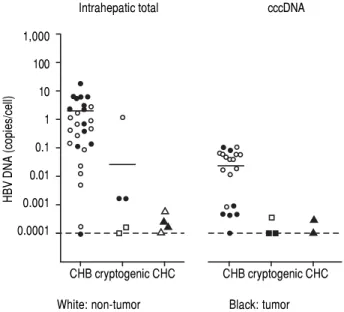
Detection of HBV DNA and cccDNA in tumor and peripheral non-tumor tissues were performed using real-time PCR. HBV DNA was detected in 14 (93.3%) patients with CHB (median, 0.6867 copies/cell; range, < 0.0001-18.12 copies/cell) and cccDNA was detected in 12 (80.0%) patients with CHB (median, 0.0398 copies/cell; range, <
Table 1.
Oligonucleotides used for real-time PCR and nested PCR identification of occult HBV infection.
Region
Round
Forward primer
Reverse primer
Probe 1
Probe 2
S
real-time PCR
ATCCTGCTGCTATGCCTCAT
GGGAAAGCCCTRCGAACCACTGAACAA
FAM-TGCACTTGTATTCCCATCCC-MGB
FAM-TGAGGCCCACTCCCATAGGA-MGB
cccDNA
real-time PCR
TCCCCGTCTGTGCCTTCTC
GCACAGCTTGGAGGCTTGA
FAM-CCGTGTGCACTTC-MGB
FAM-CCGTGAACGCCCA-MGB
S
nested PCR
1
st
AGGTATGTTGCCCGTTTGTC
AAAGCCCTACGAACCACTGA
2
nd
GTATGTTGCCCGTTTGTCCT
AAGCCCTACGAACCACTGAA
Core
nested PCR
1
st
TTCAAGCCTCCAAGCTGTGC
TCTTCCAAATTACTTCCCACCCA
2
nd
CAAGCCTCCAAGCTGTGCCT
TCTTCCAAATTACTTCCCACCCA
P
ol
nested PCR
1
st
GGAGTGTGGATTCGCACTCC
CGTCTGCGAGGCGAGGGA
2
nd
GGAGTGTGGATTCGCACTCC
CGAGGCGAGGGAGTTCTTCTTC
X
nested PCR
1
st
GGGCGCACCTCTCTTTACGC
CATGCGACGTGCAGAGGTGAAG
2
nd
TCTCCCCGTCTGTGCCTTCTC
0.0001-0.1035 copies/cell), respectively (Figure 1). HBV DNA was detected in five (22.7%) patients with cryptogenic HCC (median, 0.0002 copies/cell; range, < 0.0001-1.1735 copies/cell) and four (10.3%) patients with CHC (median, 0.0002 copies/cell; range, < 0.0001-0.0006 copies/cell), and cccDNA was detected in three (14.3%) patients with cryptogenic HCC (median, 0.0001 copies/cell; range, < 0.0001-0.0004 copies/cell) and two (5.1%) patients with CHC (median, 0.0002 copies/cell; range, < 0.0001-0.0003 copies/cell).
Detection of intrahepatic HBV DNA using a highly-sensitive in-house detection system
Detection of HBV DNA in tumor and non-tumor tis-sues was performed using a highly-sensitive in-house HBV DNA detection system, using nested PCR on four regions (S, C, Pol and X) of the HBV DNA. Five patients with CHB were positive for all four regions in non-tumor tissue (Table 3). All CHB patients were positive for at least two regions in non-tumor tissue, and 14 patients were positive for at least three regions in tumor tissue.
Table 2. Patient backgrounds
CHB Cryptogenic CHC Metastatic liver cancer
(n = 15) (n = 21) (n = 39) (n = 15)
Male : female 12 : 3 17 : 4 28 : 11 13 : 2
Mean age ± SD (yr) 52.7 ± 10.4 68.2 ± 14.9 69.8 ± 6.9 64.8 ± 11.9
Anti-HBc-positive cases (n, %) 15 (100) 5 (23.8) 21 (53.9) 0 (0)
Anti-HBs-positive cases (n, %) 1 (6.7) 5 (23.8) 12 (33.3) 0 (0)
Anti-HBe-positive cases (n, %) 9 (60.0) 1 (4.8) 8 (20.5) 0 (0)
Serum study
AST (IU/L) 46.5 ± 23.7 40.0 ± 28.4 55.8 ± 40.4 28.3 ± 11.6
ALT (IU/L) 57.9 ± 46.3 42.8 ± 36.4 57.3 ± 47.7 30.8 ± 27.2
Alb (g/dL) 4.0 ± 0.5 4.0 ± 0.3 3.9 ± 0.4 3.9 ± 0.4
T-Bil (mg/dL) 0.8 ± 0.4 0.8 ± 0.3 0.8 ± 0.3 0.6 ± 0.2
PT (s) 13.2 ± 1.1 12.6 ± 0.9 13.0 ± 0.8 12.6 ± 1.1
Plt (×104/μL) 12.7 ± 6.6 20.4 ± 7.9 14.2 ± 5.6 24.2 ± 11.7
Tumor factors
Tumor diameter (cm) 4.9 ± 3.0 5.0 ± 2.6 3.4 ± 2.7
-Multiple tumors (%) 5 (33.3) 6 (28.6) 9 (23.1)
-Cases with vascular invasion (%) 9 (60.0) 6 (28.6) 14 (35.9)
-Stage (I, II / III, IV) 8 / 7 10 / 11 29 / 10
-AFP ≥ 50 ng/mL (%) 5 (33.3) 4 (19.1) 9 (23.1)
-DCP ≥ 300 m AU/mL (%) 9 (60.0) 8 (38.1) 10 (25.6)
-Pati ents with histological evidence 0 (0) 2 (9.5) 5 (12.8) 0 (0)
of steatosis (n, %)
Patients with DM (n, %) 1 (7.1) 9 (42.9) 13 (35.1) 2 (13.3)
Patients with high inflammatory activity 3 (21.4) 1 (4.8) 26 (66.7) 0 (0)
in non-tumor tissues (n, %)
Patients with fibrosis in non-tumor 5 (35.7) 1 (4.8) 22 (56.4) 0 (0)
tissues (n, %)
Patients with Child–Pugh score >10 (n, %) 0 (0) 0 (0) 0 (0) 0 (0)
Figure 1. Figure 1.Figure 1.
Figure 1.Figure 1. Quantitative levels of intrahepatic total HBV DNA and cccDNA. Individual measurements from the non tumor liver tissue are shown as white dots and measurements from the tumor tissue are shown as black dots. CHB: chronic hepatitis B. CHC: chronic hepatitis C.
HBV DNA (copies/cell)
1,000
100
10
1
0.1
0.01
0.001
0.0001
CHB cryptogenic CHC CHB cryptogenic CHC
Eight patients with cryptogenic HCC (38.1%) and 10 with CHC (25.6%) were positive for at least one region of HBV DNA (Table 4). HBV DNA was detected in 15 non-tumor and 10 non-tumor tissues from non-CHB patients. The Pol region of HBV DNA was detected in 10 non-tumor and seven tumor tissues, while the HBx region was only detected in two non-tumor and three tumor tissues. The sequences of the nested PCR products were checked to rule out any contamination. We, therefore, defined OBI as positivity for at least one region of HBV DNA. Eighteen patients were diagnosed with OBI-positive (Figure 2).
Characteristics of OBI-positive patients
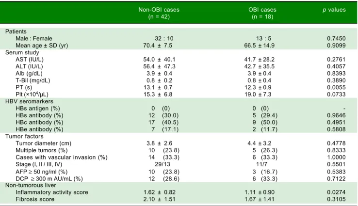
We compared the clinicopathological findings of OBI-positive and OBI-negative patients (Table 5). There was no significant difference in terms of sex or age of the pa-tients between the two groups. Prothrombin time (PT) was significantly shorter (p = 0.0055), and transaminases
Table 3. Distribution of HBV regions detectable by highly-sensitive in-house detection system in non-tumor and tumor tissues in 18 HCC patients with OBI.
Non-tumor (NT) Tumor (T)
PCR-detectable HBV Cases S C Pol X S C Pol X
regions in NT/T (n) (n)
4/1 1 + + + + +
3/4 1 + + + + + + +
3/3 1 + + + + + +
2/1 1 + + +
2/1 1 + + +
2/0 1 + +
1/3 1 + + + +
1/1 1 + +
1/0 4 +
1/0 3 +
0/4 1 + + + +
0/1 2 +
Total of cases/PCR-positive regions (n) 18 9 4 10 2 5 3 7 4
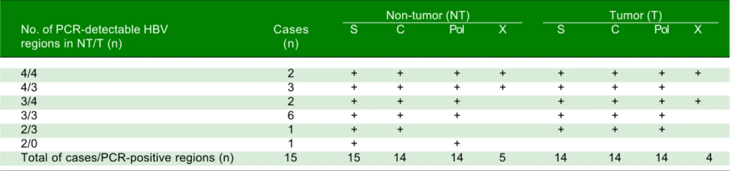
Table 4. Distribution of HBV regions detectable by highly-sensitive, in-house detection system in non-tumor and tumor tissues from 15 HCC patients with CHB
Non-tumor (NT) Tumor (T)
No. of PCR-detectable HBV Cases S C Pol X S C Pol X
regions in NT/T (n) (n)
4/4 2 + + + + + + + +
4/3 3 + + + + + + +
3/4 2 + + + + + + +
3/3 6 + + + + + +
2/3 1 + + + + +
2/0 1 + +
Total of cases/PCR-positive regions (n) 15 15 14 14 5 14 14 14 4
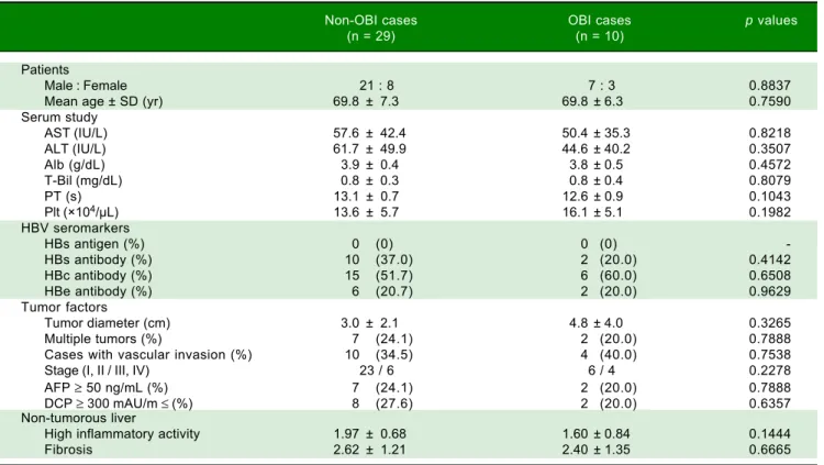
and platelets tended to be lower in OBI cases. Inflamma-tory activity score in non-tumor liver tissue was signifi-cantly lowere in OBI cases (p = 0.0274). There were no differences in HBV seromarkers, including HBsAb and HBcAb, or in tumor factors, including tumor diameter and tumor staging, between OBI-positive and -negative cases. We also compared clinicopathological findings be-tween OBI-positive and OBI-negative CHC patients (Ta-ble 6), which showed the same tendencies in terms of serological liver function markers. Similarly, OBI-posi-tive cases had lower inflammatory activity in non-tumor-ous liver (p = 0.1444).
DISCUSSION
stud-Table 5. Characteristics of OBI-positive HCC patients.
Non-OBI cases OBI cases p values
(n = 42) (n = 18)
Patients
Male : Female 32 : 10 13 : 5 0.7450
Mean age ± SD (yr) 70.4 ± 7.5 66.5 ± 14.9 0.9099
Serum study
AST (IU/L) 54.0 ± 40.1 41.7 ± 28.2 0.2761
ALT (IU/L) 56.4 ± 47.3 42.7 ± 35.5 0.4057
Alb (g/dL) 3.9 ± 0.4 3.9 ± 0.4 0.8393
T-Bil (mg/dL) 0.8 ± 0.2 0.8 ± 0.4 0.3890
PT (s) 13.1 ± 0.7 12.3 ± 0.9 0.0055
Plt (×104/μL) 15.3 ± 6.8 19.0 ± 7.3 0.0733
HBV seromarkers
HBs antigen (%) 0 (0) 0 (0)
-HBs antibody (%) 12 (30.0) 5 (29.4) 0.9646
HBc antibody (%) 17 (40.5) 9 (50.0) 0.4951
HBe antibody (%) 7 (17.1) 2 (11.7) 0.5808
Tumor factors
Tumor diameter (cm) 3.8 ± 2.6 4.4 ± 3.2 0.4778
Multiple tumors (%) 10 (23.8) 5 (26.3) 0.8333
Cases with vascular invasion (%) 14 (33.3) 6 (33.3) 1.0000
Stage (I, II / III, IV) 29/13 11/7 0.5501
AFP ≥ 50 ng/ml (%) 10 (23.8) 3 (16.7) 0.5383
DCP ≥ 300 m AU/mL (%) 12 (28.6) 6 (33.3) 0.7122
Non-tumorous liver
Inflammatory activity score 1.62 ± 0.82 1.11 ± 0.90 0.0274
Fibrosis score 2.10 ± 1.51 1.67 ± 1.41 0.3105
Figure 2. Figure 2.Figure 2.
Figure 2.Figure 2. Schematic representation of the study patients. The Number of patients with positive results for each test are shown. HCC: hepatocellular carcino-ma. CHB: chronic hepatitis B. CHC: chronic hepatitis C.
75 HCC patients and 15 metastatic liver cancer patients
15 CHB 21 cryptogenic 39 CHC 15 meta
Number of patients with:
Detectable serum HBV DNA 15 (100%) 0 (0%) 0 (0%) 0 (0%)
Detectable intrahepatic
total HBV DNA 14 (93.3%) 5 (23.8%) 4 (10.3%) 0 (0%)
Detectable cccDNA 12 (80.0%) 3 (14.3%) 2 (5.1%) 0 (0%)
Number of patients with:
Detectable HBV DNA 15 (100%) 8 (38.1%) 10 (25.6%) 0 (0%)
Real-time quantificaction of HBV DNA
Table 6. Characteristics of OBI-positive HCC patients with CHC.
Non-OBI cases OBI cases p values
(n = 29) (n = 10)
Patients
Male : Female 21 : 8 7 : 3 0.8837
Mean age ± SD (yr) 69.8 ± 7.3 69.8 ± 6.3 0.7590
Serum study
AST (IU/L) 57.6 ± 42.4 50.4 ± 35.3 0.8218
ALT (IU/L) 61.7 ± 49.9 44.6 ± 40.2 0.3507
Alb (g/dL) 3.9 ± 0.4 3.8 ± 0.5 0.4572
T-Bil (mg/dL) 0.8 ± 0.3 0.8 ± 0.4 0.8079
PT (s) 13.1 ± 0.7 12.6 ± 0.9 0.1043
Plt (×104/μL) 13.6 ± 5.7 16.1 ± 5.1 0.1982
HBV seromarkers
HBs antigen (%) 0 (0) 0 (0)
-HBs antibody (%) 10 (37.0) 2 (20.0) 0.4142
HBc antibody (%) 15 (51.7) 6 (60.0) 0.6508
HBe antibody (%) 6 (20.7) 2 (20.0) 0.9629
Tumor factors
Tumor diameter (cm) 3.0 ± 2.1 4.8 ± 4.0 0.3265
Multiple tumors (%) 7 (24.1) 2 (20.0) 0.7888
Cases with vascular invasion (%) 10 (34.5) 4 (40.0) 0.7538
Stage (I, II / III, IV) 23 / 6 6 / 4 0.2278
AFP ≥ 50 ng/mL (%) 7 (24.1) 2 (20.0) 0.7888
DCP ≥ 300 mAU/m ≤ (%) 8 (27.6) 2 (20.0) 0.6357
Non-tumorous liver
High inflammatory activity 1.97 ± 0.68 1.60 ± 0.84 0.1444
Fibrosis 2.62 ± 1.21 2.40 ± 1.35 0.6665
ies. Among 60 HCC cases, excluding patients with CHB, 18 (30.0%) were diagnosed as OBI-positive. Ten of 38 CHC cases (25.6%) and eight out of 21 cryptogenic cases (38.1%) were also diagnosed as OBI-positive. No patients with metastatic liver cancer were OBI-positive. In a previ-ous study from Hong Kong, one of six patients with CHC and HCC (16.7%) and 24 of 33 with cryptogenic HCC (72.7%) were reported to be OBI-positive.[8] In CHC pa-tients with HCC, 15 out of 80 cases in Taiwan (20.0%), three out of 91 in the United States (3.3%), and 45 out of 73 in Italy (61.6%) were OBI-positive.7-9,11 These differ-ences are thought to reflect differdiffer-ences in the causes of in-fection and the inin-fection rates of HBV and HCV, and differences in HBV genotypes among different areas. Be-cause of the geographical distance and major HBV geno-types B and C, it was reasonable that the OBI rates in Hong Kong and Taiwan were consistent with those in the current study. The fact that the HBV DNA detection rate in non-tumor tissue was higher than in tumor tissue was compatible with the results of previous reports.
The sensitivities and specificities of HBV detection ap-pear to differ greatly among studies, but this is not sur-prising given that the HBV DNA detection methods used (real-time PCR or nested PCR), and the regions of HBV DNA detected differ among studies. In most studies, OBI cases were defined by positive detection of at least two re-gions of HBV DNA, to prevent the occurrence of false
positive results due to contamination. However, we checked the sequences of the nested PCR products to rule out the possibility of contamination, and we, therefore, defined OBI cases as those positive for at least one region of HBV DNA.
HBV is a double-stranded DNA virus, the DNA se-quence of which is highly variable. We designed the prim-ers based on full-length sequences of HBV DNA in the NCBI database, using lesions with few reported varia-tions. We also referred to studies that performed whole-genome sequencing of HCC patients and identified integration sites of HBV DNA into the host genome, and designed the primers based on these reported integration sites.13,14 However, as shown in table 5, the positivity rates varied between HBV DNA lesions, with the positivity rates of HBc and HBx lesions being lower than others. This may suggest that the HBV responsible for OBI con-tains variations of HBc and HBx DNA sequences that dif-fer from those in the NCBI database. Further studies are needed to clarify this.
report-ed that HBcAb- and HBsAb-positivity were 70.0% and 60.0%, respectively, in OBI-positive operated cases of HCC. Furthermore, 10.0% of OBI-positive HCC cases were negative for all serum HBV markers.8 Further stud-ies are needed to clarify the relationship between HBV-re-lated antibodies and OBI.
In this study, inflammation in non-tumor tissue was significantly lower, and serum liver function markers tended to be better among OBI-positive patients. This suggests that factors not involving inflammation of the background liver may be involved in carcinogenesis. Fur-ther studies are needed to clarify the relationship between OBI and liver carcinogenesis.
ABBREVIATIONS
• ALT: alanine aminotransferase. • AST: aspartate aminotransferase. • CHB: chronic hepatitis B. • CHC: chronic hepatitis C.
• HALT-C: Hepatitis C Antiviral Long-term Treatment against Cirrhosis.
• HBcAb: anti-hepatitis B core antibody. • HBcAg: hepatitis B core antigen.
• HBeAb: anti-hepatitis B envelope protein. • HBsAb: anti-hepatitis B surface antibody. • HBsAg: hepatitis B surface antigen. • HBV: hepatitis B virus.
• HCC: hepatocellular carcinoma. • OBI: occult hepatitis B virus infection. • PCR: polymerase chain reaction. • PT: prothrombin time.
• TERT: telomerase reverse transcriptase.
FINANCIAL SUPPORT
The Ministry of Health Labour and Welfare, Health Labour Sciences Research. Grant (H23-Kanen-Ippan-003). The Ministry of Health Labour and Welfare, Health Labour Sciences Research. Grant (H24-Kanen-Ippan-004).
CONFLICTS OF INTEREST
The authors have no conflicts of interest.
REFERENCES
1. Parkin DM, Bray F, Ferlay J, Pisani P. Global cancer statis-tics, 2002. CA: a cancer journal for clinicians 2005; 55: 74-108.
2. Lai CL, Ratziu V, Yuen MF, Poynard T. Viral hepatitis B. Lan-cet 2003; 362: 2089-94.
3. Ikeda K, Marusawa H, Osaki Y, Nakamura T, Kitajima N, Ya-mashita Y, Kudo M, et al. Antibody to hepatitis B core anti-gen and risk for hepatitis C-related hepatocellular carcinoma: a prospective study. Annals of internal medicine 2007; 146: 649-56.
4. Ohki T, Tateishi R, Goto E, Sato T, Masuzaki R, Imamura J, Goto T, et al. Influence of anti-HBc seropositivity on the risk of hepatocellular carcinoma in HCV-infected patients after adjusting for confounding factors. Journal of viral hepatitis 2010; 17: 91-7.
5. Torbenson M, Thomas DL. Occult hepatitis B. The Lancet in-fectious diseases 2002; 2: 479-86.
6. Raimondo G, Pollicino T, Cacciola I, Squadrito G. Occult hepatitis B virus infection. Journal of hepatology 2007; 46: 160-70.
7. Chang ML, Lin YJ, Chang CJ, Yeh C, Chen TC, Yeh TS, Lee WC, et al. Occult and Overt HBV Co-Infections Independently Predict Postoperative Prognosis in HCV-Associated Hepato-cellular Carcinoma. PloS one 2013; 8: e64891.
8. Wong DK, Huang FY, Lai CL, Poon RT, Seto WK, Fung J, Hung IF. Occult hepatitis B infection and HBV replicative ac-tivity in patients with cryptogenic cause of hepatocellular carcinoma. Hepatology 2011; 54: 829-36.
9. Pollicino T, Squadrito G, Cerenzia G, Cacciola I, Raffa G, Craxi A, Farinati F. Hepatitis B virus maintains its pro-onco-genic properties in the case of occult HBV infection. Gas-troenterology 2004; 126: 102-10.
10. Squadrito G, Cacciola I, Alibrandi A, Pollicino T, Raimondo G. Impact of occult hepatitis B virus infection on the outcome of chronic hepatitis C. Journal of hepatology 2013; 59: 696-700. 11. Lok AS, Everhart JE, Di Bisceglie AM, Kim HY, Hussain M, Morgan TR. Occult and previous hepatitis B virus infection are not associated with hepatocellular carcinoma in United States patients with chronic hepatitis C. Hepatology 2011; 54: 434-42.
12. Kitab B, Ezzikouri S, Alaoui R, Nadir S, Badre W, Trepo C, Chemin I. Occult HBV infection in Morocco: from chronic hepatitis to hepatocellular carcinoma. Liver international: of-ficial journal of the International Association for the Study of the Liver 2014.
13. Fujimoto A, Totoki Y, Abe T, Boroevich KA, Hosoda F, Nguy-en HH, Aoki M, Hosono N. Whole-gNguy-enome sequNguy-encing of liver cancers identifies etiological influences on mutation patterns and recurrent mutations in chromatin regulators. Nature ge-netics 2012; 44: 760-4.
14. Sung WK, Zheng H, Li S, Chen R, Liu X, Li Y, Lee NP. Ge-nome-wide survey of recurrent HBV integration in hepato-cellular carcinoma. Nature genetics 2012; 44: 765-9. 15. Kim HE, Kim DG, Lee KJ, Son JG, Song MY, Park YM, Kim JJ.
Frequent amplification of CENPF, GMNN and CDK13 genes in hepatocellular carcinomas. PloS ONE 2012; 7: e43223.
Correspondence and reprint request: Ken Shirabe, M.D., Ph.D, F.A.C.S.,
Department of Surgery and Science, Graduate School of Medical Sciences, Kyushu University, 3-1-1 Maidashi,
Higashi-ku, Fukuoka 812-8582, Japan. Tel: +81-92-642-5466; Fax: +81-92-642-5482;