Effects of bile acid sequestration on
hepatic steatosis in obese mice
Nancy Solís,* Margarita Pizarro,* Pablo Quintero,* Juan Pablo Arab,* Arnoldo Riquelme,* Oslando Padilla,** Gonzalo Carrasco,*** Carlos J. Pirola,**** Silvia Sookoian,**** Marco Arrese*
* Departamento de Gastroenterología, Escuela de Medicina, Pontificia Universidad Católica de Chile, Santiago, Chile ** Departamento de Salud Pública, Pontificia Universidad Católica de Chile, Santiago, Chile.
*** Hospital Parroquial de San Bernardo, San Bernardo, Chile.
**** Institute of Medical Research (IDIM), University of Buenos Aires, National Council of Scientific and Technological Research (CONICET), Autonomous City of Buenos Aires 1427, Argentina.
ABSTRACT
Background. Bile acid sequestration (BAS) with resins has shown antidiabetic effects in both humans and animals. Since hepatic steatosis is commonly associated with type 2 diabetes mellitus and the effects of BAS on steatosis have not been explored in detail, we evaluated the effects of cholestyramine (CTM) admi-nistration on fatty liver development in the leptin-deficient obese mice. Aim. To study the effects of BAS on fatty liver development in obese (ob/ob) mice. Material and methods. 4 week-old ob/ob mice (B6.V-Le-pob/J, n = 4-6 per group) were fed with or without CTM (control group) during 8 weeks. Serum and biliary parameters, glucose tolerance test (GTT), hepatic triglyceride content, liver histology and hepatic gene expression of relevant genes related to bile secretion, lipid and glucose metabolism were assessed. Results. Control 12-week-old mice exhibited marked obesity and hepatic steatosis. CTM administration expectedly determined a marked de-repression of 7-α-hydroxylase and decreased biliary bile acid secretion as well as improved GTT. CTM feeding showed no effects on hepatic triglyceride content or in the degree of steatosis on liver histology. CTM was associated with increased levels of serum alanine-aminotransferase. Conclusion. Although CTM administration positively affects glucose tolerance it does not prevent hepatic steatosis development in obese mice. Moreover, CTM feeding was associated to liver enzyme elevation in this model of NAFLD. Thus, the effects BAS on NAFLD need to be specifically addressed since this therapy might not be beneficial for this condition.
Key words. NAFLD. Obese mice. Bile acids. Steatosis. Fatty liver. Cholestyramine.
Correspondence and reprint request: Marco Arrese, M.D.
Departamento de Gastroenterología, Escuela de Medicina, Pontificia Universidad Católica de Chile.
Marcoleta 367, 833-0024 Santiago, Chile. Tel.: 56-2-3543820. Fax: 56-2-6397780 E-mail: marrese@med.puc.cl
Manuscript received: May 15, 2013. Manuscript accepted: July 09, 2013.
INTRODUCTION
Non-alcoholic fatty liver disease (NAFLD) is now considered the most common form of liver disease worldwide and its presence is associated with an
in-creased liver-related mortality.1 NAFLD
encompas-ses a spectrum of histological abnormalities ranging from simple steatosis to non-alcoholic steatohepatitis (NASH), which is characterized by steatosis plus ne-croinflammatory changes and various degrees of
he-patic fibrosis.2 Patients with NASH are considered
to have a more aggressive form of the disease and
carry the risk of developing cirrhosis and
hepatocel-lular carcinoma.3
The pathogenesis of NAFLD is not fully understo-od but it is associated with obesity and insulin
re-sistance (IR).4,5 IR results in alterations on deposits
of lipids and lipolysis in insulinosensitive tissues, which induces an increased flow of fatty acids from
adipose tissue to the liver causing steatosis.6 The
ac-cumulation of triglycerides in the liver determines several alterations, including resistance to insulin action in this organ and oxidative stress that is as-sociated with cell damage, necrosis and inflamma-tion, determining the potential development of
NASH and disease progression.6 At present time,
li-festyle interventions and behavior therapy, together with drugs used to treat the associated diseases of the metabolic syndrome (hypertension, diabetes, or dyslipidemia), represent the main therapeutic
approach available to the clinician.1 In addition,
or pioglitazone administration. Of note, these thera-pies have been mainly tested in non-diabetic patients even though type 2 diabetes mellitus (T2DM) pa-tients are at higher risk of developing hepatic stea-tosis and NASH compared with non-diabetic
individuals.7-9 In fact, in the subgroup of diabetic
patients NAFLD tends to be more frequent and
more severe.7 Indeed, a good metabolic control of
T2DM could be beneficial for both simple steatosis and NASH and therefore optimal antidiabetic stra-tegies should be attempted.
Recently, bile acid sequestration (BAS) using anionic exchange resins such as cholestyramine (CTM) and colesevelam has been shown to be effec-tive improving glycemic control in both T2DM
pa-tients and experimental animals.10,11 Resins prevent
bile acid reabsorption in the terminal ileum and pro-mote their fecal excretion through the formation of non-absorbable complexes that escape from the effi-cient transport system located in the ileal
entero-cyte.11 One of the main mechanism invoked to explain
the antidiabetic action of BAS is an increased pro-duction of glucagon-like peptide-1 (GLP-1) from enteroendocrine L-cells of the intestinal epithelium through the activation of the G-protein associated
receptor TGR5.12 Independently of the mechanisms,
theoretically BAS should have a beneficial effect on hepatic steatosis due to its potential effects on insu-lin resistance or glucose metabolism although this has not been directly tested. The present study assessed the hepatic effects of BAS using CTM on fatty liver development in a genetic rodent model of NAFLD.
MATERIAL AND METHODS
Animals and treatment
Four-week-old male ob/ob mice (B6.V-Lepob/J) were purchased from Jackson Laboratory (Maine, USA). In selected experiments, chow-fed C57BL6 male mice of 12 weeks of age were used as controls. Obese mice were fed a standard rodent chow (con-trol group) or chow supplemented with 3% w/w CTM from 4 to 12 weeks of age. Each group of ani-mals (n = 4-6) were housed in transparent polycar-bonate cages subjected to 12 h light/darkness cycles under a temperature of 21 °C and a relative humidi-ty of 50%. All procedures were performed according to the local ethics review committee on animal expe-riments. At 12 weeks of age animals were anestheti-zed with an intraperitoneally administered dose of a mixture of xylazine (10 mg/kg) and ketamine (100
mg/kg). Subsequently, blood samples were taken and livers were removed, frozen in liquid nitrogen and stored at -80 °C until analyzed. Also, before eu-thanasia, bile collection was carried out after can-nulation of the common bile duct through the gallbladder, with a PE-10 polyethylene tube as
pre-viously described.13
Histological studies
Sections (5-mm thick) from the right lobe of all mice livers were routinely fixed in 10% formalin and embedded in paraffin. Then 4-mm thick sections were stained with hematoxylin/eosin. An investigator blin-ded to experimental groups evaluated the slides and assigned a score for steatosis, inflammation and
fibrosis as described.14 The scores were given as it
follows.
Steatosis:
• Grade 0: none present.
• Grade 1: steatosis of < 25% of parenchyma. • Grade 2: steatosis of 26-50% of parenchyma. • Grade 3: steatosis of 51-75% of parenchyma. • Grade 4: steatosis of > 76% of parenchyma.
Inflammation:
• Grade 0: no inflammatory foci.
• Grade 1: < 5 inflammatory foci per high power field (hpf).
• Grade 2: > 5 inflammatory foci/hpf.
Analytical procedures
Bile flow was measured gravimetrically and total biliary bile acids were quantitated by the
3-alpha-hydroxysteroid dehydrogenase method.15,16 Total
bi-liary phospholipids, cholesterol and glutathione were
measured by standard methods.17 Serum cholesterol
and triglycerides were measured using kits from Hu-man (Wiesbaden, GerHu-many), serum alanine amino-transferase (ALT) was quantited with Kovalent kit (Río de Janeiro, Brazil), serum bile salts were measu-red with Randox kit (Antrim, UK) and liver
triglyce-rides were assessed according to Carr, et al.18
Glucose tolerance test
blood glucose from tail was measured with One Touch glucometer (Johnson & Johnson Medical) at 0, 15, 30,
60, 90 and 120 min as described by Ozcan, et al.19
Quantitative real-time PCR
Real-time PCR technique was used to quantify mRNA expression of selected genes and to assess the hepatic response to BAS. We measured the gene
ex-pression of cholesterol 7-α-hydroxylase (Cyp7a1) and
mRNA levels of the orphan nuclear receptor short heterodimer partner (Shp). We also measured key
genes of hepatic bile acid transport (the Na+
-tauro-cholate cotransporting polypeptide Ntcp and the bile salt export pump Bsep), and of both lipid (the trans-cription factor Srebp-1c) and carbohydrate metabo-lism (phosphoenolpyruvate carboxykinase, Pepck). Total RNA was isolated from whole-liver tissue using Speed Vacuum Total RNA Isolation System (Promega Corporation, Madison, WI). RNA was quantified by measur-ing absorption at 260 nm on a Nanodrop spectrophotometer ND-1000. First-strand cDNA was synthesized from 1 µg of total RNA with random hexamer primers using the ImProm-II™
Reverse Transcription System (Promega Corpora-tion, Madison, WI). The real-time PCR contained, in a final volume of 20 µL, cDNA, TaqMan® Universal PCR Master Mix 2x and TaqMan® MGB probes, FAM® dye-labeled (Applied Biosystems, CA). Pri-mers designed by Primer Express Software (Applied Biosystems, CA) were used and PCR was carried out in 96-well plates. The relative amounts of all mR-NAs were calculated using the comparative
thres-hold cycles (ΔCT) method. 18S mRNA was used as
the invariant control for all experiments.
Statistics
All results are expressed as mean ± standard deviation (SD). A two-tailed non-paired Student’s t-test was used to compare differences between groups. For statistical analysis of glucose tolerance test curves we compared the area under the curve with the nonparametric Mann-Whitney test, consi-dering one tail exact significance. Areas under cur-ves were calculated using the trapezoidal rule. Values were considered significantly different
when the P value ≤ 0.05.
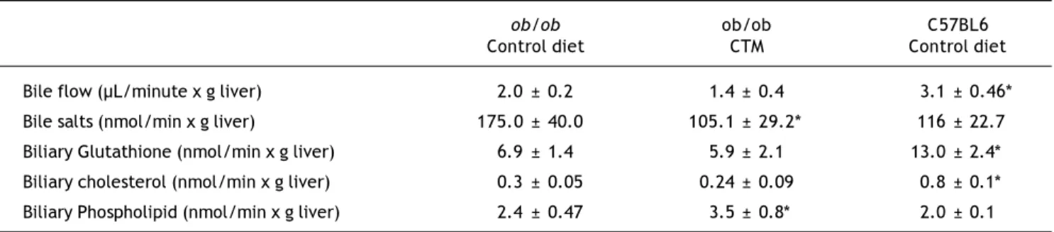
Table 2. Effect of oral administration of cholestyramine (CTM) on hepatobiliary parameters in ob/ob mice.
ob/ob ob/ob C57BL6
Control diet CTM Control diet
Bile flow (µL/minute x g liver) 2.0 ± 0.2 1.4 ± 0.4 3.1 ± 0.46* Bile salts (nmol/min x g liver) 175.0 ± 40.0 105.1 ± 29.2* 116 ± 22.7 Biliary Glutathione (nmol/min x g liver) 6.9 ± 1.4 5.9 ± 2.1 13.0 ± 2.4* Biliary cholesterol (nmol/min x g liver) 0.3 ± 0.05 0.24 ± 0.09 0.8 ± 0.1* Biliary Phospholipid (nmol/min x g liver) 2.4 ± 0.47 3.5 ± 0.8* 2.0 ± 0.1
Experimental groups are described in Material and Methods. Data are mean ± SD (n = 4-6).*p < 0.05 compared with chow-fed ob/ob mice. Table 1. Effects of oral administration of cholestyramine (CTM) on body/liver weight and serum parameters in ob/ob mice.
ob/ob ob/ob C57Bl6
Control diet CTM Control diet
Body weight (g) 45.1 ± 1.9 42.7 ± 1.3 24.1 ± 1.1*
Liver weight (g) 2.1 ± 0.16 2.1 ± 0.2 1.1 ± 0.04*
Body/liver weight 21.3 ± 1.0 20.9 ± 1.6 21.9 ± 1.3
ALT (IU/L) 102.7 ± 26.9 327.4 ± 86.1* 47.5 ± 21.6*
Serum bile acids (µmol/L) 11.4 ± 4.4 32.9 ± 4.6* 7.6 ± 0.1 Serum triglycerides (mg/dL) 35.3 ± 9.9 34.6 ± 2.7 36.4 ± 4.0 Serum cholesterol (mg/dL) 156.5 ± 25.3 168 ± 14.4 87.3 ± 9.3* Serum Insulin (ng/mL) 3.0 ± 0.8 3.5 ± 1.9 0.49 ± 0.1*
RESULTS
Body and liver weight, serum biochemistry, hepatic triglyceride content and liver histology of chow-fed obese mice
Ob/ob mice exhibit a greater body and liver weight compared to normal C57BL6 at week 12 of life and increased serum levels of ALT, insulin and cholesterol (Table 1). Liver histology assessment at different time points (4, 7, 8 and 12 week-old mice) showed that steatosis development started at week 8, reaching maximum severity at week 12 as it has
been previous described.20 In line with this finding,
hepatic triglyceride content in 12 weeks-old obese mice was significantly higher compared to C57BL6 mice (111 ± 19 mg/g liver vs. 12.5 ± 4.5 mg/g liver, p < 0.05).
Effect of oral administration of CTM on steatosis in ob/ob mice
Feeding 4 week-old ob/ob mice during 8 weeks a chow diet supplemented with CTM was not associa-ted with changes in body weight, liver weight and serum levels of triglycerides, cholesterol and insulin (Table 1). No differences were found in the grades
assigned by the pathologist to steatosis or inflam-mation in livers from the experimental groups. Of note, serum ALT levels were significant increased by three-fold in CTM-fed mice.
Hepatobiliary parameters were also determined to better explore the effects of experimental diet and gi-ven the relationship between obesity, insulin resis-tance and altered bile secretory function described
in the literature by our group and others.21,22 As
shown in table 2, 12 weeks-old ob/ob mice exhibited cholestasis with a significantly reduced bile flow. Administration of CTM decreased biliary excretion of bile acids and increased biliary phospholipid ex-cretion. Besides, ob/ob mice showed a significant de-crease in biliary glutathione excretion which was not affected by CTM feeding.
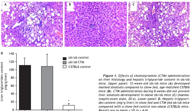
Representative histological evaluation of liver samples after 8 weeks of experimental diet revealed that administration of CTM had no effect on steato-sis development in obese ob/ob mice (Figure 1). De-termination of hepatic triglyceride content revealed that obese mice had a nine-fold increase in this va-riable compared to non-obese mice. CTM adminis-tration had no influence on liver triglycerides (Figure 1).
Figure 2 shows results obtained in studies of glu-cose tolerance (GTT) in different experimental
Figure 1. Effects of cholestyramine (CTM) administration on liver histology and hepatic triglyceride content in ob/ob mice. Upper panel: 12-week-old ob/ob mice (A) developed marked steatosis compared to chow-fed, age-matched C57Bl6 mice (B). CTM administration during 8 weeks did not prevent liver steatosis development in obese ob/ob mice (C) (haema-toxylin-eosin stain, 20 x). Lower panel: D. Hepatic triglyceri-des content (mg/g liver) in chow-fed and CTM-fed ob/ob mice compared with a chow-fed control non-obese (C57BL6) mice. Results are as mean ± SD (n = 4-6).
140
120
100
80
60
40
20
0
Liver triglycerides content
(mg/g liver)
A B C
ob/ob control ob/ob CTM C57BL6 control
groups. Analysis of the area under the curve of the different groups shows that ob/ob mice exhibited a marked intolerance to glucose compared with the control group (one-tail significance p = 0.01) and CTM was able to significantly reduce this parameter after 8 weeks of treatment (p = 0.01).
Effect of oral administration of CTM on hepatic gene expression
Assessment of the expression of genes that are known to be regulated by BAS such as CYP7A1 and SHP is shown in figure 3. As expected, CTM feeding was associated with a marked induction of Cyp7a1 and to a reduction of Shp mRNA levels, which is consistent with an effect of deactivation of the nu-clear receptor FXR determined by a decrease in the bile acid pool size. On the other hand, CTM did not influence the expression of genes involved in hepa-tic bile acid transport (Ntcp and Bsep), lipogenesis (Srebp-1c) or carbohydrate metabolism (Pepck).
DISCUSSION
The use of BAS using resins such as CTM or cole-sevelam has been proposed as a potential new ap-proach to the management of T2DM based in both clinical and experimental studies showing that BAS is able to lower plasma glucose and glycosylated he-moglobin levels as well as to improve tissue glucose
metabolism.23,25 However, long-term effects of BAS
in T2DM patients are still insufficiently characteri-zed. Also, specifically with regard to NAFLD, the use of BAS has not been well addressed. The latter is relevant since the prevalence of NAFLD in T2DM patients is considerable with a higher prevalence of
NASH, the aggressive form of the disease.26 Thus,
BAS therapeutic potential as well as safety in this specific patient population needs to be explored. In this context, the present study evaluated the hepatic effects of CTM on NAFLD development in
leptin-deficient ob/ob mice, a widely used obese mouse
model of NAFLD.
The results of this work show that BAS did not produce any positive effects on liver steatosis deve-lopment in the ob/ob mice. Neither histology nor hepatic triglyceride content was influenced by CTM administration. Our data is in agreement with
observations made in other animal studies.27,28
Indeed, Herrema, et al.27 reported that BAS are able
to induce the lipogenic pathway in an FXR- and
LXRα-dependent manner leading to the increase of
liver triglyceride droplets. Other studies have observed
Figure 3. Effects of cholestyramine (CTM) administration on hepatic expression of selected genes: Na+
-taurocholateco-transporting polypeptide (Ntcp); bile salt export pump (Bsep); Cholesterol 7 a-hydroxylase (Cyp7a1); sterol regula-tory element binding transcription factor 1 (Srebp-1c); Phos-phoenolpyruvate carboxykinase (Pepck); short heterodimeric partner (Shp). Results are means ± SE (n=4-6). * p<0.05 com-pared to ob/ob control.
Figure 2. Effects of cholestyramine (CTM) administration on glucose tolerance in ob/ob mice. A. Plasma glucose con-centrations during the intraperitoneal glucose tolerance test (1 g/kg) following fasting for 12 h in C57BL6, control ob/ob and ob/ob treated with CTM mice. Results are means ± SD (n = 4-6). B. Areas under curves were calculated using the trapezoidal rule. * One-tail significance p = 0.01.
600
400
200
0
Serum glucose
(mg/dL)
0 30 60 90 120
Time (minutes)
2.8
2.4
2.0
1.6
1.2
0.8
0.4
0.0
RQ/18S
Ntcp Bsep Cyp7a1 Pepck Srebp-1c Shp ob/ob control ob/ob CTM C57BL6 mice
*
*
ob/ob control ob/ob CTM C57BL6 mice 15
10
5
0
AUC
(g/dL 120 min)
*
*
ob/ob control ob/ob CTM C57BL6 mice A
beneficial effects of BAS on hepatic steatosis in
gene-tically diabetic mice.29,30 These differences could be
explained in part by differences in the experimen-tal models. It has been established that BAS produ-ce malabsorption of dietary fatty acids in the
intestine due to impaired micelle formation.28 Since
Matsumoto, et al. and Kobayashi, et al. fed mice a high-fat diet, CTM might have prevented the absorp-tion of the fat content of the diet and therefore an important part of the caloric input. On the other hand, it could be possible that the effect of CTM in diabetic mice fed chow diet, as used in the Herrema, et al. and Meissner, et al. reports and in the current study, is lost since carbohydrates are the major part of the composition of this diet. It is known that high intake of carbohydrates in obese and diabetic pa-tients may lead to increase liver steatosis through the activation of de novo lipogenesis in
hepato-cytes.31 Thus, it is suggested that the action of BAS
in liver steatosis may depend on the macronutrients composition of the diet. The use of genetic rather than dietary models is likely more accurate to test the effects of BAS on hepatic steatosis.
Noteworthy, 8 weeks of CTM treatment produced a significant increase in serum levels of ALT and bile acids implying the occurrence of liver injury in ob/ob mice. These observations are in line with a re-cent report in humans where colesevelam treatment increased serum ALT levels, being this outcome as-sociated with a modest increase in liver fat content when measured by a sensitive magnetic resonance
technique.32 These authors suggest that BAS may
lead to a compensatory increase in bile acid synthe-sis and fatty acid synthesynthe-sis in the liver although we did not observed changes in these two compensatory mechanisms judging by gene expression analysis. A recent study in mice had shown that CTM feeding tended to increase hepatic levels of cytotoxic bile acids such as chenodeoxycholic acid and lithocholic acid and also decrease hepatic levels of the
hydro-philic bile acid beta-muricholic acid.33 These
chan-ges in the bile acid pool might be related to liver damage in an injury-prone organ such as the
steato-tic liver.34 Thus, if long-term CTM administration is
harmful in the setting of hepatic steatosis warrants further study.
The current work also found a significant impro-vement in glucose tolerance in CTM-treated mice in agreement with previous reports. In this regard, it has been proposed that BAS may act increasing the metabolic glucose clearance by peripheral tissues
while hepatic glucose output remains unaffected.28
Also, some studies have demonstrated that BAS are
able to induce the expression of GLP-1 by
stimula-tion of L-cells in the terminal ileum or colon.35,36
Fi-nally, it has been recently suggested that increased plasma cholecystokinin concentrations induced by BAS treatment may contribute to postprandial
glu-cose control via a delay in gastric emptying.37 These
mechanisms could explain the beneficial effects ob-served by CTM in the present study on reducing blood glucose levels. Why the improvement in GTT does not translate in fatty liver improvement is un-clear. We aimed to explore this assessing the hepatic gene expression of selected relevant genes. CTM treatment evoked the expected effects on mRNA levels of Cyp7a1 and Shp, likely due to a deactivation of the nuclear receptor FXR. Decreased activation of SHP could result in relative activation of liver
receptor homolog-1 and liver-x receptor α, which
ultimately results in increased triglyceride synthesis. Of note, no changes were seen in the expression of major bile acid transporters Ntcp and Bsep after CTM feeding.
Among the limitations of the present study it should be kept in mind that, in spite of the
agree-ment with some human data,32 these findings
cannot be directly extrapolated to humans since mice and men exhibit multiple differences in biliary
physiology.38,39 Also, giving the emerging role of
bile acids in energy metabolism the explanation of the dissociation between the positive impact on glucose metabolism and the lack of effect in the liver could be related to effects in other organs such as brown adipose tissue and skeletal muscle which could have a role in amelioration of glucose
metabolism.40
In summary, BAS with CTM reduces glucose plas-ma levels in obese, diabetic mice. However, the re-sults obtained in this work demonstrate that BAS does not ameliorate hepatic steatosis and can also exert some deleterious effects in the liver in some circumstances. Thus, our findings and those from
Le, et al.32 suggest that BAS might not be beneficial
for the treatment of NAFLD and its long-term safe-ty in this regard needs to be addressed. This is par-ticularly important considering the proposed role of BAS in the treatment of diabetic patients a popula-tion at-risk of liver complicapopula-tions due to the increa-sed prevalence of NAFLD and its potentially
progressive form NASH.26,41
ABBREVIATIONS
• ALT: serum alanine aminotransferase.
• BSEP: bile salt export pump. • CTM: cholestyramine.
• CYP7A1: cholesterol 7-alpha-hydroxylase. • GLP-1: glucagon-like peptide-1.
• GTT: glucose tolerance test.
• IR: insulin resistance.
• NAFLD: non-alcoholic fatty liver disease. • NASH: non-alcoholic steatohepatitis.
• NTCP: Na/Taurocholate Cotransporting Poly-peptide.
• SHP: small heterodimer partner.
• SREBP-1C: sterol regulatory element-binding protein-1c.
• T2DM: type 2 diabetes mellitus.
• TGR-5: G protein-coupled bile acid receptor 1.
FINANCIAL SUPPORT
This study was carried out with support of grants from the Fondo Nacional de Investigación Científica y Tecnológica (FONDECYT, project 1110455) and the Comisión Nacional de Investigación, Ciencia y Tecnología (CONICYT, project ACT 79).
REFERENCES
1. Chalasani N, Younossi Z, Lavine JE, Diehl AM, Brunt EM, Cusi K, Charlton M, et al. The diagnosis and management of non-alcoholic fatty liver disease: practice guideline by the American Gastroenterological Association, American Association for the Study of Liver Diseases, and American College of Gastroenterology. Gastroenterology 2012; 142: 1592-609.
2. Kleiner DE, Brunt EM. Nonalcoholic fatty liver disease: pathologic patterns and biopsy evaluation in clinical research. Semin Liver Dis 2012; 32: 3-13.
3. Bellentani S, Marino M. Epidemiology and natural history of non-alcoholic fatty liver disease (NAFLD). Ann Hepatol 2009; 8(Suppl. 1): S4-S8.
4. Mendez-Sanchez N, Arrese M, Zamora-Valdes D, Uribe M. Current concepts in the pathogenesis of nonalcoholic fatty liver disease. Liver Int 2007; 27: 423-33.
5. Feldstein AE. Novel insights into the pathophysiology of nonalcoholic fatty liver disease. Semin Liver Dis 2010; 30: 391-401.
6. Trauner M, Arrese M, Wagner M. Fatty liver and lipotoxici-ty. Biochim Biophys Acta 2010; 1801: 299-310.
7. Cusi K. Nonalcoholic fatty liver disease in type 2 diabetes mellitus. Curr Opin Endocrinol Diabetes Obes 2009; 16: 141-9.
8. Younossi ZM, Gramlich T, Matteoni CA, Boparai N, McCullough AJ. Nonalcoholic fatty liver disease in patients with type 2 diabetes. Clin Gastroenterol Hepatol 2004; 2: 262-5. 9. Arrese M. Nonalcoholic fatty liver disease: liver disease:
an overlooked complication of diabetes mellitus. Nat Rev Endocrinol 2010; 6: 660-1.
10. Younk LM, Davis SN. Evaluation of colesevelam hydrochlo-ride for the treatment of type 2 diabetes. Expert Opin Drug Metab Toxicol 2012; 8: 515-25.
11. Zarrinpar A, Loomba R. Review article: the emerging terplay among the gastrointestinal tract, bile acids and in-cretins in the pathogenesis of diabetes and non-alcoholic fatty liver disease. Aliment Pharmacol Ther 2012. 12. Harach T, Pols TW, Nomura M, Maida A, Watanabe M,
Auwerx J, Schoonjans K. TGR5 potentiates GLP-1 secre-tion in response to anionic exchange resins. Sci Rep 2012; 2: 430.
13. Arrese M, Pizarro M, Solis N, Accatino L. Adaptive regula-tion of hepatic bile salt transport: role of bile salt hydro-phobicity and microtubule-dependent vesicular pathway. J Hepatol 1997; 26: 694-702.
14. Ibanez P, Solis N, Pizarro M, Aguayo G, Duarte I, Miquel JF, Accatino L, et al. Effect of losartan on early liver fi-brosis development in a rat model of nonalcoholic steato-hepatitis. J Gastroenterol Hepatol 2007; 22: 846-51. 15. Talalay P. Enzymic analysis of steroid hormones. Methods
Biochem Anal 1960; 8: 119-43.
16. Turley SD, Dietschy JM. Re-evaluation of the 3 alpha-hydroxysteroid dehydrogenase assay for total bile acids in bile. J Lipid Res 1978; 19: 924-8.
17. Anderson ME. Determination of glutathione and glutathione disulfide in biological samples. Methods Enzymol 1985; 113: 548-55.
18. Carr TP, Andresen CJ, Rudel LL. Enzymatic determination of triglyceride, free cholesterol, and total cholesterol in tissue lipid extracts. Clin Biochem 1993; 26: 39-42. 19. Ozcan U, Yilmaz E, Ozcan L, Furuhashi M, Vaillancourt E,
Smith RO, Gorgun CZ, et al. Chemical chaperones reduce ER stress and restore glucose homeostasis in a mouse mo-del of type 2 diabetes. Science 2006; 313: 1137-40. 20. Candia R, Riquelme A, Baudrand R, Carvajal CA, Morales M,
Solis N, Pizarro M, et al. Overexpression of 11beta-hydroxysteroid dehydrogenase type 1 in visceral adipose tissue and portal hypercortisolism in non-alcoholic fatty li-ver disease. Liver Int 2012; 32: 392-9.
21. Geier A, Dietrich CG, Grote T, Beuers U, Prufer T, Fraun-berger P, Matern S, et al. Characterization of organic anion transporter regulation, glutathione metabolism and bile formation in the obese Zucker rat. J Hepatol 2005; 43: 1021-30.
22. Pizarro M, Balasubramaniyan N, Solis N, Solar A, Duarte I, Miquel JF, Suchy FJ, et al. Bile secretory function in the obese Zucker rat: evidence of cholestasis and al-tered canalicular transport function. Gut 2004; 53: 1837-43.
23. Beysen C, Murphy EJ, Deines K, Chan M, Tsang E, Glass A, Turner SM, et al. Effect of bile acid sequestrants on gluco-se metabolism, hepatic de novo lipogenesis, and choleste-rol and bile acid kinetics in type 2 diabetes: a randomised controlled study. Diabetologia 2012; 55: 432-42.
24. Holst JJ, McGill MA. Potential new approaches to modifying intestinal GLP-1 secretion in patients with type 2 diabetes mellitus: focus on bile acid sequestrants. Clin Drug Inves-tig 2012; 32: 1-14.
25. Ooi CP, Loke SC. Colesevelam for type 2 diabetes mellitus. Cochrane Database Syst Rev 2012; 12: CD009361.
26. Smith BW, Adams LA. Nonalcoholic fatty liver disease and diabetes mellitus: pathogenesis and treatment. Nat Rev Endocrinol 2011; 7: 456-65.
28. Meissner M, Herrema H, van Dijk TH, Gerding A, Havinga R, Boer T, Muller M, et al. Bile acid sequestration reduces plasma glucose levels in db/db mice by increasing its me-tabolic clearance rate. PLoS One 2011; 6: e24564. 29. Matsumoto K, Yokoyama S. Gene expression analysis on
the liver of cholestyramine-treated type 2 diabetic model mice. Biomed Pharmacother 2010; 64: 373-8.
30. Kobayashi M, Ikegami H, Fujisawa T, Nojima K, Kawabata Y, Noso S, Babaya N, et al. Prevention and treatment of obesity, insulin resistance, and diabetes by bile acid-bin-ding resin. Diabetes 2007; 56: 239-47.
31. Browning JD, Baker JA, Rogers T, Davis J, Satapati S, Bur-gess SC. Short-term weight loss and hepatic triglyceride reduction: evidence of a metabolic advantage with dieta-ry carbohydrate restriction. Am J Clin Nutr 2011; 93: 1048-52.
32. Le TA, Chen J, Changchien C, Peterson MR, Kono Y, Patton H, Cohen BL, et al. Effect of colesevelam on liver fat quantified by magnetic resonance in nonalcoholic stea-tohepatitis: a randomized controlled trial. Hepatology 2012; 56: 922-32.
33. Zhang Y, Klaassen CD. Effects of feeding bile acids and a bile acid sequestrant on hepatic bile acid composition in mice. J Lipid Res 2010; 51: 3230-42.
34. Yang SQ, Lin HZ, Lane MD, Clemens M, Diehl AM. Obesity increases sensitivity to endotoxin liver injury: implications for the pathogenesis of steatohepatitis. Proc Natl Acad Sci USA 1997; 94: 2557-62.
35. Shang Q, Saumoy M, Holst JJ, Salen G, Xu G. Coleseve-lam improves insulin resistance in a diet-induced obesi-ty (F-DIO) rat model by increasing the release of GLP-1. Am J Physiol Gastrointest Liver Physiol 2010; 298: G419-G424.
36. Zema MJ. Colesevelam hydrochloride: evidence for its use in the treatment of hypercholesterolemia and type 2 dia-betes mellitus with insights into mechanism of action. Core Evid 2012; 7: 61-75.
37. Marina AL, Utzschneider KM, Wright LA, Montgomery BK, Marcovina SM, Kahn SE. Colesevelam improves oral but not intravenous glucose tolerance by a mechanism indepen-dent of insulin sensitivity and beta-cell function. Diabetes Care 2012; 35: 1119-25.
38. Hofmann AF, Hagey LR. Bile acids: chemistry, pathoche-mistry, biology, pathobiology, and therapeutics. Cell Mol Life Sci 2008; 65: 2461-83.
39. Arrese M, Ananthanarayanan M. Mice and men: are they bile-ologically different? Hepatology 2001; 33: 1551-3.
40. Zarrinpar A, Loomba R. Review article: the emerging interplay among the gastrointestinal tract, bile acids and incretins in the pathogenesis of diabetes and non-alcoho-lic fatty liver disease. Aliment Pharmacol Ther 2012; 36: 909-21.