R E V I S I Ó N
Recibido: 16-03-2012 Aceptado: 11-10-2012
Correspondence to: Luis Jesuino de Oliveira Andrade - Rua Nações Unidas, 511 - Centro Zip code: 45.600-673 - Itabuna - Bahia - Brazil
Telephone: +55 (73) 3212-1511 luis_jesuino@yahoo.com.br
Molecular Markers in Thyroid Cancer
Marcadores moleculares en el cáncer de tiroides
Andrade LJO 1,Melo PRS 1,Neto WA1,Andrade CS1,Margotto MAS1,França LS1,Bittencourt AMV 2
1Department of Health - Santa Cruz State University, Ilhéus, Brazil. 2University of Bahia - School of Medicine, Salvador, Brazil
ABSTRACT
Advances in thyroid molecular biology studies are providing not only insight into thyroid development of diseases but may offer diagnostic and therapeutic potential in thyroid cancer.
Objective: To describe the molecular aspects of thyroid cancer reported in international scientific literature, as well as their indications and epidemiological impact, through a descriptive bibliographical review. Method: Articles were searched in the EMBASE, and PubMed/Medline databases. Inclusion criteria were: publication between 1985 and 2012; keywords “molecular” AND “markers” AND “thyroid” AND “cancer”. With these documents in hands, an analytical reading was done and the papers organized by themes. Results: Out of the 609 documents found, 85 were selected to present information on several molecular, epidemiological, and practices-related aspects of thyroid cancer. Articles were assessed and classified accord-ing to predetermined categories, especially number and types of markers analyzed. Among the most relevant information, we can mention the indications for use of molecular markers in thyroid cancer, which are found in more than 70 % of papillary and follicular thyroid carcinomas. The use of these markers will probably improve the diagnosis of malignancy in thyroid nodules and it may also allow individualization of surgical therapy and postoperative management.
Conclusion: This study shows the importance of knowledge of molecular aspects of thyroid cancer, which allows identification of the correlation between specific mutations and phenotypic characteristics of thyroid cancer, which is essential for its prognosis. Rev Argent Endocrinol Metab 50:84-98, 2013
No financial conflicts of interest exist.
Key words: molecular genetics, carcinogen markers, thyroid cancer
RESUMEN
Los avances en biología molecular de la tiroides no solo proporcionan una visión sobre el desarrollo de la enfermedad de la tiroides, sino que además pueden ofrecer posibilidades diagnósticas y terapéuticas para el cáncer de tiroides.
Objetivo: Describir los aspectos moleculares del cáncer de tiroides presentados en la literatura científica internacional, así como sus indicaciones y el impacto epidemiológico, a través de una revisión bibliográfica descriptiva.
folicu-lar de la tiroides. El empleo de estos marcadores probablemente mejore el diagnóstico de malignidad en los nódulos tiroideos, y quizás permita individualizar el tratamiento quirúrgico y el seguimiento posoperatorio. Conclusión: Este estudio muestra la importancia del conocimiento de aspectos moleculares del cáncer de tiroides, que permite la identificación de la correlación entre las mutaciones específicas y las características fenotípicas del cáncer de tiroides, esencial para su pronóstico. Rev Argent Endocrinol Metab 50:84-98, 2013
Los autores declaran no poseer conflictos de interés.
Palabras clave: genética molecular, marcadores cancerígenos, cáncer de tiroides
1. INTRODUCTION
In the last decades, we have seen major develop-ments in molecular biology and genetics, and the accumulation of new data offers the promise of im-portant future clinical applications. The advances in thyroid molecular biology studies are providing not only insight into thyroid development of the diseases but too may offer an accurate diagnosis, and therapeutic potential in the thyroid cancer(1). One of the most important objectives of genetic markers of thyroid cancer is nable accurate diag-nosis, the possible identification of individuals at greatest risk in order to allow better management and prognosis.
Thyroid cancer is the most prevailing endo-crine malignancy, and its incidence presents a growing tendency during the last decades in the all world. The thyroid gland presents a wide spectrum of tumors derived from follicular cells that range from well differentiated, papillary and follicular carcinoma, usually carrying a good prognosis, to the clinically aggressive, poorly differentiated and undifferentiated thyroid car-cinoma. The genetic predisposition to thyroid cancer appears to consist of a variety of genetic mutations of low to moderate penetrance genes, interacting with each other and with the envi-ronment. In thyroid cancer there is a dramatic difference in the incidence, aggressiveness, and death rate by gender, given that the thyroid cancer is 2-4 times more frequent in women than men, and the molecular basis for gender disparity is poorly understood(2).
The carcinogen markers in thyroid gland are found in more than 70 % of papillary and follicu-lar thyroid carcinomas, and will likely improve the diagnosis of malignancy in thyroid nodules as well as treatment individualized surgical, and postoperative appropriate follow-up.
The goal of paper was to conduct a descriptive bibliographical review addressing the molecular aspects of thyroid cancer, emphasizing the
impor-tance of the new molecular biology techniques in diagnosis, and prognosis of thyroid cancer.
2. METHOD
2.1. Sample delimitation
The following international journal databases were reviewed: EMBASE and PubMed/Medline. Selection of these databases was based on the wide range of journals covered by each of them and our goal was to provide an overview of the scientific production devoted to the topic over the timeframe under analysis. The following inclusion criteria were considered during the review: articles published between January 1985 and July 2012; use of the keywords “molecular” AND “markers” AND “thyroid” AND “cancer” entered into the ad-vanced search form; and availability of an abstract in English. Articles were assessed and classified according to predetermined categories, especially types of markers analyzed. With these documents in hands, an analytical reading was done and the papers organized by themes.
3. RESULTS AND DISCUSSION
Out of the 609 documents found, 85 studies were selected due they contained information about the several molecular biology aspects of thyroid cancer.
3.1. Thyroid cancer
equal measure. The mortality are two times higher in female subjects(3,4).
Majority of thyroid carcinomas are derived of follicular cells, and are classified as well differen-tiated, poorly differentiated and undifferentiated thyroid carcinoma. The diagnosis of thyroid cancer is made on the basis of a biopsy of a thyroid nodule or after the nodule removal by surgery. According to the histological features the thyroid cancers are divided into papillary carcinoma, follicular carcinoma, medullary thyroid carcinoma (origi-nate from parafolicullar cells), anaplastic thyroid carcinoma (derived from follicular epithelial cells), primary lymphoma of the thyroid, and primary sarcoma of the thyroid(5). Mutations and epigenetic alterations in poorly differentiated and undifferen-tiated thyroid carcinoma are far from being totally clarified. Whereas that poorly differentiated and undifferentiated thyroid carcinoma may derive from well differentiated thyroid carcinomas, it is expected that some poorly differentiated and undifferentiated thyroid carcinomas would target genetic alterations that are typical of papillary and follicular carcinoma.
The papillary carcinoma represent 80 % of all thyroid malignancies, followed by follicular carcinoma that represents approximately 15 %, medullary thyroid 3 %, and anaplastic thyroid carcinoma 2 % carcinoma of cases, while primary lymphoma of the thyroid, and primary sarcoma of the thyroid are very rare(6,7).
Several factors influence the etiology of cancer thyroid drawing attention the genetic aspects and environmental factors interaction in individuals at risk. The main risk factors for thyroid cancer are: gender and age; geographic and ethnic vari-ability; environmental factors emphasizing previ-ous exposure to ionizing radiation, age at the time of irradiation, previous history of benign thyroid disease, iodine in the food, body mass index, and hormonal factors. Genetic factors are also included emphasizing: familial history and associated dis-eases such as familial polyposis of colon, Cowden’s disease, and Carney’s complex; genetic alterations, detected by molecular biology studies mainly the activation of oncogenes such as serine/threonine-kinase B-type Raf serine/threonine-kinase (BRAF), rat sarcoma viral oncogene (RAS), rearranged during trans-fection (RET), and neurotrophic tyrosine kinase receptor type I (NTRK1), the silencing of tumor suppressor genes such as phosphatase and tensin homologue deleted on chromosome ten (PTEN),
and the tumor protein p53 gene (TP53), that will be discussed later(8).
Clinically unrecognized thyroid cancer has been seen in 4-35.6 % of autopsies, which is far greater than prevalence of diagnosed cancer (9). The major-ity of thyroid cancer diagnosis is incidental, and rise in your incidence it’s influenced by diagnostic technology and medical surveillance practices. Af-ter the discovery of a thyroid nodule by palpation or incidentally by imaging method the diagnosis of thyroid cancer is made on the basis of a biopsy by fine needle aspiration or after the nodule removal by surgery(10).
Generally, treatments include: surgery, radioac-tive iodine treatment, thyroid hormone suppres-sion therapy(11).
Individuals with progressive differentiated thyroid cancer that are not responsive to standard treatment require additional therapy. The neck dissection, external beam radiation, and chemo-therapy should be considered in the setting of metastatic disease especially if cancer threatens vital neck structures(12).
Several kinase inhibitors, especially those tar-geting vascular endothelial growth factor receptors (VEGFR) and/or RET have already demonstrated promising activity in differentiated and medullary thyroid cancers. Recently new pharmacological agents (Axitinib, Motesanib Diphosphate, Pazo-panib, Sorafenib, Sunitinib, Thalidomide, and Lenalidomide) for the treatment of advanced thyroid cancer have emerged, whose mechanism of action would be block constitutive activation of the mitogen-activated protein kinase (MAPK) and/ or phosphatidylinositol-3-kinase (PI3K) pathway and VEGFR(13).
Tools such as molecular genetics to detect the aggressiveness of the cancer, can aid the recommendation of most appropriate therapy. However, the surgery remains the appropriate treatment, with later judicious use of radioac-tive iodine.
3.2. Molecular biology in thyroid cancer
Various molecular mutational alterations have been discovered in thyroid cancer in the last years, emphasizing by example BRAF and RAS genes (KRAS, HRAS, NRAS) as well as RET-PTC and receptor tyrosine kinases (TRK) rear-rangements, all of which are able to activate the MAPK pathway are found in papillary thyroid carcinoma (PTC), and RAS mutations or paired box gene 8-peroxisome proliferator/activated receptor gamma (PAX8-PPARγ) rearrangement are identified in follicular carcinomas. The lipid kinase phosphatidylinositol-3-kinase (PI3K/ AKT) signaling pathway occurs in higher preva-lence in less differentiated thyroid carcinomas. Additional mutations involve the TP53 and catenin β 1 (CTNNB1) genes occur in poorly differentiated and anaplastic carcinomas, and point mutations located in the RET gene occur in medullary thyroid carcinomas, both familiar and sporadic. Some of the markers including galectin-3, Hector Battifora Mesothelial cell (HBME-1), human telomerase reverse transcrip-tase (hTERT), telomerase, microRNA, and mi-croarray and multigene assays have shown great promise as an adjunct to lesions with cellular atypia, suspicious cytology, and demonstrating a follicular pattern(14-18).
The major molecular alterations known in thy-roid cancer are being introduced into pathology practice, although no one marker has proven to be a panacea. However, the use of molecular markers is expected to improve significantly the accuracy of cancer diagnosis and allow more individualized surgical and postsurgical management of patients with thyroid cancer.
3.3. Molecular mechanisms of thyroid cancer
3.3.1. Model of thyroid cancer
Two theories have been described in thyroid cancer pathogenesis, the fetal cell carcinogenesis theory and the more common, multistep carci-nogenesis theory. The multistep carcicarci-nogenesis theory is now the accepted model in many hu-man cancers, including thyroid cancer. The early events of tumor formation are the consequence of activation of either various growth factors or the proto-oncogenes. This results in the formation of differentiated thyroid cancers like the papillary, follicular or Hürthle cell cancers, and anaplast carcinoma(19) (Figure 1).
The molecular genetic basis of thyroid carcino-genesis is not well understood. Mouse models have contributed to elucidate thyroid tumorigenesis and provide tools to develop novel diagnostic ap-proaches and therapies. There are three general approaches for inducing tumorigenesis: carcino-genic compounds, implantation of tumor cells via a subcutaneous or orthtotopic route in immu-nocompromised mice, or genetically engineered models(20,21).
Over the past decade, genetically engineered mouse models of thyroid cancer facilitate analy-sis of the roles of specific genetic mutations in thyroid tumorigenesis. Mouse models, as previ-ously mentioned, have consistently shown that RET-PTC rearrangements can initiate thyroid carcinogenesis(22). Several molecular forms have been identified that differ according to the 5´ partner gene involved in the rearrangement, with RET-PTC1 and RET-PTC3 being the most common. Transgenic RET-PTC3 mice develop PTC, similar to the human solid variant of PTC. Likewise, FVB/N strain transgenic mice expressing thyroid-targeted RET-PTC1 tyrosine kinase consistently developed bilateral PTC with similar histopathologic features to the human disease(23). However, RET-PTC mouse models have expanded our ability to understand the initiators of PTC(24).
A genetically engineered mouse model of follicu-lar thyroid carcinoma was created by introduction of a dominant-negative mutation, although this mouse model offers a reasonably good recapitula-tion of human follicular carcinoma arising from hyperplastic goiter, it differs in that these mice have elevated circulating thyroid hormones levels due to production by the tumor cells, while human follicular thyroid carcinomas are infrequently functional(25).
Experimentally, anaplastic thyroid carcinomas were developed in transgenic mice Tg-SV40 LT, however few survived long enough to develop metastases, while invasion of the trachea led to dyspnea and death in 90 % with 22 weeks of age(26).
Experimental studies in transgenic mice from a single CGRP-RETC634R model of medullary carci-noma, developed papillary carcinomas, which were interpreted as evidence that activated RET can transform thyroid follicular cells in addition to C cells, highlighting the role of modifier genes in dis-ease phenotype whose results can be unexpected(27). Thus, it was demonstrated that the mutation of the RET proto-oncogene can lead to constitutive activation in neuroendocrine cells, such as thyroid C cells, leading to medullary thyroid carcinoma and/or the MEN syndromes in humans.
One clinical question that may be addressed by mouse models is how much thyrotropin (TSH) contributes to limiting aggressive thyroid cancer progression, because higher TSH levels also led to increased oncogene expression which is not similar to human papillary thyroid cancer. Molecular stud-ies in this direction are under way(28).
Therefore, mouse models of thyroid cancer are important not only to elucidate thyroid car-cinogenesis, as also as for developing therapeutic interventions.
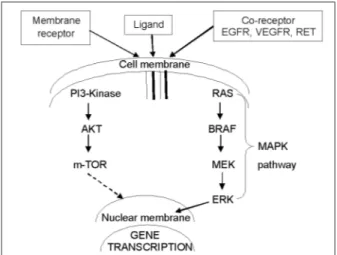
3.3.2. Signaling pathways in thyroid cancer
Mitogenic signaling pathways have been described in the thyroid cells, which are influenced by vari-ous stimulatory and inhibitory hormones, growth factors and neurotransmitters. At a molecular level, the signaling pathway plays a critical role in thyroid cancer development and progression.
The main signaling pathways involved in thy-roid carcinogenesis are the MAPK and PI3K-AKT pathways however several other signaling path-ways are currently being tested for thyroid mo-lecular signaling and cancer cell biology (Figure 2). The MAPK cascade is a classical conserved intracellular signaling pathway involved in the regulation of cellular proliferation, differentiation, apoptosis and survival. Constitutive activation and deregulation of this pathway has been found in numerous human malignancies. Evidence of MAPK activation is most compelling in thyroid cancer development. The activation of MAPK signaling pathway in the thyroid cancer is initi-ated by growth factor binding to receptor tyrosine
kinases that dimerize and the result this activation is propagated by autophosphorylation of tyrosine residues in the intracellular compartment(29). The MAPK signaling pathways play an important role in transmission of cell signals through transduc-tion systems to cell nucleus, where they influence the expression of genes that regulate important cellular processes including cell growth, prolifera-tion, apoptosis, and differentiaprolifera-tion, already men-tioned above. The MAPK pathway seems to play an important role in thyroid cancer being activated by genetic events involving RAS and BRAF(30).
The PI3K are a family of related intracellular signal transducer enzymes capable of phosphory-lating the inositol ring of phosphatidylinositol. While the AKT protein family plays an important role in mammalian cellular signaling. The forma-tion of PI3K/AKT pathway is fundamental in regu-lating cell growth, proliferation, and survival(31).
Genetic alterations involving the PI3K/AKT signaling pathway occurs in thyroid tumors, es-pecially in less differentiated thyroid carcinomas, are rare in well-differentiated thyroid cancer(32).
Therefore, determining of regulatory pathways in thyroid cancer progression at primary and meta-static sites, apart from signaling pathways quoted, represents a challenge important, and Identify key mechanisms of cancer progression remains a key goal in advancing therapeutic options for patients with thyroid cancer.
3.4. Molecular diagnostics of thyroid cancer
uses the information to distinguish between nor-mal, precancerous, and cancerous tissues at the molecular level.
The detection of mutations and other diagnostic markers in thyroid cancer must be by to those labo-ratory techniques that provide highly accurate, re-producible, and clinically relevant information and the molecular techniques currently contribute to the diagnosis. The expression of certain oncogenes is related to thyroid carcinogenesis and tumor pro-gression. Molecular studies performed in the last years, implemented by new methodologies, have elucidated in part the molecular mechanisms un-derlying thyroid cancer. The application of molecu-lar methods has led to the development of highly sensitive markers for the diagnosis of new cases of thyroid cancer, in the evaluation of patients for recurrent disease, also to determining prognosis, and for early detection of residual and recurrent disease in postoperative follow-up of patients. For these reasons, the application of molecular analysis to better characterize the thyroid nodule has been an area of intense interest for studies.
The main molecular markers of thyroid can-cer are mainly represented by the activation of oncogenes such as BRAF, RAS, NTRK1 and the silencing of tumor suppressor genes such as PTEN and TP53, PI3K/AKT, CTNNB1, RET-PTC and PAX8-PPARγ, besides mutation in the thyrotro-pin receptor (TSHR). Based upon several studies, molecular markers can potentially improve the diagnostic accuracy of thyroid cancer, as will be described hereinafter.
3.4.1. MicroRNAs and thyroid cancer
MicroRNAs is a class of endogenous small noncod-ing RNA molecules that regulate gene expression, and play a significant role in regulate major pro-cesses, such as cell differentiation, proliferation/ growth, mobility and apoptosis in diverse cancer-related biological processes. Current data support its use as a diagnostic tool of highly accurate and prognosis definition for thyroid cancer(2). The thyroid carcinomas are characterized by aberrant expression of microRNAs, which are non-coding RNAs of 20-24 nucleotides in length evolutionarily conserved and control gene expression at the post-transcriptional level(33). Currently, several studies identified microRNA signatures in thyroid carci-nomas for to evaluate how they modulate thyroid cancer progression.
Microarray profiling is a technique that has been used to systematically detect the differential expression of microRNAs in cancer and control samples, but different melting temperatures of short-length microRNAs and the high sequence consistency between microRNA family members can lead to false positive microarray results(34).
The microRNA deregulation plays role impor-tant in thyroid cancer development. The role of microRNAs in papillary thyroid carcinoma were analyzed in some studies coming to the conclusion that the upregulation of five specific microRNAs, particularly miR-146b, miR-221, and miR-222, and the subsequent downregulation of KIT receptor seem to be involved in papillary thyroid carcinoma pathogenesis, and sequence changes in microRNA target genes can contribute to their regulation(35).
There are few studies evaluating the role of microRNAs in follicular thyroid carcinoma, where were investigated if microRNAs are differentially expressed between human follicular thyroid carci-nomas and follicular adecarci-nomas testing two high-density microRNA expression arrays(36). Another study compared microRNA expression profiles of principal types of thyroid cancers, showing that miR-155, miR-187, miR-221, miR-222, and miR-224 resulted in being highly over expressed in conventional follicular thyroid carcinoma and, miR-183, miR-187, miR-197, miR-221, miR-222, while miR-339 were over expressed in the follicular thyroid carcinoma oncocytic variants(37).
Few studies have analyzed the role of microR-NAs in anaplastic thyroid carcinoma. It was analyzed the miRNA expression of anaplastic thyroid carcinoma using a micrRNA microarray chip, finding a significant down-expression of miR-26a, miR-30a-5p, miR-30d, and miR-125b in anaplastic thyroid carcinoma compared to normal thyroid samples(38). More recently, was identified two miRNAs, miR-30 and miR-200, which were significantly down-expressed in anaplastic thy-roid carcinoma suggesting that miR-200 as a key factor of the epithelial phenotype in cancer cells that could be used as therapeutic agent to reduce the rates of invasive and metastatic thyroid car-cinomas(39).
Moreover, microRNAs are possible targets for a therapy of cancer by positively modulation of the expression of specific microRNAs using expression vectors and/or inhibiting microRNA expression by transfecting specific 35).
3.4.2. Molecular markers of thyroid cancer
· RET-PTC: The RET proto-oncogene, located on
chromosome 10q11.2, encodes a membrane recep-tor of extracellular domain with a tyrosine kinase activity. RET exhibits an oncogenic potential and plays a important role in thyroid human cancers. In PTC, genomic rearrangements juxtapose the RET kinase and COOH-terminus encoding do-mains (exons 11–21) to unrelated genes, thereby creating dominantly transforming oncogenes called RET/PTC(40).
RET-PTC oncogenes are believed to play an important role in the pathogenesis of a significant subset of PTC, in particular those arising after radiation exposure, and in pediatric cancers (41). There at least 15 different RET-PTC rearrange-ments, the most of rearrangements do not seem to have an important role in the pathogenesis of PTC, however the RET-PTC1 and RET-PTC3 are the most common ones. RET-PTC1 is formed by fusion with the H4 gene, and RET-PTC3 by fusion with the ELE1 gene(40-42).
RET-PTC is involved in the pathogenesis of thyroid cancer through of the activation of this oncogene at early stages of tumor development, thyroid specific everexpression of either RET-PTC1 or RET-PTC3 in transgenic mice model indicating that these oncoproteins can recreate the PTC, ionizing radiation results in expression of RET-PTC in short time, suggesting a direct role for radiation in the illegitimate recombination of RET, and direct double-strand DNA breakage re-sulting in illegitimate reciprocal recombination(43). RET-PTC rearrangement was described in thyroid tumours associated with radiation exposure this was observed in the contaminated areas after Chernobyl explosion with a higher prevalence of RET-PTC1(44).
The different types of RET-PTC chromosomal rearrangements, between the 3’ portion of the RET gene and the 5’ portion of an unrelated gene produce an aberrant RET-PTC protein with ligand-independent activation of its intracellular tyrosine kinase domain. This phenomenon has been frequently described in small PTC and can
to be an early event in thyroid tumorigenesis. The variant aggressive PTC subtype, is closely associ-ated with the RET-PTC3 oncogene, whereas the classic variant is associated with RET-PTC1(42,45).
RET/PTC rearrangements have so far been identified only in thyroid lesions, and in particular in PTC. Most studies show that RET-PTC rear-rangements are rare or absent in benign follicular adenomas, and absent in follicular and medullary carcinomas. Somatic rearrangements of the RET proto-oncogene have been detected in 3-60 % of sporadic PTC. The prevalence of RET-PTC in PTC varies significantly in different studies, in most series with sporadic PTC, RET-PTC1 is the most common type, comprising up to 60-70 % of the rearrangements, whereas RET-PTC3 accounts for 20-30 %(46).
RET-PTC oncogene relationship with radiation exposure has been established, the chromosomal basis for its activation has been clarified trans-forming and signaling properties have been char-acterized in the thyroid cancer.
· NTRK1: The NTRK1 proto-oncogene is
locat-ed on the q arm of chromosome 1 and encodes the high affinity transmembrane receptor for nerve growth factor. Constitutive activation of NTRK1 has been detected in several tumors types. The fusion of the NTRK1 TK domain to 5′ sequences with at least three different genes (TPM3 gene, TPR gene and TFG gene), leads to oncogenic activation of NTRK1. Somatic rearrangements of NTRK1, producing chimeric oncogenes (activated version of the proto-oncogene was generated by a somatic intrachromosomal rearrangement fusing the tyrosine kinase domain of NTRK1 with 5¢ sequences of the non-muscular tropomyosin gene) with constitutive tyrosine kinase activity, has been frequently found in a consistent fraction of PTC(47). However, PTC arising in patients with a history of exposure to elevated levels of ionizing irradia-tion do not carry these known abnormalities. In the same way NTRK1 rearrangements are rare in cases of sporadic papillary thyroid carcinomas(48). It was observed that the frequency of NTRK1 rear-rangements in post-Chernobyl papillary thyroid tumors is equivalent to that of sporadic tumors(49).
carcinomas, showing the involvement of NTRK1 and of p75 in the etiopathogenesis of papillary thyroid carcinomas(50).
Study carried out to characterize the sequence of the genomic regions involved in the NTRK1 activating rearrangements, showed that the dif-ferent breakpoints occurred in intronic regions of genes, with presence of some recombinogenic ele-ments including palindromes, direct and inverted repeats and Alu sequences(51).
The somatic rearrangements of the NTRK1 gene in papillary thyroid carcinomas has frequency does that not exceeds 12 %. However, studies Ital-ian showed a frequency of NTRK1 activation in spontaneous papillary thyroid carcinomas ranging from 15 to 25 %, whereas French and Japanese studies the frequency was less than 5 %. This dif-ference may be the consequence of geographical factors(49,52).
Therefore, rearrangements of NTRK1 provide a useful model for studying the molecular basis of thyroid carcinogenesis, although occurring less frequently that other oncogenes.
· PTEN: The PTEN was identified in 1997 as
a tumor-suppressor gene located on 10q23.3, is a member of the protein tyrosine phosphatase family and, following activating mutations or amplifications of the genes encoding the effectors proteins of PI3K/AKT pathway, inhibits PI3K signaling, thereby reducing the level of activated AKT. Structurally, PTEN protein is composed of an N-terminal dual specificity phosphatase-like enzyme domain and a C-terminal regulatory do-main, which binds to phospholipid membranes (53). The dysregulation of PI3K/AKT signaling pathway contributes for many cancers in man with action antiapoptotic. Recent findings have demonstrated that PTEN also plays a critical role in DNA dam-age repair and DNA damdam-age response (54). The importance of PTEN catalytic activity in its tumor suppressor function is underscored by fact that the majority of PTEN missense mutations detected in tumor specimens target the phosphatase domain and, cause a loss in PTEN phosphatase activity. These data suggest that genetic loss of PTEN is sufficient to induce thyroid cancer in vivo.
The role of PTEN as a tumor suppressor gene has been well established in thyroid cancer, and 3 %-10 % of individuals with differentiated thyroid cancer are carrying a germ line PTEN mutation, whereas mutations or deletions in PTEN are rare
events in sporadic thyroid cancer(55). Perhaps the strongest evidence supporting a role for PI3K signaling in the development of thyroid cancer is Cowden syndrome, where the thyroid cancer can be identified in approximately two-thirds of affected individuals for this syndrome(56).
In this way, the oncogenic power of loss of PTEN seems to lie in its cooperation with genetic alterations of the PI3K/AKT pathway in the tu-morigenesis of thyroid cancer.
· TP53: The TP53 gene is located on
chromo-some 17p13.1 and consist of 11 exons, coding for a nuclear phosphoprotein which can bind to specific DNA sequences, and acts as a transcription factor. The TP53 is recognized as a tumor-suppressor gene because it encodes protein p53 participat-ing in the processes of cell-cycle arrest in the G1 phase of the cell cycle via DNA repair, and also in apoptosis(57). Inactivation of TP53 in immortalised cells results in a marked instability of chromosome structure, including translocations, deletions, telo-meric associations, and ring chromosomes(58). The TP53 gene is frequently affected by loss of alleles and by point mutations in almost all cancers. A principle of this function for cancer progression is provided by the fact that mice without functional TP53 gene breed and develop (almost) normally but die at an early age from multiple cancers(59). Over 20,000 alterations in TP53 have been dis-covered in human tumors, and the role of TP53 in cancer has been studied, and the presence of a TP53 mutation may be predictive of the tumor response to treatment and patient survival(60).
About half of patients with anaplastic cancer have a previous or coexistent differentiated thyroid carci-noma, with evidence of dedifferentiation from more differentiated tumors often associated with loss of the TP53 tumor suppressor protein(61).
Therefore, TP53 encoding p53 plays a central role in the response to DNA damage through its ability to activate DNA repair, arrest cell cycle progression or induce apoptosis, and thus controls the genomic stability an important factor to avoid malignant transformation of cells.
· PI3K/AKT: There are several classes of PI3Ks,
among which class I is the best characterized and most important in human tumorigenesis. The PI3K is a lipid kinase what generates a messen-ger essential for the translocation of AKT to the plasma membrane. Akt is a Ser/Thr kinase and three types of its isoforms are found in human tis-sues AKT-1, AKT-2, and AKT-3, being that AKT-1, and AKT-2 are the most abundant and important in thyroid cancer. Activation of AKT plays a pivotal role fundamental in cellular functions such as cell proliferation and survival by phosphorylating a variety of substrates. The PI3K/AKT signaling pathway PI3K/AKT play an important role in transmission of cell signals through transduction systems to cell nucleus, where they influence the expression of genes that regulate important cellular processes: cell growth, proliferation and apoptosis. Therefore, a major consequence of acti-vating PI3K/AKT signaling is the inhibition of cell cycle inhibitors. Genetic and epigenetic alterna-tions, concerning PI3K/AKT signaling pathways, contribute to their activation and interaction in consequence of malignant cell transformation. Thus, the alterations to the PI3K/AKT signaling pathway are frequent in human cancer(62).
The genetic alterations in the PI3K/AKT path-way, represents major advance in understand-ing the molecular mechanism of thyroid cancer. Studies showed that the PIK3CA amplification is associated with overexpression of the PIK3CA protein and phosphorylation of AKT, suggesting that this genetic alteration of the PIK3CA gene is functional and represents a relevant mechanism for the activation of the PI3K/AKT pathway in thyroid cancer(63). Other studies show that aber-rant activation of the pathway plays a fundamen-tal role in thyroid tumorigenesis, particularly in follicular thyroid cancer and aggressive thyroid cancer, such as anaplastic thyroid cancer(64).
Genetic alternations involving the PI3K/AKT pathway has been found in thyroid cancers such as anaplastic thyroid cancer and metastatic tumors from radioactive iodine refractory papillary thyroid cancer, particularly in the later stages of cancer progression(16). Genetic alterations involving the PI3K/AKT signaling pathway in thyroid tumors are smaller in well-differentiated thyroid cancer and have higher prevalence in less-differentiated thyroid carcinomas. The PI3K/AKT signaling pathway alterations in thyroid cancer occurs in approximately 7 % of follicular thyroid cancer, and 15 % of anaplastic thyroid carcinoma(8).
· CTNNB1 (Catenin β-1): The CTNNB1 is a
dominantly acting cancer gene located on chro-mosome 3p22-p21.3 is composed of 16 exons, and has two main functions in cell regulation - as a cadherin-mediated adhesion regulator and as a mediator of WNT/CTNNB1 signaling.
The CTNNB1 play a crucial role in carcino-genesis, transcriptional targets of over-expressed CTNNB1 were observed in malignant neoplasm but were absent in the case of benign thyroid tis-sues. Mutation of the CTNNB1 results in disrup-tion of a large number of cellular funcdisrup-tions that may be important in various forms of carcinomas development.
The CTNNB1 exon 3 mutations and nuclear catenin β-1 localization are found almost exclu-sively in poorly differentiated and anaplastic car-cinomas, were demonstrated frequently in 0-25 % of poorly differentiated thyroid carcinomas and up to 66 % of undifferentiated thyroid carcinomas, but not in well-differentiated thyroid carcino-mas. These observations indicate that CTNNB1 mutation contributes to progression towards poorly differentiated or undifferentiated thyroid carcinomas(17).
It has been reported that CTNNB1 is related with the activation of the WNT-signaling path-way. The name WNT is a portmanteau of Wg and Int, which in turn are the abbreviations for two homologous genes: Wg (Wingless) and Int (Integration). The WNT-signaling pathway plays a number of crucial roles in the development of organism. Malfunctions of this pathway lead to various diseases including cancer.
stabilizing mutations and/or aberrant CTNNB1 nuclear localization are present in 80 % of anaplas-tic thyroid carcinoma. CTNNB1 nuclear localiza-tion is accompanied by its cellular redistribulocaliza-tion with marked decrease of the CTNNB1 membrane bound fraction(65).
Activation of WNT signaling involves the inhi-bition of CTNNB1 degradation by proteasomes, which leads to its accumulation in the nucleus. Mutations in components that regulate CTNNB1 turnover lead to an excessive accumulation of CTNNB1 in the nucleus. This nuclear accumula-tion can result in an adequately high concentraaccumula-tion of CTNNB1 in the nucleus of tumor cells(66).
The reduction of CTNNB1 bound to the cell sur-face has been demonstrated in thyroid carcinoma, the biological and clinical relevance of CTNNB1 dysregulation in thyroid neoplasia is primarily unknown(67).
Therefore, CTNNB1 exon 3 mutations shows interesting differences between poorly and un-differentiated tumors, and can be a marker for aggressive tumor phenotypes among neoplasms of thyroid follicular cell derivation. Thus, the analysis of CTNNB1 expression or mutation status may be very useful to objectively subtype thyroid neoplasms and more accurately predict diagnosis.
· PAX8-PPARγ: The fusion of the PAX8 gene
with the PPARγ gene arises of t(2;3)(q13;p25) translocation, where the fusing the 5′ portion of the thyroid-specific transcription factor gene PAX8 in frame with the entire translated reading frame of the PPARγ nuclear receptor gene, resulting in strong overexpression of the chimeric PAX8-PPARγ protein (42).
The pathogenesis by which PAX8-PPARy causes follicular thyroid tumor is not definite clearly. Opinions differ as to how the fusion product PAX8-PPARy is oncogenic. The initial mechanism prob-able would that the fusion protein can function as a dominant-negative suppressor of PPARy-induced gene transcription which confers anti-apoptotic properties, and that the fusion protein would retain at least some functional properties of its individual components(68). Other mechanism would that the fusion product can disruptor PAX8 tran-scriptional activity resulting in deregulation of expression of its target thyroid-specific genes such as thyroglobulin, thyroperoxidase, and sodium-iodide symporter, where transcriptional
activa-tion of target genes by recruiting transcripactiva-tional coactivators to cognate DNA-binding sites in a ligand- dependant manner. A third mechanism would be activation of genes not related either to the wild-type PPARγ or to the PAX8 pathways(69).
PAX8-PPARγ rearrangement has been found in 30% to 40% of follicular carcinomas, in 2%-13% of follicular adenomas, and with lower prevalence in oncocytic carcinomas suggesting these injuries to be in situ forms of follicular carcinomas(42). PAX8-PPARγ has been found in the follicular variant of papillary carcinoma, with a reported prevalence as high of 38 %, although in most populations, the prevalence of PAX8-PPARγ in this tumor type is much lower, probably less than 5%, and its prevalence in childhood is observed in about one-fifth of patients(70).
Tumors expressing PAX8/PPARc rearrange-ments occur at a younger age and are small in size with a solid growth pattern or nests and more frequently have vascular invasion(42,71).
Ultimately, PAX8-PPARγ accelerates cell growth, reduces rates of apoptosis and permits anchorage independent and contact uninhibited growth of a thyroid cell line.
· RAS: The RAS mutations are common in
The RAS proteins exist in an inactive form that is bound to guanosine diphosphate and an active form that exhibits guanosine triphosphatase activity. The RAS transmits signals originating from tyrosine kinase membrane receptors to a cascade of MAPK, activating the transcription of target genes involved in cell proliferation, survival, and apoptosis(75).
The RAS mutation occurs in approximately 10-20% of papillary cancers, 25-30% of poorly dif-ferentiated carcinoma, and 40-50% of anaplastic carcinoma and 40-50% of follicular carcinoma(42).
Therefore, the RAS family oncogenes are im-portant regulators of cell growth and have a role in thyroid tumor differentiation.
· BRAF: The BRAF is located on chromosome
7q24 and encodes a serine–threonine kinase, playing a critical role in cell signaling and are de-pendent of the MAPK signaling pathway, and has been implicated in human cancers, which occurs in approximately 40-45 % of papillary cancers, 10-15% of poorly differentiated carcinoma, and 20-30% of anaplastic carcinoma(42). Thus it, BRAF has occupied a central role in thyroid cancer patho-genesis since the original identification of BRAF mutations in papillary and anaplastic thyroid cancer. Activated versions of BRAF can also be gen-erated by intrachromosomal inversions that fuse the kinase domain of BRAF to the NH2-terminal portion of AKAP9, resulting in BRAF-AKAP fusion proteins are similar in structure to the RET/papil-lary thyroid cancer fusion proteins, and are found in about 11% of patients whose thyroid cancers are thought to be caused by the Chernobyl nuclear power station disaster in 1986(76).
Moreover, it is known that BRAF mutations are associated with impaired iodine trapping through the sodium iodine symporter, decreased thyroid hormone synthesis and increased TSH in vivo(30). A BRAF point mutation at codon 600 results in a valine to glutamate (V600E) has in consequence the activation of BRAFV600E resulting in a continu-ous phosphorylation of MAPK pathway effectors, and the BRAFV600E mutation is the most common genetic alteration found in thyroid cancer(77). Oth-ers rare activating mutations of BRAF described in 1% to 2 % of papillary carcinomas are the K601E point mutation, small in-frame insertion or deletion surrounding codon 600 and determining a lysine-glutamic substitution, and the AKAP9-BRAF rearrangement that is associated with previous radiation exposure(78).
BRAFV600E mutation may serve as a prognostic
marker for papillary thyroid carcinoma and has been found to be how independent predictor of treatment failure and with higher probability of tumor recurrence, even in patients with low-stage disease(78).
There is evidence that BRAF mutation testing of thyroid carcinoma might improve the diagnosis, prognostic stratification and treatment of these tu-mors, however, studies still are needed to unravel the etiologic factors in the formation of BRAF point mutations in thyroid cancer. The BRAF mutation is a prognostic marker, and can be detected pre-operatively by fine-needle-aspirated cytological materials, thereby improving risk stratification and recurrence prediction of thyroid cancer and distant metastasis(79).
Studies to evaluate the utility of ultrasound-guided fine-needle aspiration biopsy in the ad-ditional contribution of BRAF V600E mutation analysis in the detection of differentiated thyroid cancer have been performed, and the results shows a diagnostic sensitivity for thyroid cancer significantly increased by BRAF V600E mutation analysis, indicating that the screening for BRAF mutation in fine-needle aspiration biopsy samples has a relevant diagnostic potential(80).
· TSHR: The TSHR regulates thyroid growth
and differentiation at late developmental stages. TSHR molecules in the membrane are quite stable and signalling in the thyrocyte will be controlled mainly through circulating TSH levels. The ma-ture TSHR is encoded by a single gene with 10 exons. The protein contains 2 subunits, a large ectodomain also called α subunit encoded by ex-ons 1-8 and binds TSH, and intracellular domain encoded by exons 9-10 called β subunit that will interact with G proteins to initiate signaling (81). Hence, excesses or defaults in TSHR activity may play a role important in the onset, evolution, of thyroid cancer by mechanisms that including im-proper epigenetic marking of the gene, incorrect transcriptional regulation or mutations in critical domains.
thy-roid carcinomas in hyperfunctioning autonomous nodules, with a handful of those being autonomous hyperfunctioning thyroid carcinomas(82). Thus, the question of whether or not TSHR mutations involved in the onset of thyroid cancers is contro-versial. Distinct mutations in the TSHR gene have been reported, but so far none of these mutations appear to be related to the onset of thyroid cancer. Reduced TSHR expression in thyroid cancers may be secondary to ongoing de-differentiation, may happen in parallel or may cause disappearance of other differentiation markers(83).
The TSHR signalling for the onset and evolu-tion of thyroid cancer is supported by experiments with xenotransplanted cells of follicular thyroid cancer cell line(84). The TSHR coupling to G pro-teins provides the opportunity for activation of the PKA, PKC and PI3K pathways, but mutations in G proteins hardly correlate with thyroid cancers, and hot thyroid nodules with constitutively active TSHR mutants(85).
The expansion of knowledge about genetic mutations occurring in different thyroid tumors has been characterized recent years, allowing the identification of a correlation between specific mutations and phenotypic characteristics of thy-roid cancers, essential for their prognosis. Specific mutations can be reliably detected by newer tools such as molecular techniques in thyroid tissue samples that may aid clinicians in recommending the most appropriate therapy.
REFERENCES
1. Davies TF, Latif R, Minsky NC, Ma R. Clinical review: The emerging cell biology of thyroid stem cells. J Clin Endocrinol Metab. 96(9):2692-702, 2011 2. de la Chapelle A, Jazdzewski K. MicroRNAs in thy-roid cancer. J Clin Endocrinol Metab. 96(11):3326-36, 2011
3. Edwards BK, Brown ML, Wingo PA, Howe HL, Ward E, Ries LA, Schrag D, Jamison PM, Je-mal A, Wu XC, Friedman C, Harlan L, Warren J, Anderson RN, Pickle LW. Annual report to the nation on the status of cancer, 1975-2002, featuring population-based trends in cancer treatment. J Natl Cancer Inst. 97(19):1407-27, 2005
4. Harris PJ, Bible KC. Emerging therapeutics for advanced thyroid malignancies: rationale and tar-geted approaches. Expert Opin Investig Drugs. 20(10):1357-75, 2011
5. Schmid KW. Molecular pathology of thyroid tumors. Pathologe. 31(Suppl 2):229-33, 2010
6. Cooper DS, Doherty GM, Haugen BR, Kloos RT,
Lee SL, Mandel SJ, Mazzaferri EL, McIver B, Sherman SI, Tuttle RM; American Thyroid As-sociation Guidelines Taskforce. American Thy-roid Association Guidelines Taskforce. Management guidelines for patients with thyroid nodules and differentiated thyroid cancer. Thyroid. 16(2):109-42, 2006
7. De Lellis RA, Lloyd RV, Heitz PU, Eng C. Eds., World Health Organization International Classifica-tion of Tumors. Pathology and Genetics of Tumors of Endocrine Organs, IARC Press, Lyon, France, 2004 8. Giusti F, Falchetti A, Franceschelli F, Marini
F, Tanini A, Brandi ML. Thyroid cancer: current molecular perspectives. J Oncol. 2010:351679, 2010. 9. Dean DS, Gharib H. Epidemiology of thyroid
nodules. Best Pract Res Clin Endocrinol Metab. 22(6):901-11, 2008
10. Kahn C, Simonella L, Sywak M, Boyages S, Ung O, O’Connell D. Pathways to the diagnosis of thyroid cancer in New South Wales: a population-based cross-sectional study. Cancer Causes Control. 23(1):35-44, 2012
11. DeGroot LJ, Kaplan EL, McCormick M, Straus FH. Natural history, treatment, and course of papil-lary thyroid carcinoma. J Clin Endocrinol Metab. 71(2):414-24, 1990
12. Lessin L. Chemotherapy for Differentiated Papillary or Follicular Thyroid Cancer. Totowa, NJ: Humana Press; 2006
13. Costa AM, Herrero A, Fresno MF, Heymann J, Alvarez JA, Cameselle-Teijeiro J, García-Rostán G. BRAF mutation associated with other genetic events identifies a subset of aggressive pap-illary thyroid carcinoma. Clin Endocrinol (Oxf). 68(4):618-34, 2008
14. Kimura ET, Nikiforova MN, Zhu Z, Knauf JA, Nikiforov YE, Fagin JA. High prevalence of BRAF mutations in thyroid cancer: genetic evidence for constitutive activation of the RET/PTC-RAS-BRAF signaling pathway in papillary thyroid carcinoma. Cancer Res. 63(7):1454-7, 2003
15. Soares P, Trovisco V, Rocha AS, Lima J, Castro P, Preto A, Máximo V, Botelho T, Seruca R, Sobrinho-Simões M. BRAF mutations and RET/ PTC rearrangements are alternative events in the etiopathogenesis of PTC. Oncogene. 22(29):4578-80, 2003
16. Ricarte-Filho JC, Ryder M, Chitale DA, Ri-vera M, Heguy A, Ladanyi M, Janakiraman M, Solit D, Knauf JA, Tuttle RM, Ghossein RA, Fagin JA. Mutational profile of advanced primary and metastatic radioactive iodine-refractory thyroid cancers reveals distinct pathogenetic roles for BRAF, PIK3CA, and AKT1. Cancer Res. 69(11):4885–93, 2009
18. Kato MA, Fahey TJ. 3rd. Molecular markers in thyroid cancer diagnostics. Surg Clin North Am. 89(5):1139-55, 2009
19. Parameswaran R, Brooks S, Sadler GP. Mo-lecular pathogenesis of follicular cell derived thyroid cancers. Int J Surg. 8(3):186-93, 2010
20. Schweppe RE, Klopper JP, Korch C, Pugazhen-thi U, Benezra M, Knauf JA, Fagin JA, Marlow LA, Copland JA, Smallridge RC, Haugen BR. Deoxyribonucleic acid profiling analysis of 40 human thyroid cancer cell lines reveals crosscontamination resulting in cell line redundancy and misidentifica-tion. J Clin Endocrinol Metab. 93(11):4331-41, 2008 21. Ahn SH, Henderson Y, Kang Y, Chattopadhyay
C, Holton P, Wang M, Briggs K, Clayman GL. An orthotopic model of papillary thyroid carcinoma in athymic nude mice. Arch Otolaryngol Head Neck Surg. 134(2):190-7, 2008
22. Santoro M, Chiappetta G, Cerrato A, Salvatore D, Zhang L, Manzo G, Picone A, Portella G, Santelli G, Vecchio G, Fusco A. Development of thyroid papillary carcinomas secondary to tissue-specific expression of the RET/PTC1 oncogene in transgenic mice. Oncogene. 12(8):1821-6, 1996 23. Jhiang SM, Sagartz JE, Tong Q,
Parker-Thorn-burg J, Capen CC, Cho JY, Xing S, Ledent C. Targeted expression of the ret/PTC1 oncogene in-duces papillary thyroid carcinomas. Endocrinology. 137(1):375-8, 1996
24. Powell DJ Jr, Russell J, Nibu K, Li G, Rhee E, Liao M, Goldstein M, Keane WM, Santoro M, Fusco A, Rothstein JL. TheRET/PTC3 oncogene: metastatic solid-type papillary carcinomas in murine thyroids. Cancer Res. 58(23):5523-8, 1998
25. Kaneshige M, Kaneshige K, Zhu X, Dace A, Garrett L, Carter TA, Kazlauskaite R, Pank-ratz DG, Wynshaw-Boris A, Refetoff S, Wein-traub B, Willingham MC, Barlow C, Cheng S. Mice with a targeted mutation in the thyroid hormone beta receptor gene exhibit impaired growth and resistance to thyroid hormone. Proc Natl Acad Sci USA. 97(24):13209-14, 2000
26. Ledent C, Coppee F, Dumont JE, Vassart G, Parmentier M. Transgenic models for prolifera-tive and hyperfunctional thyroid diseases. Exp Clin Endocrinol Diabetes. 104 Suppl 3:43-6, 1996 27. Reynolds L, Jones K, Winton DJ, Cranston A,
Houghton C, Howard L, Ponder BA, Smith DP. C-cell and thyroid epithelial tumours and altered follicular development in transgenic mice express-ing the long isoform of MEN 2A RET. Oncogene. 20(30):3986-94, 2001
28. Knauf JA, Ma X, Smith EP, Zhang L, Mitsutake N, Liao XH, Refetoff S, Nikiforov YE, Fagin JA.. Targeted expression of BRAFV600E in thyroid cells of transgenic mice results in papillary thyroid cancers that undergo dedifferentiation. Cancer Res. 65(10):4238-45, 2005
29. Carter WB, Tourtelot JB, Savell JG, Lilien-feld H. New treatments and shifting paradigms in differentiated thyroid cancer management. Cancer Control. 18(2):96-103, 2011
30. Riesco-Eizaguirre G, Gutiérrez-Martínez P, García-Cabezas MA, Nistal M, Santisteban P. The oncogene BRAF V600E is associated with a high risk of recurrence and less differentiated papillary thyroid carcinoma due to the impairment of Na+/I- targeting to the membrane. Endocr Relat Cancer. 13(1):257-69, 2006
31. Wang Y, Hou P, Yu H, Wang W, Ji M, Zhao S, Yan S, Sun X, Liu D, Shi B, Zhu G, Condouris S, Xing M. High prevalence and mutual exclusivity of genetic alterations in the phosphatidylinositol-3-kinase/akt pathway in thyroid tumors. J Clin En-docrinol Metab. 92(6):2387-90, 2007
32. Hou P, Liu D, Shan Y, Hu S, Studeman K, Con-douris S, Wang Y, Trink A, El-Naggar AK, Tal-lini G, Vasko V, Xing M.. Genetic alterations and their relationship in the phosphatidylinositol 3-ki-nase/Akt pathway in thyroid cancer. Clin Cancer Res. 13(4):1161-70, 2007
33. Fabian MR, Sonenberg N, Filipowicz W.
Regula-tion of mRNA translaRegula-tion and stability by microR-NAs. Annu Rev Biochem. 79:351-79, 2010
34. Li X, Wang Q, Zheng Y, Lv S, Ning S, Sun J, Huang T, Zheng Q, Ren H, Xu J, Wang X, Li Y. Prioritizing human cancer microRNAs based on genes’ functional consistency between microRNA and cancer. Nucleic Acids Res. 39(22):e153, 2011 35. Marini F, Luzi E, Brandi ML. MicroRNA Role
in Thyroid Cancer Development. J Thyroid Res. 2011:407123, 2011
36. Weber F, Teresi RE, Broelsch CE, Frilling A, Eng C. A limited set of human MicroRNA is de-regulated in follicular thyroid carcinoma. J Clin Endocrinol Metab. 91(9):3584-91, 2006
37. Nikiforova MN, Tseng GC, Steward D, Diorio D, Nikiforov YE. MicroRNA expression profiling of thyroid tumors: biological significance and diagnostic utility. J Clin Endocrinol Metab. 93(5):1600-8, 2008 38. Visone R, Pallante P, Vecchione A, Cirombella
R, Ferracin M, Ferraro A, Volinia S, Coluzzi S, Leone V, Borbone E, Liu CG, Petrocca F, Troncone G, Calin GA, Scarpa A, Colato C, Tal-lini G, Santoro M, Croce CM, Fusco A. Specific microRNAs are downregulated in human thyroid anaplastic carcinomas. Oncogene. 26(54):7590-5, 2007
39. Braun J, Hoang-Vu C, Dralle H, Hüttelmaier S. Downregulation of microRNAs directs the EMT and invasive potential of anaplastic thyroid carcinomas. Oncogene. 29(29):4237-44, 2010
40. Grieco M, Santoro M, Berlingieri MT, Melillo RM, Donghi R, Bongarzone I, Pierotti MA, Della Porta G, Fusco A, Vecchio G. PTC is a novel rearranged form of the ret proto-oncogene and is frequently detected in vivo in human thyroid papillary carcinomas. Cell. 60(4):557-63, 1990 41. Santoro M, Melillo RM, Carlomagno F, Fusco A,
Vecchio G. Molecular mechanisms of RET activation in human cancer. Ann N Y Acad Sci. 963:116-21, 2002 42. Nikiforov YE. Molecular Diagnostics of Thyroid
mo-lecular genetics--role of RET/PTC and BRAF in tumor initiation. J Clin Endocrinol Metab. 89(9):4264-6, 2004 44. Bounacer A, Wicker R, Caillou B, Cailleux
AF, Sarasin A, Schlumberger M, Suárez HG. High prevalence of activating RET proto-oncogene rearrangements in thyroid tumors from patients who had received external radiation. Oncogene. 15(11):1263-73, 1997
45. Guarino V, Castellone MD, Avilla E, Melillo RM. Thyroid cancer and inflammation. Mol. Cell Endocrinol. 321(1):94-102, 2010
46. Trovisco V, Soares P, Preto A, Castro P, Máximo V, Sobrinho-Simões M. Molecular genetics of papil-lary thyroid carcinoma: great expectations. Arq Bras Endocrinol Metabol. 51(5):643-53, 2007
47. Pierotti MA, Greco A. Oncogenic rearrangements of the NTRK1/NGF receptor. Cancer Lett. 232(1):90-8, 2006
48. Weier HU, Ito Y, Kwan J, Smida J, Weier JF, Hieber L, Lu CM, Lehmann L, Wang M, Kassa-bian HJ, Zeng H, O’Brien B. Delineating Chro-mosomal Breakpoints in Radiation-Induced Papillary Thyroid Cancer. Genes (Basel). 2(3):397-419, 2011 49. Bounacer A, Schlumberger M, Wicker R,
Du-Villard JA, Caillou B, Sarasin A, et al. Search for NTRK1 proto-oncogene rearrangements in hu-man thyroid tumours originated after therapeutic radiation. Br J Cancer. 82(2):308-14, 2000
50. Russell JP, Powell DJ, Cunnane M, Greco A, Portella G, Santoro M, Fusco A, Rothstein JL. The TRK-T1 fusion protein induces neoplastic transformation of thyroid epithelium. Oncogene. 19(50):5729-35, 2000
51. Butti MG, Bongarzone I, Ferraresi G, Mondel-lini P, Borrello MG, Pierotti MA. A sequence analysis of the genomic regions involved in the re-arrangements between TPM3 and NTRK1 genes producing TRK oncogenes in papillary thyroid car-cinomas. Genomics. 28(1):15-24, 1995
52. Greco A, Miranda C, Pierotti MA. Rearrange-ments of NTRK1 gene in papillary thyroid carci-noma. Mol Cell Endocrinol. 321(1):44-9, 2010 53. Lee JO, Yang H, Georgescu MM, Di Cristofano
A, Maehama T, Shi Y, Dixon JE, Pandolfi P, Pavletich NP. Crystal structure of the PTEN tumor suppressor: implications for its phosphoinositide phosphatase activity and membrane association. Cell. 99(3):323-34, 1999
54. Al-Khouri AM, Ma Y, Togo SH, Williams S, Mustelin T. Cooperative phosphorylation of the tumor suppressor phosphatase and tensin homologue (PTEN) by casein kinases and glycogen synthase kinase 3beta. J Biol Chem. 280(42):35195-202, 2005 55. Nagy R, Ganapathi S, Comeras I, Peterson C,
Orloff M, Porter K, Eng C, Ringel MD, Kloos RT. Frequency of germline PTEN mutations in dif-ferentiated thyroid cancer. Thyroid. 21(5):505-10, 2011
56. Paes JE, Ringel MD. Dysregulation of the phos-phatidylinositol 3-kinase pathway in thyroid neopla-sia. Endocrinol Metab Clin North Am. 37(2):375-87, viii-ix, 2008
57. Lane DP. A death in the life of p53. Nature. 362(6423):786-7, 1993
58. Meyn MS. Chromosome instability syndromes: lessons for carcinogenesis. Curr Top Microbiol Im-munol. 221:71-148, 1977
59. Donehower LA, Harvey M, Slagle BL, McAr-thur MJ, Montgomery CA Jr, Butel JS, Bradley A. Mice deficient for p53 are developmentally normal but susceptible to spontaneous tumours. Nature. 356(6366):215-21, 1992
60. Hainaut P, Hollstein M. p53 and human cancer: the first ten thousand mutations. Adv Cancer Res. 77:81-137, 2000
61. Moretti F, Farsetti A, Soddu S, Misiti S, Cres-cenzi M, Filetti S, Andreoli M, Sacchi A, Ponte-corvi A. p53 re-expression inhibits proliferation and restores differentiation of human thyroid anaplastic carcinoma cells. Oncogene. 14(6):729-40, 1997 62. Brzezianska E, Pastuszak-Lewandoska D. A
minireview: the role of MAPK/ERK and PI3K/Akt pathways in thyroid follicular cell-derived neoplasm. Front Biosci. 16:422-39, 2011
63. Liu Z, Hou P, Ji M, Guan H, Studeman K, Jensen K, Vasko V, El-Naggar AK, Xing M. Highly prevalent genetic alterations in receptor tyrosine kinases and phosphatidylinositol 3-kinase/ Akt and mitogen-activated protein kinase pathways in anaplastic and follicular thyroid cancers. J Clin Endocrinol Metab. 93(8):3106-16, 2008
64. Xing M. Recent advances in molecular biology of thyroid cancer and their clinical implications. Oto-laryngol Clin North Am. 41(6):1135-46, ix, 2008 65. Weinberger PM, Adam BL, Gourin CG, Moretz
WH 3rd, Bollag RJ, Wang BY, Liu Z, Lee JR, Terris DJ. Association of nuclear, cytoplasmic ex-pression of galectin-3 with beta-catenin/Wnt-path-way activation in thyroid carcinoma. Arch Otolar-yngol Head Neck Surg. 133(5):503-10, 2007
66. Jung CK, Jung JH, Lee A, Lee YS, Choi YJ, Yoon SK, Lee KY. Diagnostic use of nuclear beta-catenin expression for the assessment of endometrial stromal tumors. Mod Pathol. 21(6):756-63, 2008 67. Huang SH, Wu JC, Chang KJ, Liaw KY, Wang
SM. Expression of the cadherin-catenin complex in well-differentiated human thyroid neoplastic tissue. Thyroid. 9(11):1095-103, 1999
68. Kroll TG, Sarraf P, Pecciarini L, Chen CJ, Mueller E, Spiegelman BM, Fletcher JA. PAX8-PPARgamma1 fusion oncogene in human thyroid carcinoma [corrected]. Science 289(5484):1474, 2000 69. Reddi HV, McIver B, Grebe SK, Eberhardt NL.
The paired box-8/peroxisome proliferator-activated receptor-gamma oncogene in thyroid tumorigenesis. Endocrinology. 148(3):932-5, 2007
70. Nikiforova MN, Biddinger PW, Caudill CM, Kroll TG, Nikiforov YE. PAX8-PPARgamma re-arrangement in thyroid tumors: RT-PCR and im-munohistochemical analyses. Am J Surg Pathol. 26(8):1016-23, 2002
proliferator-activated receptor gamma rearrangement in fol-licular thyroid tumors. J Clin Endocrinol Metab. 88(9):4440-5, 2003
72. Garcia-Rostan G, Zhao H, Camp RL, Pollan M, Herrero A, Pardo J, Wu R, Carcangiu ML, Costa J, Tallini G. Ras mutations are associated with ag-gressive tumor phenotypes and poor prognosis in thyroid cancer. J Clin Oncol. 21(17):3226-35, 2003 73. Namba H, Rubin SA, Fagin JA. Point mutations
of ras oncogenes are an early event in thyroid tu-morigenesis. Mol Endocrinol. 4(10):1474-9, 1990 74. Giehl K. Oncogenic Ras in tumour progression and
metastasis. Biol Chem. 386(3):193-205, 2005 75. Barbacid M. Ras genes. Annu Rev Biochem.
56:779-827, 1987
76. Ciampi R, Knauf JA, Kerler R, Gandhi M, Zhu Z, Nikiforova MN, Rabes HM, Fagin JA, Nikiforov YE. Oncogenic AKAP9-BRAF fusion is a novel mechanism of MAPK pathway activation in thyroid cancer. J Clin Invest. 115(1):94-101, 2005 77. Lee JH, Lee ES, Kim YS. Clinicopathologic
sig-nificance of BRAF V600E mutation in papillary carcinomas of the thyroid: A meta-analysis. Cancer. 110(1):38-46, 2007
78. Kebebew E, Weng J, Bauer J, Ranvier G, Clark OH, Duh QY, Shibru D, Bastian B, Griffin A. The prevalence and prognostic value of BRAF mu-tation in thyroid cancer. Ann Surg. 246(3):466-70; discussion 470-1, 2007
79. Xing M, Westra WH, Tufano RP, Cohen Y, Rosenbaum E, Rhoden KJ, Carson KA, Vasko V, Larin A, Tallini G, Tolaney S, Holt EH, Hui P, Umbricht CB, Basaria S, Ewertz M, Tufaro AP,
Califano JA, Ringel MD, Zeiger MA, Sidransky D, Ladenson PW. BRAF mutation predicts a poorer clinical prognosis for papillary thyroid cancer. J Clin Endocrinol Metab. 90(12):6373-9, 2005
80. Rossi M, Buratto M, Bruni S, Filieri C, Tagliati F, Trasforini G, Rossi R, Beccati MD, Degli Uberti EC, Zatelli MC. Role of Ultrasonographic/ Clinical Profile, Cytology, and BRAF V600E Muta-tion EvaluaMuta-tion in Thyroid Nodule Screening for Malignancy: A Prospective Study. J Clin Endocrinol Metab. 97(7):2354-61, 2012
81. Loosfelt H, Pichon C, Jolivet A, Misrahi M, Caillou B, Jamous M, Vannier B, Milgrom E. Two-subunit structure of the human thyrotropin receptor. Proc Natl Acad Sci U S A. 89(9):3765-9, 1992 82. Russo D, Arturi F, Schlumberger M, Caillou B,
Monier R, Filetti S, Suárez HG. Activating muta-tions of the TSH receptor in differentiated thyroid carcinomas. Oncogene. 11(9):1907-11, 1995
83. Fricke-Otto S, Pfarr N, Mühlenberg R, Pohlenz J. Mild congenital primary hypothyroidism in a Turkish family caused by a homozygous missense thyrotropin receptor (TSHR) gene mutation (A593 V). Exp Clin Endocrinol Diabetes. 113(10):582-5, 2005
84. Hoffmann S, Maschuw K, Hassan I, Wunderlich A, Lingelbach S, Ramaswamy A, Hofbauer LC, Zielke A. Functional thyrotropin receptor attenu-ates malignant phenotype of follicular thyroid cancer cells. Endocrine. 30(1):129-38, 2006