Predictive value of neutrophil to lymphocyte ratio for the presence of
coronary artery ectasia in patients with aortic aneurysms
Hiroki Ikenaga
a, Satoshi Kurisu
a,⁎
, Noriaki Watanabe
a, Takashi Shimonaga
a, Tadanao Higaki
a,
Toshitaka Iwasaki
a, Hiroto Utsunomiya
a, Naoya Mitsuba
a, Ken Ishibashi
a, Yoshihiro Dohi
a, Yukihiro Fukuda
a,
Katsuhiko Imai
b, Taijiro Sueda
b, Yasuki Kihara
aaDepartment of Cardiovascular Medicine, Hiroshima University Graduate School of Biomedical and Health Sciences, Hiroshima, Japan bDepartment of Cardiovascular Surgery, Hiroshima University Graduate School of Biomedical and Health Sciences, Hiroshima, Japan
a b s t r a c t
a r t i c l e i n f o
Article history:
Received 27 May 2014
Received in revised form 5 August 2014 Accepted 10 August 2014
Available online 15 August 2014
Keywords:
Coronary artery ectasia Aortic aneurysms
Neutrophil to lymphocyte ratio
Background:Both aortic aneurysms and coronary artery ectasia (CAE) frequently coexist and are associated with more pronounced inflammation. Neutrophil to lymphocyte ratio (NL ratio) is widely used as a marker of infl am-mation. However, relation between CAE and NL ratio in patients with aortic aneurysms is not fully understood. This study was undertaken to assess relation between CAE and NL ratio in patients with aortic aneurysms. Methods:This study consisted of 93 consecutive patients with aortic aneurysms (AA group) and 79 patients without aortic aneurysms who had angiographically normal coronary arteries as the control group. Moreover, patients with aortic aneurysms were classified into two groups based on the presence of CAE; CAE (+) group (n = 44) and CAE (−) group (n = 49). We compared blood chemical parameters in both groups.
Results:In the AA group, 44 patients (47.3%) had CAE. The AA group had a significantly higher NL ratio than the control group (2.93 ± 1.43 vs. 2.45 ± 1.05, p = 0.027). Furthermore, the CAE (+) group had a significantly higher NL ratio than the CAE (−) group (3.39 ± 1.67 vs. 2.52 ± 1.04, pb0.01). Multivariate logistic regression analysis revealed that the high NL ratio was an independent predictor for CAE in patients with aortic aneurysms (odds ratio 1.76, 95% confidence interval 1.24–2.69, p = 0.001).
Conclusions:Patients with aortic aneurysms had a significantly higher NL ratio than those without aortic aneu-rysms. Furthermore, the NL ratio might predict the presence of CAE in patients with aortic aneuaneu-rysms. © 2014 The Authors. Published by Elsevier Ireland Ltd. This is an open access article under the CC BY-NC-ND
license (http://creativecommons.org/licenses/by-nc-nd/3.0/).
1. Introduction
Aortic aneurysms are an important cardiovascular disease and a complex disease with both genetic and environmental factors contributing to the disease process. In practice, established risk factors for aortic aneurysms include advancing age, male gender, smoking, hypertension and atherosclerosis[1]. In addition, aortic aneurysms are associated with more pronounced inflammation and elevation of in-flammatory markers in patients with aortic aneurysms is widely recog-nized[2]. Also, coronary artery disease is the most important cause of morbidity and mortality in patients with aortic aneurysms, including during the postoperative period of aortic aneurysms[3]. However, it is difficult to predict morbidity and mortality only by evaluating coronary artery stenosis[4]. On the other hand, it was reported that coronary artery ectasia (CAE) was frequently observed in patients with aortic
aneurysms[5,6]and CAE regardless of aortic aneurysms may be a form of atherosclerosis with more active inflammatory[7–11]. White blood cell (WBC) count and related parameters are markers of infl am-mation in cardiovascular disease[12,13]. Especially, neutrophil to lymphocyte ratio (NL ratio) has been shown to have the greatest predic-tive value for poor outcomes in patients with coronary artery disease [13]. However, the relation between CAE and NL ratio in patients with aortic aneurysms is not fully understood. This study was undertaken to assess the relation between CAE and NL ratio in patients with aortic aneurysms.
2. Methods
2.1. Study patient
In total, 1735 patients underwent coronary angiograms from January 2011 to October 2013 at Hiroshima University Graduate School of Biomedical and Health Sciences. Of these, we excluded patients accompanied with acute coronary syndrome and chronic infl amma-tory disease. In addition, we excluded patients with traumatic, ⁎ Corresponding author at: Department of Cardiovascular Medicine, Hiroshima
University Graduate School of Biomedical and Health Sciences, 1-2-3 Kasumi, Minami-ku, Hiroshima 734-8551, Japan. Tel./fax: +81 82 257 1602.
E-mail address:[email protected](S. Kurisu).
http://dx.doi.org/10.1016/j.ijchv.2014.08.002
2214-7632/© 2014 The Authors. Published by Elsevier Ireland Ltd. This is an open access article under the CC BY-NC-ND license (http://creativecommons.org/licenses/by-nc-nd/3.0/).
Contents lists available atScienceDirect
IJC Heart & Vessels
inflammatory, infective and congenital aortic aneurysms. We have retrospectively analyzed 93 consecutive patients with aortic aneu-rysms (AA group) and 79 patients without aortic aneuaneu-rysms who had angiographically normal coronary arteries and no ischemia on myocardial perfusion scintigram or the treadmill exercise test as the control group (control group). Moreover, patients with aortic an-eurysms were classified into two groups based on the presence of CAE; CAE (+) group (n = 44) and CAE (−) group (n = 49). Coronary angiograms were performed due to the presence of anginal chest pain or the evaluation of coronary artery before aortic aneurysm sur-gery. Angiographically normal coronary arteries were defined as no reduction of the internal diameter of the coronary arteries. Aortic an-eurysms were defined as a circumferential or local enlargement (the diameter was increased to a degree at least 1.5-fold greater than nor-mal (exceeding 45 mm in the thoracic region and 30 mm in the ab-dominal region)) or protrusion of a part of the aortic wall[14,15]. The maximum minor-axis diameter was used in principle for plain and early contrast-enhanced computed tomography images[16]. Es-timated glomerularfiltration rate (eGFR) was calculated using the Japanese equations from serum creatinine, and chronic kidney dis-ease (CKD) was thought to be present if it wasb60 ml/min per 1.73 m2. Informed consent was obtained from each patient. This study was approved by the ethical committee at Hiroshima Universi-ty Graduate School of Biomedical and Health Sciences.
2.2. Laboratory measurements
Blood samples were collected from the ante-cubital vein by an atraumatic puncture just before the coronary angiography in the postabsorptive state and were sent to the central laboratory of our hospital within 1 h after collection. Venous blood was collected in a tube containing K3 EDTA for the measurement of hematologic indices in all patients. Hematologic indices were evaluated from a complete blood count analysis performed at the central laboratory of our hospital.
2.3. Angiographic evaluation
Coronary angiograms were obtained and evaluated according to standard techniques using 4 Fr catheter. Using quantitative coronary an-giographic (QCA) analysis (QCA-CMS v.6.0, Medis, Leiden, NL), coronary artery stenosis was defined as the 50% reduction of the internal diame-ter of the coronary ardiame-teries compared to normal, non-ectatic segments. Coronary artery disease (CAD) severity was assessed by the number of diseased vessels [the right coronary artery (RCA), left anterior descend-ing (LAD), or left circumflex (LCX) coronary artery]. The left main coronary artery (LMCA) was considered a two-vessel disease. Coronary artery ectasia (CAE) was defined as coronary artery with the diameter of the ectatic segment being more than 1.5-fold larger compared with an adjacent healthy reference segment[17,18].
2.4. Statistical analysis
Standard statistical methods were used in this study. Significant dif-ferences were tested using theχ2test for categorical variables. Normally distributed continuous variables were presented as mean and standard deviation (SD). Unpaired Student'sttest or Wilcoxon rank-sum test when appropriate was used for continuous variables. Univariate and multivariate logistic regression analyses were used to identify inde-pendent predictors of the presence of CAE in patients with aortic aneu-rysms, adjusting blood chemical parameters. The univariable predictors with a p value of less than 0.1 were entered into a multivariate model. In addition, to adjust for selection bias, propensity scores for each patient were estimated with logistic regression, with CAE (+) as the outcome. Eighteen baseline clinical variables were chosen for imputation and der-ivation of propensity scores, based on clinical relevance and ability to correct for differences between CAE (+) and CAE (−) groups. The
JMP statistical package (version 11.0, SAS Institute, Inc. Cary, NC, USA) was used for all statistical tests. A significance level of 0.05 was used and two-tailed tests were applied.
3. Results
In the AA group, there were 30 (32.3%) patients with ascending tho-racic aneurysms and 63 (67.7%) patients with descending thotho-racic or abdominal aneurysms. The baseline characteristics of the study patients are shown inTable 1. Age, the frequencies of hypertension and previous myocardial infarction (MI) were significantly higher in the AA group than in the control group (age; 72.8 ± 8.8 years vs. 67.1 ± 10.9 years, pb0.001, hypertension; 82.8% vs. 69.6%, p = 0.04, previous MI; 6.5% vs. 0%, p = 0.02, respectively). The frequencies of diabetes mellitus was significantly lower in the AA group than in the control group (18.3% vs. 35.4%, p = 0.01). There was no significant difference in other baseline clinical variables between the AA group and the control group. The AA group had a significantly higher C-reactive protein level and D-dimer level than the control group (C-reactive protein; 1.03 ± 2.97 mg/dl vs. 0.19 ± 0.29 mg/dl, pb0.01, D-dimer; 6.73 ± 7.67μg/ml vs. 1.03 ± 1.59μg/ml, pb0.01, respectively). The AA group had a significantly higher NL ratio than the control group (2.93 ± 1.43 vs. 2.45 ± 1.05, p = 0.027). There was no significant difference in other blood chemical parameters between the AA group and the control group.
Angiographic characteristics of the study patients are shown in Table 2. Forty-three of 93 patients (46.2%) in the AA group had a signif-icant coronary artery stenosis. In the AA group, the prevalence of coro-nary artery stenosis and CAD severity were significantly higher in the patients with descending thoracic or abdominal aneurysms than those
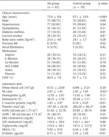
Table 1
Baseline characteristics between the patients with AA and those without AA. AA group
(n = 93)
Control group (n = 79)
p value
Clinical characteristics
Age (years) 72.8 ± 8.8 67.1 ± 10.9 b0.001 Male 75 (80.7%) 55 (69.6%) 0.09 Hypertension 77 (82.8%) 55 (69.6%) 0.04 Dyslipidemia 41 (44.1%) 41 (51.9%) 0.31 Diabetes mellitus 17 (18.3%) 28 (35.4%) 0.01 Current smoker 28 (30.1%) 23 (29.1%) 0.87 Body mass index (kg/m2
) 23.4 ± 4.0 24.1 ± 3.7 0.28 Previous MI 6 (6.5%) 0 (0%) 0.02 Atrialfibrillation 9 (9.7%) 5 (6.3%) 0.42 Medication
Aspirin 28 (30.1%) 19 (24.1%) 0.37
β-Blocker 28 (30.1%) 16 (20.3%) 0.13 Ca-blocker 51 (54.8%) 41 (51.9%) 0.70 ACE-I/ARB 56 (60.2%) 36 (45.6%) 0.06 Statin 28 (30.1%) 23 (29.1%) 0.43 Diuretic 11 (11.8%) 12 (15.2%) 0.52 LVEF (%) 60.4 ± 7.8 61.7 ± 7.2 0.16
Laboratory data
White blood cell (103
/μl) 6721 ± 2258 6396 ± 2121 0.30 NL ratio 2.93 ± 1.43 2.45 ± 1.05 0.027 Hemoglobin (g/dl) 12.95 ± 2.07 13.42 ± 1.90 0.11 Hematocrit (%) 38.33 ± 5.70 39.58 ± 5.11 0.13 C-reactive protein (mg/dl) 1.03 ± 2.97 0.19 ± 0.29 b0.01 Platelet (mm3
/μl) 187.30 ± 62.26 200.29 ± 66.37 0.48 eGFR (ml/min per 1.73 m2
) 61.02 ± 22.78 65.80 ± 28.74 0.08 eGFRb60 ml/min per 1.73 m2
with ascending thoracic aneurysms (coronary artery stenosis; 55.5% vs. 26.7%, pb0.01, CAD severity; 1-/2-/3-vessel disease; 30.1/14.3/11.1% vs. 20.0/6.7/0%, pb0.02). Forty-four of 93 patients (47.6%) in the AA group had CAE. The prevalence of CAE in the AA group was 32.6% in LAD, 26.1% in the LCX and 34.8% in RCA in the AA group. Also, in each
ectatic artery, the prevalence of diffuse CAE was 56.6% in LAD, 87.5% in LCX and 90.6% in RCA.
The baseline characteristics between the CAE (+) group and the CAE (−) group are shown inTable 3. There was no significant difference in the ratio of ascending thoracic aneurysms. Prescription ofβ-blocker was significantly higher in the CAE (+) group than in the CAE (−) group (40.9% vs. 20.4%, p = 0.03). There was no significant difference in other baseline clinical variables between the CAE (+) group and the CAE (−) group. The CAE (+) group had a significantly higher WBC level and triglyceride level than the CAE (−) group (WBC; 7149 ± 1937 103/μl vs. 6338 ± 2469 103/μl, p = 0.04, triglyceride; 147.4 ± 61.4 mg/dl vs. 121.7 ± 64.6 mg/dl, p = 0.02, respectively). Re-lation between NL ratio and CAE in patients with aortic aneurysms is shown inFig. 1. The CAE (+) group had a significantly higher NL ratio than the CAE (−) group (3.39 ± 1.67 vs. 2.52 ± 1.04, pb0.01). Univar-iate and multivarUnivar-iate logistic regression analyses to identify indepen-dent predictors of the presence of CAE in patients with aortic aneurysms are shown inTable 4. Univariate analysis revealed that the NL ratio and triglyceride were associated with CAE in patients with aortic aneurysms (NL ratio; odds ratio [OR] 1.65, 95% confidence inter-val [CI] 1.18–2.44, p = 0.002, triglyceride; OR 1.01, 95% CI 1.01–1.02, p = 0.049, respectively). To detect the independent effect of the NL ratio for the presence of CAE in patients with aortic aneurysms, the NL ratio as well as triglyceride and high-density lipoprotein cholesterol was incorporated to multivariate logistic regression analysis. Multivari-ate logistic regression analysis revealed that the high NL ratio was an independent predictor for CAE in patients with aortic aneurysms (OR 1.76, 95% CI 1.24–2.69, p = 0.001). Logistic regression analysis of the NL ratio for CAE after adjustment for propensity scores is shown in Table 4. After adjusting for ascending thoracic aneurysms, age, male, hy-pertension, dyslipidemia, diabetes mellitus, current smoker, body mass index, previous MI, atrialfibrillation, left ventricular ejection fraction, use of aspirin,β-blocker, Ca-blocker, angiotensin-converting enzyme inhibitor (ACE-I), angiotensin II receptor blocker (ARB), statin and diuretic using propensity scores, the NL ratio was an independent pre-dictor for CAE in patients with aortic aneurysms (OR 1.56, 95% CI 1.10–2.40, p = 0.01).
4. Discussion
The majorfinding of the present study was that patients with aortic aneurysms had a significantly higher NL ratio than those without aortic aneurysms. Furthermore, among the AA group, patients with CAE had a significantly higher NL ratio than those without CAE and the high NL ratio might predict the presence of CAE. To our knowledge, this is the Table 2
Angiographic characteristics between the patients with AA and those without AA. AA group
(n = 93)
Control group (n = 79) Coronary stenosis 43 (46.2%) –
LMCA stenosis 8 (8.6%) –
LAD stenosis 16 (17.2%) –
LCX stenosis 13 (14.0%) –
RCA stenosis 26 (28.0%) –
CAD severity
1-Vessel disease 25 (26.9%) –
2-Vessel disease 11 (11.9%) –
3-Vessel disease 7 (7.5%) –
Coronary ectasia 44 (47.3%) –
LAD ectasia 30 (32.6%) –
Diffuse 17 (18.5%) –
LCX ectasia 24 (26.1%) –
Diffuse 21 (22.8%) –
RCA ectasia 32 (34.8%) –
Diffuse 29 (31.5%) –
LMCA = left main coronary artery, LAD = left anterior descending coronary artery, LCX = left circumflex coronary artery, RCA = right coronary artery, CAD = coronary artery disease.
Table 3
Baseline characteristics between the patients with CAE and those without CAE in the AA group.
CAE (+) group (n = 44)
CAE (−) group (n = 49)
p value
Clinical characteristics
Ascending thoracic aneurysms 11 (25.0%) 19 (38.8%) 0.15 Age (years) 72.5 ± 9.8 73.2 ± 8.0 0.92 Male 36 (81.8%) 39 (79.6%) 0.79 Hypertension 39 (88.6%) 38 (77.6%) 0.15 Dyslipidemia 21 (47.7%) 20 (40.8%) 0.50 Diabetes mellitus 7 (15.9%) 10 (20.4%) 0.31 Current smoker 11 (25.0%) 17 (34.7%) 0.87 Body mass index (kg/m2
) 23.7 ± 3.7 23.1 ± 3.7 0.50 Previous MI 3 (6.8%) 3 (6.1%) 0.89 Atrialfibrillation 6 (13.6%) 3 (6.1%) 0.22 Medication
Aspirin 15 (34.1%) 13 (26.5%) 0.43
β-Blocker 18 (40.9%) 10 (20.4%) 0.03 Ca-blocker 28 (63.6%) 23 (46.9%) 0.11 ACE-I/ARB 31 (70.5%) 25 (51.0%) 0.06 Statin 24 (54.6%) 18 (36.7%) 0.08 Diuretic 5 (11.4%) 6 (12.2%) 0.90 LVEF (%) 60.0 ± 6.9 60.1 ± 8.6 0.55
Laboratory data
White blood cell (103
/μl) 7149 ± 1937 6338 ± 2469 0.04 NL ratio 3.39 ± 1.67 2.52 ± 1.04 b0.01 Hemoglobin (g/dl) 13.25 ± 2.12 12.68 ± 2.00 0.21 Hematocrit (%) 39.27 ± 5.69 37.51 ± 5.65 0.13 C-reactive protein (mg/dl) 1.39 ± 4.01 0.72 ± 1.53 0.99 Platelet (mm3
/μl) 197.71 ± 59.91 177.96 ± 63.44 0.12 eGFR (ml/min per 1.73 m2
) 60.60 ± 26.83 61.39 ± 18.68 0.94 eGFRb60 ml/min per 1.73 m2
17 (38.6%) 24 (50.0%) 0.32 HDL cholesterol (mg/dl) 48.4 ± 13.1 53.2 ± 15.1 0.12 LDL cholesterol (mg/dl) 113.9 ± 28.4 107.1 ± 29.4 0.35 Triglyceride (mg/dl) 147.4 ± 61.4 121.7 ± 64.6 0.02 HbA1c (%) 6.02 ± 0.46 5.85 ± 0.57 0.13 D-dimer (μg/ml) 6.10 ± 7.25 7.34 ± 8.12 0.42 MI; myocardial infarction, ACE-I; angiotensin-converting enzyme inhibitor, ARB; angio-tensin II receptor blocker, LVEF; left ventricular ejection fraction, NL ratio; neutrophil to lymphocyte ratio, eGFR; estimated glomerularfiltration rate, HDL; high-density lipopro-tein, LDL; low-density lipoprolipopro-tein, HbA1c; hemoglobin A1c. P values were two-tailed, and pb0.05 was considered statistically significant.
first report to present a relation between NL ratio and CAE in patients with aortic aneurysms.
Previous studies have shown that there are some differences in the pathobiology between descending thoracic or abdominal aortic aneu-rysms and ascending thoracic aneuaneu-rysms[19,20]. Descending thoracic or abdominal aortic aneurysms are characterized by a decrease in the number of smooth muscle cells in the aortic media layer and fragmenta-tion of the extracellular matrix of the aorta at the site of the aneurysms because of inflammation, tissue remodeling and upregulation of matrix metalloproteinases (MMPs)[21]. Ascending thoracic aneurysms are characterized by medial necrosis, mucoid infiltration, cyst formation with elastin degradation and vascular smooth muscle cell apoptosis [19]. Although there are some differences in the pathobiology between descending thoracic or abdominal aortic aneurysms and ascending thoracic aneurysms, inflammatory response has an influence on the formation of aortic aneurysms[22]. In the present study, the AA group had a significantly higher NL ratio and C-reactive protein level than the control group. The result was conclusive evidence that the inflammation exists in patients with aortic aneurysms and consistent with results of previous studies[23]. On the other hand, the pathophys-iology of CAE remains unclear. However, Virmani et al. had reported that the main histological features of CAE were lipid deposition with foam cells,fibrous caps and extensive destruction of musculoelastic elements of the media[24]. Additionally, several studies reported that both abdominal aortic aneurysms and thoracic aortic aneurysms were associated with CAE and shared common histological mechanisms to develop aneurysms with CAE[5,6]. Same as aortic aneurysms, CAE has atherosclerotic risk factors[1,10]. Although the mechanism of aortic an-eurysms and CAE is variable, patients with aortic anan-eurysms or CAE demonstrated more pronounced inflammation[2,7–11]. The relation between CAE and inflammatory markers in patients with aortic rysms has not been reported. Regardless of the presence of aortic aneu-rysms, CAE was associated with increased inflammatory markers such as C-reactive protein, interleukin-6, tumor necrosis factor-alpha, and MMPs[8–11]. In addition, it was demonstrated that patients with CAE had infiltration of the media layer by inflammatory cells in ectatic coro-nary segments[7]. This may be related to the chronic inflammatory response occurring in CAE. Furthermore, it was reported that CAE was
strongly associated with the NL ratio[25]. This mechanism was that neutrophils cause damage to the tissue and may play an important role in CAE by secreting elastase, MMPs and oxygen free radicals[26, 27]. As a result of this chronic inflammatory response, coronary arterial walls are weakened potentially resulting in CAE. In this study, the CAE (+) group had a significantly higher WBC level and NL ratio than the CAE (−) group. It was suggested that the inflammatory response was stronger in the CAE (+) group than in the CAE (−) group and this may eventually lead to a high WBC level and NL ratio in the CAE (+) group.
Patients with CAE present poor long-term cardiac outcomes[28]. CAE has been shown to decrease coronaryflow velocity[29]. In addi-tion, the extent of CAE was correlated with coronaryflow velocity and associated with a history of myocardial infarction independent of coexisting significant coronary stenosis[28]. And persistent slow coro-nary bloodflow is associated with an increased risk of stent thrombosis [30]. It was reported that preoperative NL ratio appears to be a signifi -cant predictor of both 30-day mortality and long-term outcome in open aortic aneurysm surgery and myocardial infarction was the main cause of perioperative mortality for aortic aneurysms[31]. For these reasons, it has a clinical importance to assess NL ratio and detect CAE in patients with aortic aneurysms in clinical decisions regarding man-agement of these patients to avoid myocardial infarction and stent thrombosis. Also, it was reported that ACE-I, ARB and statin reduced MMPs and significantly inhibited infiltration of macrophages into the aortic wall, accompanied by a reduction of protein expression of intercellular adhesion molecule-1[32–34]. Because these drugs reduce systemic inflammation, more use of these drugs would be an attractive strategy as medical treatment to prevent progression to CAE and cardio-vascular event in patients with a high NL ratio before aortic aneurysm surgery.
5. Study limitations
Major limitation of this study is a small sample size. In addition, this study is a retrospective analysis. Accurate assessment of coronary artery stenosis may be difficult in the presence of coronary ectasia due to uncertainty in identifying the reference part of the vessel. Cytokines related to inflammation were not investigated. We did not perform an analysis of the prognostic value of the NL ratio in CAE. Although the formation of aortic aneurysms and CAE is a slow, chronic condition, we only evaluated a single NL ratio in this study.
6. Conclusions
The presence of aortic aneurysms was associated with a higher inci-dence of angiographic CAE and high NL ratio. Furthermore, the NL ratio might predict the presence of CAE in patients with aortic aneurysms. Thesefindings suggested that CAE might need to be evaluated in pa-tients with both aortic aneurysms and high NL ratio. Further studies should be advocated to investigate the more clinical prognostic value of NL ratio for patients with aortic aneurysms to reduce cardiovascular event and long-term outcome of patients with both aortic aneurysms and CAE with high NL ratio.
Conflict of interest
None declared.
Acknowledgments
None. Table 4
Independent predictors of coronary artery ectasia in patients with aortic aneurysms. Univariate
OR 95% CI
p value Multivariate OR 95% CI
p value
White blood cell (103
/μl) 0.99 (0.99–1.01) 0.79
NL ratio 1.65 (1.18–2.44) 0.002 1.76 (1.24–2.69) 0.001 Hemoglobin (g/dl) 1.15 (0.94–1.43) 0.18
Hematocrit (%) 1.06 (0.83–1.15) 0.13 C-reactive protein (mg/dl) 1.09 (0.94–1.37) 0.26 Platelet (mm3
/μl) 1.00 (0.99–1.02) 0.12 eGFR (ml/min per
1.73 m2
)
0.99 (0.98–1.02) 0.86 eGFRb60 ml/min per
1.73 m2
1.52 (0.28–1.64) 0.32
HDL cholesterol (mg/dl) 0.98 (0.94–1.01) 0.09 0.99 (0.95–1.02) 0.50 LDL cholesterol (mg/dl) 1.00 (0.99–1.02) 0.26
Triglyceride (mg/dl) 1.01 (1.01–1.02) 0.049 1.00 (0.99–1.01) 0.07 HbA1c (%) 1.91 (0.83–5.01) 0.13
D-dimer (μg/ml) 0.98 (0.92–1.01) 0.49 After adjustment for
propensity scoresa
NL ratio 1.56 (1.10–2.40) 0.01
OR; odds ratio, CI; confidence interval, NL ratio; neutrophil to lymphocyte ratio, eGFR; estimated glomerularfiltration rate, HDL; high-density lipoprotein, LDL; low-density lipoprotein, HbA1c; hemoglobin A1c.
aAdjusted for ascending thoracic aneurysms, age, male, hypertension, dyslipidemia,
diabetes mellitus, current smoker, body mass index, previous myocardial infarction, atrial
References
[1]Scott RA, Wilson NM, Ashton HA, Kay DN. Influence of screening on the incidence of ruptured abdominal aortic aneurysm: 5-year results of a randomized controlled study. Br J Surg 1995;82:1066–70.
[2]Vainas T, Stassen FR, de Graaf R, et al. C-reactive protein in peripheral arterial disease: relation to severity of the disease and to future cardiovascular events. J Vasc Surg 2005;42:243–51.
[3]Bub Guilherme L, Greenberg Roy K, Mastracci Tara M, et al. Perioperative cardiac events in endovascular repair of complex aortic aneurysms and association with preoperative studies. J Vasc Surg 2011;53:21–7.
[4]Shin S, Kwon TW, Cho YP, Lee JY, Park H, Han Y. Preoperative cardiac evaluation by dipyridamole thallium-201 myocardial perfusion scan provides no benefit in patients with abdominal aortic aneurysm. World J Surg 2013;37:2965–71.
[5]Stajduhar KC, Laired JR, Rogan KM, Wortham DC. Coronary arterial ectasia: increased prevalence in patients with abdominal aortic aneurysm as compared to occlusive atherosclerotic peripheral vascular disease. Am Heart J 1993;125:86–92.
[6]Papadakis MC, Leontiadis E, Manginas A, et al. Frequency of coronary artery ectasia in patients undergoing surgery for ascending aortic aneurysms. Am J Cardiol 2004; 94:1433–5.
[7]Antoniadis AP, Chatzizisis YS, Giannoglou GD. Pathogenetic mechanisms of coronary ectasia. Int J Cardiol 2008;130:335–43.
[8]Aydin M, Tekin IO, Dogan SM, et al. The levels of tumor necrosis factor-alpha and interleukin-6 in patients with isolated coronary artery ectasia. Mediators Inflamm 2009:106–45.
[9]Dogan A, Tuzun N, Turker Y, Akcay S, Kaya S, Ozaydin M. Matrix metalloproteinases and inflammatory markers in coronary artery ectasia: their relationship to severity of coronary artery ectasia. Coron Artery Dis 2008;19:559–63.
[10]Finkelstein A, Michowitz Y, Abashidze A, Miller H, Keren G, George J. Temporal association between circulating proteolytic, inflammatory and neurohormonal markers in patients with coronary ectasia. Atherosclerosis 2005;179:353–9.
[11]Li JJ, Nie SP, Qian XW, Zeng HS, Zhang CY. Chronic inflammatory status in patients with coronary artery ectasia. Cytokine 2009;46:61–4.
[12]Horne BD, Anderson JL, John JM, et al. Which white blood cell subtypes predict increased cardiovascular risk? J Am Coll Cardiol 2005;45:1638–43.
[13]Núñez J, Núñez E, Bodí V, et al. Usefulness of the neutrophil to lymphocyte ratio in predicting long-term mortality in ST segment elevation myocardial infarction. Am J Cardiol 2008;101:747–52.
[14]Johnston KW, Rutherford RB, Tilson MD, Shah DM, Hollier L, Stanley JC. Suggested standards for reporting on arterial aneurysms: Subcommittee on Reporting Standards for Arterial Aneurysms, AdHoc Committee on Reporting Standards, Society for Vascular Surgery and North American Chapter, International Society for Cardiovascular Surgery. J Vasc Surg 1991;13:452–8.
[15]Thompson RW, Geraghty PJ, Lee JK. Abdominal aortic aneurysms: Basic mechanisms and clinical implications. Curr Probl Surg 2002;39:110–230.
[16]Hirose Y, Hamada S, Takamiya M, Imakita S, Naito H, Nishimura T. Aortic aneurysms: growth rates measured with CT. Radiology 1992;185:249–52.
[17]Swaye PS, Fisher LD, Litwin P, et al. Aneurysmal coronary artery disease. Circulation 1983;67:134–8.
[18]Demopoulos V, Olympios C, Fakiolas C, et al. The natural history of aneurysmal coronary artery disease. Heart 1997;78:136–41.
[19]Beckman JA. Aortic aneurysms: pathophysiology, epidemiology, and prognosis. In: Creager MA, Dzau VJ, Loscalzo J, editors. Vascular medicine. Philadelphia, Pa: Saunders Elsevier Inc; 2006.
[20]Elefteriades John A, Pomianowski Pawel. Practical genetics of thoracic aortic aneurysm. Prog Cardiovasc Dis 2013;56:57–67.
[21]Thompson RW. Reflections on the pathogenesis of abdominal aortic aneurysms. Cardiovasc Surg 2002;10:389–94.
[22]Ross R. Atherosclerosis-an inflammatory disease. N Engl J Med 1999;340:115–26.
[23]Kuivaniemi H, Platsoucas CD, Tilson III MD. Aortic aneurysms: an immune disease with a strong genetic component. Circulation 2008;117:242–52.
[24]Virmani R, Robinowitz M, Atkinson JB, Forman MB, Silver MD, McAllister HA. Acquired coronary arterial aneurysms: an autopsy study of 52 patients. Hum Pathol 1986;17:575–83.
[25]Işık T, Ayhan E, Uyarel H, et al. Association of neutrophil to lymphocyte ratio with presence of isolated coronary artery ectasia. Turk Kardiyol Dern Ars 2013;41:123–30.
[26]Baldus S, Heeschen C, Meinertz T, et al. Myeloperoxidase serum levels predict risk in patients with acute coronary syndromes. Circulation 2003;108:1440–5.
[27]Dollery CM, Owen CA, Sukhova GK, Krettek A, Shapiro SD, Libby P. Neutrophil elas-tase in human atherosclerotic plaques: production by macrophages. Circulation 2003;107:2829–36.
[28]Zografos TA, Korovesis S, Giazitzoglou E, et al. Clinical and angiographic characteris-tics of patients with coronary artery ectasia. Int J Cardiol 2013;167:1536–41.
[29]Papadakis MC, Manginas A, Cotileas P, et al. Documentation of slow coronaryflow by the TIMI frame count in patients with coronary ectasia. Am J Cardiol 2001;88:1030–2.
[30]Lüscher Thomas F, Steffel Jan, Franz Franz R, et al. Drug-eluting stent and coronary thrombosis biological mechanisms and clinical implications. Circulation 2007;115: 1051–8.
[31]Appleton ND, Bailey DM, Morris-Stiff G, Lewis MH. Neutrophil to lymphocyte ratio predicts perioperative mortality following open elective repair of abdominal aortic aneurysms. Vasc Endovascular Surg 2014;48(4):311–6.
[32]Fujiwara Y, Shiraya S, Miyake T, et al. Inhibition of experimental abdominal aortic aneurysm in a rat model by the angiotensin receptor blocker valsartan. Int J Mol Med 2008;22:703–8.
[33]Sapienza P, Borrelli V, Sterpetti AV, Dinicola S, Tartaglia E, di Marzo L. Dose-dependent effect of rosuvastatin in the regulation of metalloproteinase expression. Ann Vasc Surg 2011;25:823–9.