PERMANYER www.permanyer.com
ORIGINAL ARTICLE Rev Inves Clin. 2015;67:199-206
Prognostic Value of Dobutamine Stress
Echocardiography in Predicting Major
Cardiac Events in Patients with
Metabolic Syndrome
Martha Hernández-González
1*, Sergio Solorio
2, Manuel Crisóstomo Solís-Jiménez
2,
Leticia Rodríguez-Mariscal
3, Blanca Murillo-Ortíz
2and Sergio Vazquez-Olvera
21Department of Internal Medicine, Hospital General de Zona No.21; 2Clinical Research Unit, Unidad Médica de Alta
Especialidad No.1; 3Cardiology Department, Unidad Médica de Alta Especialidad No.1, Instituto Mexicano del Seguro
Social IMSS, León, Guanajuato, Mexico
Corresponding author:
*Martha Hernández-González Department of Internal Medicine Hospital General de Zona No.21. IMSS Coral, 101, Col. San Rafael
Leon, Gto
E-mail: [email protected] Received for publication: 24-03-2015 Accepted for publication: 02-06-2015
ABSTRACT
Background: The metabolic syndrome increases cardiovascular mortality. We report the mid-term prognostic value of
dobutamine echocardiography for metabolic syndrome. Patients and methods: A dobutamine echocardiography protocol was
performed in patients aged 18 years of age or older who suffered from chest pain and who were followed-up for two years. The patients were divided in two groups, with and without metabolic syndrome. Statistical analyses were performed using ROC curves and survival analysis; the Begg-Greenes method was used to correct for verification bias. We included 128 patients, 66 with metabolic syndrome and 62 without. Results: Forty-one patients with metabolic syndrome and 36 without had positive
dobutamine echocardiography test results (p = 0.77). Coronary artery disease was found in 27 patients with metabolic syndrome and in 29 without metabolic syndrome; percutaneous revascularization was required in 24 and 26 patients, re-spectively (p = 0.29). Cardiovascular events occurred in 28 patients during follow-up (19 in metabolic syndrome vs. 9 in non-metabolic syndrome; p = 0.17). The odds ratio of major cardiovascular events in the metabolic syndrome group was 5.8 (95% CI: 1.74-19.60); in the control group it was 8.6 (95% CI: 2.53-29.59). Conclusion: Dobutamine echocardiography for
metabolic syndrome has high sensitivity but is not a determining factor for mid-term prognosis. (REV INVES CLIN. 2015;67:199-206) Corresponding author: Martha Hernández-González, [email protected]
Key words: Dobutamine echo test. Metabolic syndrome. Coronary artery disease. Diagnostic test
.r
e
hsi
l
b
u
p
e
ht
f
o
n
ois
si
mr
e
p
ne
tti
r
w r
oir
p
e
ht
t
u
o
hti
w
g
ni
y
p
oc
ot
o
h
p r
o
de
c
u
d
or
pe
r
e
b
ya
m
n
oit
aci
l
b
u
p s
i
ht
f
o t
ra
p
o
N
BACKGROUND
The increased prevalence of ischemic heart disease in recent decades mainly affects the economically active population, in which there is a high prevalence of risk factors such as dyslipidemia, diabetes, and hyperten-sion, and obesity takes a prominent place. Cardiovascu-lar diseases represent one of the most common causes of morbidity and mortality worldwide1,2.
Many recent studies have demonstrated the relation-ship between the metabolic syndrome (MS) and car-diovascular mortality, even though it is difficult to compare them due to differences in cohorts studied and diagnostic criteria for MS3. Coronary mortality is
three-times higher in patients with MS, even in the ab-sence of diabetes mellitus or previous cardiovascular disease. The components of MS account for 18% of the risk of cardiovascular disease4,5, and this risk
in-creases exponentially when more than three MS com-ponents are associated6.
Stress echocardiography has demonstrated its useful-ness in the evaluation of coronary artery disease. Do-butamine echocardiography (doDo-butamine echo) is used for the diagnosis of ischemic heart disease7, identifying
patients with a poor prognosis after myocardial in-farction8 and medically treated angina9. It also has an
adequate sensitivity and specificity in detecting myo-cardial viability10. However, it is not known whether
its performance is different in MS patients and in pa-tients with suspected ischemic heart disease. We re-port on the performance and medium-term prognos-tic value of dobutamine echo in patients with MS and patients with suspected ischemic heart disease.
MATERIAL AND METHODS
Patient selection
Between January 2010 and February 2013, stress echo-cardiography was performed in patients over 18 years of age, regardless of gender, who had experienced typical chest pain (functional class II or III of the Ca-nadian Cardiovascular Society) within the past three months, with or without electrocardiographic chang-es. We did not include patients with unstable angina, electrocardiogram changes suggestive of acute myo-cardial infarction (ST-segment elevation of more than
2 mV or presence of Q wave), high creatine kinase-MB enzyme flow curve, history of acute myocardial in-farction, positive stress test, revascularization or catheterization, diagnosis of heart failure, cardiomy-opathy or uncontrolled hypertension. All patients un-derwent pharmacological stress echocardiography with dobutamine (dobutamine echo), with follow-up of two years after the study. The end points were major adverse cardiac events (MACE): angina, heart failure, stroke, need for revascularization, or death. The patients who were candidates for a dobutamine echo test were divided into two groups depending on their status as carriers of MS. For MS diagnosis, we used the definition of the National Cholesterol Educa-tion Program Adult Treatment Panel III (NCAP ATP-III). According to this definition, MS is present if three or more of the following five criteria are met: waist circumference > 90 cm (men) or 80 cm (women), blood pressure > 130/85 mmHg, fasting triglyceride level > 150 mg/dl, fasting high-density lipoprotein cho-lesterol levels < 40 mg/dl (men) or 50 mg/dl (women), and fasting blood sugar > 100 mg/dl11.
In those patients with positive stress echo, cardiac catheterization was performed within 30 days. The study design and data collection were planned pro-spectively once the project was approved by the Eth-ics and Research Committee of the National Commis-sion of Scientific Research. All patients gave written informed consent before the protocol began.
Dobutamine echocardiography
All echocardiographic studies were performed using a Philips Sonos 7500 (Bothell, Washington), before car-diac catheterization, by two echocardiography experts with over 10 years of experience in this area and unaware of the research hypothesis. Dobutamine echo was performed according to a standard protocol12,13,
starting with a dose of 10 µg/kg for three minutes, and increasing the dose by 10 µg/kg every three min-utes until reaching 85% of the theoretical maximum heart rate (220 minus age in years), without exceed-ing 40 µg/kg. If the expected heart rate was not achieved, we added 1 mg of atropine. The electrocar-diogram and blood pressure were monitored continu-ously during the study at each stage of the test and five minutes post-drug administration. The analyzed images were acquired from apical 4-chamber and short-axis parasternal views, with the patient in left lateral
.r
e
hsi
l
b
u
p
e
ht
f
o
n
ois
si
mr
e
p
ne
tti
r
w r
oir
p
e
ht
t
u
o
hti
w
g
ni
y
p
oc
ot
o
h
p r
o
de
c
u
d
or
pe
r
e
b
ya
m
n
oit
aci
l
b
u
p s
i
ht
f
o t
ra
p
o
N
position, allowing comparison of the different views at different stages. Left ventricular regional contrac-tility was assessed according to the recommendations of the American Society of Echocardiography14; each of
the 16 segments was classified according to regional mobility: normal, hypokinetic, akinetic, and dyskinetic. We considered the following criteria for terminating the dobutamine stress echo test before completing the infu-sion protocol: (i) unequivocal presence of abnormal re-gional contractility, (ii) decrease in absolute or relative systemic blood pressure > 30 mmHg, and (iii) any of the circumstances listed as criteria for terminating the stress test. The test was considered positive for ischemia if there was a new regional anomaly in a segment with normal contractility, and/or deterioration with respect to the degree of dyssynergia in a segment with abnor-mal baseline, provided it was not akinetic or dyskinetic15.
Cardiac catheterization
The indication for coronary angiography was in ac-cordance with the clinical criteria used by our group: angina and heart failure or the presence of myocar-dial ischemia or viability demonstrated by dobutamine echo. The invasive study was performed in a General Electric Cath Lab Advantx (Wisconsin, USA) by the Judkins technique, with selective angiography and left ventriculography in conventional projections. The de-gree of stenosis in each major coronary artery was estimated using the measurements obtained with an automated caliper using at least two projections. We considered as significant those lesions ≥ 70% of ves-sel diameter and ≥ 50% of the left main trunk.
All images were stored in digital media (CD) and were interpreted by hemodynamic experts with over 10 years of experience who were unaware of the research hypothesis.
Follow-up
Monitoring of patients was performed monthly or bi-monthly by their respective clinical cardiologists in the outpatient cardiology hospital where the study was con-ducted. Those patients who did not attend their ap-pointment or who had negative results on dobutamine echo were referred to their primary care unit, con-tacted by telephone by medical personnel involved in the project, and given an appointment at the research unit for cardiovascular clinical evaluation, which was
repeated bimonthly until at least 18 months of follow-up. For analysis purposes, we considered as MACE during follow-up any of the following: cardiac death, hospitaliza-tion for heart failure, nonfatal myocardial infarchospitaliza-tion or unstable angina, and need for revascularization (angio-plasty or surgery). In all cases, the event was confirmed by clinical evaluation of the patient and/or review of his clinical history. All events were considered in patients
with more than one event. This study was approved by the Ethics and Research Committee and all the pa-tients signed an informed consent.
Statistical analysis
We performed an exploratory analysis of all numeric variables to determine the characteristics of the dis-tribution curve. Using 2 × 2 tables, we obtained the level of inter- and intra-observer agreement in 20 sub-jects who were not included in the study.
The comparison of qualitative variables between pa-tients with and without MS was done by chi-square test, while the quantitative variables were compared using Student’s t test for independent samples with normal distribution, or the Mann-Whitney U test for samples with non-normal distribution.
Bayesian analysis was used to estimate sensitivity, specificity, positive predictive value, and negative pre-dictive value of the test in patients with and without MS; the comparison of proportions between the two groups was done using chi-square test.
The comparison of the diagnostic performance be-tween one group and another was done using ROC curves, while the area under the ROC curve was calcu-lated through Wilcoxon test, and the Begg and Greenes method was used to correct for verification bias16.
To evaluate the occurrence of MACE in the group with MS and in the group without MS, we used the Kaplan-Meier method and compared the groups by log rank analysis.
We obtained the likelihood of MACE by analyzing the risk factors in patients with and without MS with positive stress test results.
All inferential statistical analyses were considered significant when p < 5%.
.r
e
hsi
l
b
u
p
e
ht
f
o
n
ois
si
mr
e
p
ne
tti
r
w r
oir
p
e
ht
t
u
o
hti
w
g
ni
y
p
oc
ot
o
h
p r
o
de
c
u
d
or
pe
r
e
b
ya
m
n
oit
aci
l
b
u
p s
i
ht
f
o t
ra
p
o
N
We calculated the power of the test by considering the proportion of event-free patients in each group with negative stress echocardiography test results (42.9% in the control group vs. 59.8% in the MS group). For the bilateral hypothesis, the power of the test was 88.07% and for the unilateral assumption, the power was 93.29%. Thus, we believe that our results are reliable and that the equality of the groups with respect to the presence of MACE was not due to the sample size.
The statistical software used for analysis was SPSS 22.0 and Med Calc 13.1.
RESULTS
Clinical features
A total of 133 patients met the selection criteria, 63 without and 70 with MS; of them, one patient in the first group and four patients in the second were excluded because of inconclusive stress echocardiogram. There
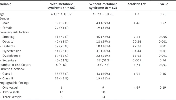
were no significant differences between groups with re-spect to the clinical variables of interest, except for those that by definition are part of the MS (Table 1).
Diagnostic performance
of dobutamine echocardiography
The adequate levels of inter- and intra-observer agree-ment for diagnosis in the case of positive dobutamine stress test were k = 0.81.
Forty-one patients with MS and 36 without MS had positive pharmacological stress echo, with no signifi-cant differences between groups (p = 0.77).
Cardiac catheterization revealed significant coronary artery disease (blockage of ≥ 50% of the lumen) in at least one of the coronary segments in 27 patients with MS and 29 patients without the syndrome; per-cutaneous revascularization was necessary in 24 and 26 of those patients, respectively; there was no sta-tistical difference between groups (p = 0.29).
Table 1. Baseline clinical characteristics and comparison between patients with and without metabolic syndrome*
Variable With metabolic
syndrome (n = 66) Without metabolic syndrome (n = 62) Statistic t/z P value
Age 63.15 ± 10.17 60.73 ± 10.98 1.3 0.21 Gender
– Male – Female
39 (59%) 27 (41%)
43 (69%) 19 (31%)
1.46 0.22
Coronary risk factors – Smoking
– Obesity – Diabetes – Hypertension – Dyslipidemia – Sedentary
31 (47%) 42 (63%) 52 (78%) 64 (96%) 57 (86%) 40 (61%)
45 (72%) 18 (29%) 10 (16%) 31 (50%) 32 (51%) 37 (59%
7.64 20.26 47.78 34.44 16.62 0.005
0.005 0.001 0.001 0.001 0.001 0.94 Number of risk factors 5 (4-6)† 3 (2-4)† 6.74 0.001
Current functional – Class II
– Class III
38 (58%) 28 (42%)
43 (69%) 19 (31%)
1.91 0.16
Angiographic findings – One vessel – Two vessels – Three vessels
6 16
8
9 10 14
4.69 0.19
*Metabolic syndrome criteria according to NCEP-III. According to the NCEP ATP III definition, metabolic syndrome is present if three or more of the following five criteria are met: waist circumference > 90 cm (men) or 80 cm (women), blood pressure > 130/85 mmHg, fasting triglyceride level > 150 mg/dl, fasting high-density lipoprotein cholesterol level < 40 mg/dl (men) or 50 mg/dl (women) and fasting blood sugar > 100 mg/dl. †Values are expressed as median and interquartile range. The inferential analysis by Mann-Whitney U considered as significant p values < 5%.
.r
e
hsi
l
b
u
p
e
ht
f
o
n
ois
si
mr
e
p
ne
tti
r
w r
oir
p
e
ht
t
u
o
hti
w
g
ni
y
p
oc
ot
o
h
p r
o
de
c
u
d
or
pe
r
e
b
ya
m
n
oit
aci
l
b
u
p s
i
ht
f
o t
ra
p
o
N
Five patients with MS and six without the syndrome with positive dobutamine echo did not accept coronary angiography; however, all were followed up to assess the occurrence of any major cardiovascular event.
For analysis purposes, we considered the findings on se-lective coronary angiography as a gold standard or refer-ence test for those patients with positive results in do-butamine echo. To correct for verification bias in subjects with negative results in the stress test, and therefore not catheterized, we used the Bayesian method developed by Begg-Greenes. According to our results, the sensitiv-ity and negative predictive value of dobutamine echo was significantly higher in patients with MS (sensitiv-ity 0.81 vs. 0.63 with MS and without MS, respectively, p = 0.03; specificity 0.57 vs. 0.53 with MS and without MS, respectively, p = 0.7; positive predictive value 0.66 vs. 0.73 with MS and without MS, respectively, p = 0.5; negative predictive value 0.75 vs. 0.41 with MS and without MS, respectively, p = 0.0003). How-ever, there were no differences between groups when comparing the areas under the ROC curve (AUC: 0.60; 95% CI: 0.41-0.73 for the group without MS vs. AUC: 0.70; 95% CI: 0.50-0.80 for the MS group; p = 0.47)
Follow-up
Loss to follow-up for the entire cohort was 5.4%, three for the group with MS and four for the control group, with no significant differences (p = 0.09). There were no differences between groups regarding the follow-up time (8.53 ± 3.9 months for the MS group vs. 8.24 ± 3.86 months for the group without MS; p = 0.69).
We recorded at least one cardiovascular event during follow-up in 28 patients; although there were more adverse events in patients with MS, there were no sig-nificant differences between groups (19 in MS group vs. nine in control group; p = 0.17). In the group with MS, 11 patients had angina, five needed revascularization, one had heart failure and two died; none had a docu-mented heart attack. In the control group, the events were: three patients with angina, two with document-ed myocardial infarction and two who nedocument-eddocument-ed revas-cularization, one with heart failure and one death.
We compared the occurrence of MACE in patients with positive results in dobutamine echo to look for evidence of the predictive power of the test for each group. In the MS group, 47.7% of patients with negative results for myocardial ischemia or viability on the stress echo-cardiogram were event-free during follow-up compared with 16.1% of those with positive results, and the dif-ference was statistically significant (p = 0.0004; Fig. 1 A). In addition, there was a statistically significant difference between the results of dobutamine echo in patients without MS; of those without MS and nega-tive dobutamine echo, 29.9% were event-free during follow-up, compared with 17% of those with positive dobutamine echo test results (p = 0.0087; Fig. 1 B).
Furthermore, we analyzed whether there were differ-ences in the proportion of event-free patients between carriers of MS and the control groups according to the results of echocardiography: 23.6% of carriers of MS were event-free during follow-up, compared to 10.7% of the control patients with positive results for stress
Figure 1. Follow-up of patients according to metabolic syndrome. A: with metabolic syndrome. B: without metabolic syndrome
Negative dobutamine echo
Follow-up time (months) 10
20 30 40 50 60 70 80 90 100
Positive dobutamine echo
MA
CE-fr
ee surviv
al (%)
15 10
5 0
Negative dobutamine echo
Follow-up time (months) 0
20 30 40 50 60 70 80 90 100
Positive dobutamine echo
MA
CE-fr
ee surviv
al (%)
10
16 12 14 10
4 6 8
2
A B
.r
e
hsi
l
b
u
p
e
ht
f
o
n
ois
si
mr
e
p
ne
tti
r
w r
oir
p
e
ht
t
u
o
hti
w
g
ni
y
p
oc
ot
o
h
p r
o
de
c
u
d
or
pe
r
e
b
ya
m
n
oit
aci
l
b
u
p s
i
ht
f
o t
ra
p
o
N
echocardiography, with no significant differences (p = 0.28; Fig. 2 A). There were no significant differences between groups when the test result was negative (59.8% event-free in the MS group vs. 42.9% in the control group; p = 0.67; Fig. 2 B).
Finally, we calculated the risk of MACE in patients with positive results in dobutamine echo. According to our results, the overall risk for the control group was 8.6 (95% CI: 2.53-29.59) and 5.8 for those with MS (95% CI: 1.74-19.60).
DISCUSSION
The results obtained in our study showed that phar-macological stress echocardiography with dobutamine (dobutamine echo) is a useful test for diagnosis of ischemic heart disease in MS patients in whom there is a high suspicion of cardiovascular disease, with a high sensitivity and negative predictive value compared with those obtained in patients without MS, for whom the results are similar to those obtained by other investiga-tors. The findings obtained from the analysis of the ROC curves confirmed that the test is useful in both groups17. Kamelesh, et al. reported similar findings, but
they used stress-imaging studies to assess the preva-lence of CAD, they did not perform cardiac catheter-ization, and only 4% of their patients were women18.
Diabetic patients have increased morbidity and higher rates of cardiovascular events such as silent ischemia and myocardial infarction compared to non-diabetics19.
The presence of MS (combined with central obesity, hypertension, and hyperglycemia) contributes to the increased risk of cardiovascular events and death from these causes20 since MS affects mainly
large-to-me-dium caliber vessels21. In our study, the occurrence of
major cardiovascular events was more frequent in car-riers of MS, most of whom also had an established diagnosis of diabetes mellitus.
During follow-up, there was a lower proportion of event-free patients among those with positive dobu-tamine echo test results, and even among those with MS and negative dobutamine echo.
Patients with and without MS behaved in a similar manner, independently of having positive or negative stress test results, with no statistically significant dif-ferences. This effect has already been observed and is consistent with that reported by other authors in patients with diabetes mellitus, in whom it was ob-served that using the results of dobutamine echo test to plan their management could be confusing due to the propensity of the patients to experience mural abnormalities in the absence of a previous infarction, the presence of microvascular ischemia, and the ac-celerated rate of progression that can limit the long-term negative predictive value of normal dobutamine echo test in these patients22. This is known by
coro-nary angiography in patients who were considered candidates for dobutamine echo, or through subsequent monitoring and analysis of the stress test results cor-rected for verification bias with the Begg and Greenes method, which has proven useful in other studies23.
Figure 2. Follow-up of patients according to dobutamine echocardiography test result. A: positive dobutamine echo test. B: nega-tive dobutamine echo test
Without MS
Follow-up time (months) 0
20 30 40 50 60 70 80 90 100
With MS
MA
CE-fr
ee surviv
al (%)
10
15 10
5 0
Without MS
Follow-up time (months) 20
30 40 50 60 70 80 90 100
With MS
MA
CE-fr
ee surviv
al (%)
15 10
5 0
A B
.r
e
hsi
l
b
u
p
e
ht
f
o
n
ois
si
mr
e
p
ne
tti
r
w r
oir
p
e
ht
t
u
o
hti
w
g
ni
y
p
oc
ot
o
h
p r
o
de
c
u
d
or
pe
r
e
b
ya
m
n
oit
aci
l
b
u
p s
i
ht
f
o t
ra
p
o
N
The MS is considered a marker of cardiovascular disease; Al Badarin, et al. found, in a cohort of pa-tients with abnormal dobutamine echo test results, that the presence of MS slightly increases the prev-alence of coronary artery disease, but not signifi-cantly, compared to patients without MS. These findings are similar to ours, where no significant difference was observed between patients with and without MS with positive test results; perhaps the number of diabetic patients included influenced the test result because patients with diabetes and/or glucose intolerance are more likely to have coronary artery disease24.
Other studies with diabetic patients have found that when these patients are candidates for dobutamine echo, they usually have other comorbidities that make them candidates for it and that favor an increased prevalence of stroke, peripheral vascular disease, and neuropathy. Thus, the dobutamine echo test can pre-dict a greater number of cardiac events in this group of patients, who are at a higher risk compared to those who are not candidates for the test and have fewer comorbidities22. The inability to perform
ex-ercise in patients with diabetes mellitus and the in-dication of dobutamine echo test constitute a mark-er not only of a high pretest probability of coronary heart disease, but also generally of poor prognosis, which may be true for the group of patients with MS in this study because about 80% of them had diabetes mellitus25,26.
The results of this study support the diagnostic use of dobutamine echo test in patients with MS in the study protocol for coronary heart disease due to its high sensitivity compared to that reported in the gen-eral population, although it is not a determining factor in establishing the mid-term prognosis of these pa-tients. Patients with extensive abnormalities in multi-vessel distribution according to dobutamine echo, which are more frequent in patients with MS, are at increased risk of death from myocardial infarction, which may justify the need for coronary angiography and myocardial revascularization.
DECLARATION OF INTEREST
The authors declare no conflicts of interest.
ACKNOWLEDGEMENTS
This study and/or authorship was supported by a grant from the Consejo Nacional de Ciencia y Tecnología del Estado de Guanajuato (CONCYTEG, grant number 09-38-K662-091).
REFERENCES
1. Velázquez O, Barinagarrementería F, Rubio F, Verdejo J, Méndez MA, Violante R. [Morbidity and mortality by ischemic heart disease and stroke in Mexico. 2005]. Arch Cardiol Mex. 2007;77:31-9. 2. García-Castillo A, Jerjes-Sánchez C, Martínez Bermúdez P, et al.
RENASICA II, Mexican Registry of Acute Coronary Syndromes. Arch Cardiol Mex. 2005;75(Suppl 1):S6-19.
3. Grima Serrano A, León Latre M, Ordoñez Rubio B. [Metabolic syndrome as a cardiovascular risk factor]. Rev Esp Cardiol. 2005;5(Suppl D):16-20.
4. Lakka HM, Laaksonen DE, Lakka TA, et al. The metabolic syn-drome and total and cardiovascular disease mortality in middle-aged men. JAMA. 2002;288:2709-16.
5. Ninomiya JK , L’Italien G, Criqui MH, Whyte JL, Gamst A, Chen RS. Association of the metabolic syndrome with history of myo-cardial infarction and stroke in the Third National Health and Nutrition Examination Survey. Circulation. 2004;109:42-46. 6. Klein BE, Klein R, Lee KE. Components of the metabolic
syn-drome and risk of cardiovascular disease and diabetes in Beaver Dam. Diabetes Care 2002;25:1790-4.
7. Sawada SG, Segar DS, Ryan T. Echocardiographic detection of coronary artery disease during dobutamine infusion. Circulation. 1991;83:1604-14.
8. Sicari R, Picano E, Landi P. Prognostic value of dobutamine-at-ropine stress echocardiography early after acute myocardial infarction. Echo Dobutamine International Cooperative (EDIC) Study. J Am Coll Cardiol. 1997;29:254-60.
9. Sitges M, Paré C, Azqueta M, et al. Feasibility and prognostic value of dobutamine-atropine stress echocardiography early in unstable angina. Eur Heart J. 2000;21:1063-71.
10. Pierard LA, DeLandsherre CM, Berthe C, Rigo P, Kulbertus HE. Identification of viable myocardium by echocardiography during dobutamine infusion in patients with myocardial infarction after thrombolytic therapy: comparison with positron emission to-mography. J Am Coll Cardiol. 1990;15:1021-31.
11. Grundy SM, Brewer HB, Cleeman JI, Smith SC, Lenfant C; American Heart Association; National Heart, Lung, and Blood Institute. Def-inition of metabolic syndrome. Report of the National Heart, Lung, and Blood Institute/American Heart Association conference on scientific issues related to definition. Circulation. 2004;109:433-8. 12. Douglas PS, Khandheria B, Stainback RF, et al. ACCF/ASE/ACEP/ AHA/ASNC/SCAI/SCCT/SCMR 2008 appropriateness criteria for stress echocardiography: a report of the American College of Car-diology Foundation Appropriateness Criteria Task Force, American Society of Echocardiography, American College of Emergency Phy-sicians, American Heart Association, American Society of Nu-clear Cardiology, Society for Cardiovascular Angiography and Interventions, Society of Cardiovascular Computed Tomogra-phy, and Society for Cardiovascular Magnetic Resonance en-dorsed by the Heart Rhythm Society and the Society of Critical Care Medicine. J Am Coll Cardiol. 2008;51:1127-47.
13. Douglas PS, Hendel RC, Cummings JE, et al. ACCF/ACR /AHA/ ASE/ASNC/HRS/NASCI/RSNA/SAIP/SCAI/ SCCT /SCMR 2008 Health Policy Statement on Structured Reporting in Cardiovas-cular Imaging. Circulation. 2009;119:187-200.
14. ACCF/ASE/AHA/ASNC/HFSA/HRS/SCAI/SCCM/SCCT/SCMR 2011 Appropriate Use Criteria for Echocardiography. A Report of the American College of Cardiology Foundation Appropriate Use Criteria Task Force, American Society of Echocardiography, American Heart Association, American Society of Nuclear Car-diology, Heart Failure Society of America, Heart Rhythm Soci-ety, Society for Cardiovascular Angiography and Interventions, Society of Critical Care Medicine, Society of Cardiovascular Computed Tomography, Society for Cardiovascular Magnetic Resonance American College of Chest Physicians. J Am Soc Echocardiogr. 2011;24:229-67.
15. Arnese M, Fioretti PM, Cornel JH, Postma-Tjoa J, Reijs AE, Roelandt JR. Akinesis becoming diskinesis during high-dose dobutamine
.r
e
hsi
l
b
u
p
e
ht
f
o
n
ois
si
mr
e
p
ne
tti
r
w r
oir
p
e
ht
t
u
o
hti
w
g
ni
y
p
oc
ot
o
h
p r
o
de
c
u
d
or
pe
r
e
b
ya
m
n
oit
aci
l
b
u
p s
i
ht
f
o t
ra
p
o
N
stress echocardiography: a marker of myocardial ischemia or a mechanical phenomenon? Am J Cardiol. 1994;73:896-9. 16. Begg CB, Greenes RA. Assessment of diagnostic tests when
disease verification is subject to selection bias. Biometrics. 1983;39:207-15.
17. Kuntz KM, Fleischmann KE, Hunink MGM, Douglas PS. Cost-ef-fectiveness of diagnostic strategies for patients with chest pain. Ann Intern Med. 1999;130:709-18.
18. Kemelesh M, Campbell S, Ligler L, Meda M, Eckert GJ, Sawada S. Metabolic syndrome does not predict an increased risk of coro-nary disease in patients with traditional risk factors referred for stress imaging study. Metab Syndr Relat Disord. 2010;8:223-8. 19. Mak KH, Moliterno DJ, Granger CB, et al. Influence of diabetes
mellitus on clinical outcome in the thrombolytic era of acute myocardial infarction. GUSTO-I Investigators. Global Utilization of Streptokinase and Tissue Plasminogen Activator for Occlud-ed Coronary Arteries. J Am Coll Cardiol. 1997;30:171-9. 20. Franco O, Massaro J, Civil J, et al. Trajectories of entering the
metabolic syndrome. The Framingham Heart Study. Circulation. 2009;120:1943-50.
21. Cull CA, Jensen CC, Retnakaran R, Holman R. Impact of the met-abolic syndrome on macrovascular and microvascular outcomes in Type 2 diabetes mellitus. Circulation. 2007;116:2119-26. 22. Biagini E, Elhendy A, Bax JJ, Schinkel AF, Poldermans D. The use
of stress echocardiography for prognostication in coronary ar-tery disease: an overview. Curr Opin Cardiol. 2005;20:386-94. 23. Miller TD, Hodge DO, Christian TF, et al. Effects of adjustment
for referral bias on the sensitivity and specificity of single pho-ton emission computed tomography for diagnostic of coronary artery disease. Am J Med. 2002;112:290-7.
24. Al Badarin FJ, From AM, McCully RB, Lopez-Jimenez F. Likelihood of obstructive coronary disease in metabolic syndrome patients with abnormal stress echocardiography. In J Cardiol. 2011;152:207-11. 25. Florkowski C, Scott R, Coope P, Moir C. Predictors of mortality
from type 2 diabetes mellitus in Canterbury, New Zealand; a ten-year cohort study. Diabetes Res Clin Pract. 2001;53:113-20. 26. Chaowalit N, Arruda A, McCully R, et al. Dobutamine stress
echocardiography in patients with diabetes mellitus: Enhanced prognostic prediction using a simple risk score. J Am Coll Car-diol. 2006;47;1029-36.
.r
e
hsi
l
b
u
p
e
ht
f
o
n
ois
si
mr
e
p
ne
tti
r
w r
oir
p
e
ht
t
u
o
hti
w
g
ni
y
p
oc
ot
o
h
p r
o
de
c
u
d
or
pe
r
e
b
ya
m
n
oit
aci
l
b
u
p s
i
ht
f
o t
ra
p
o
N