P
www.permanyer.com Rev Invest Clin. 2015;67:46-53
Oxidative Stress in Patients with
Rheumatoid Arthritis
Adolfo García-González
1,2, Ramón Gaxiola-Robles
1,2* and Tania Zenteno-Savín
11Centro de Investigaciones Biológicas del Noroeste, S.C.; 2Hospital General de Zona No. 1, Instituto Mexicano del Seguro
Social, La Paz, Baja California Sur, Mexico
Corresponding author:
*Ramón Gaxiola Robles
Centro de Investigaciones Biológicas del Noroeste, S.C. Planeación Ambiental y Biomedicina
Instituto Politécnico Nacional 195 Playa Palo de Santa Rita Sur
La Paz, C.P. 23096, Baja California Sur, Mexico
E-mail: [email protected] Received for publication: 20-08-2014 Accepted for publication: 10-12-2014
ABSTRACT
Background: Rheumatoid arthritis is an autoimmune disease of unknown etiology, characterized by articular inflammation.
Oxidative damage induced by reactive oxygen species has been related to the pathophysiology of rheumatoid arthritis in several studies, although results have been inconsistent and contradictory. Objective: To determine oxidative stress markers in patients
with rheumatoid arthritis. Methods: Descriptive cross-sectional study in rheumatoid arthritis patients and healthy controls. In
peripheral blood samples from all study subjects, lipid peroxide (thiobarbituric acid reactive substances) and protein carbonyl levels were quantified as oxidative damage markers; superoxide dismutase and glutathione peroxidase activities, glutathione concentration, and the reduced glutathione/oxidized glutathione ratio were analyzed as antioxidant defense indicators. Mann-Whitney U tests were run. Statistical significance (a) was 0.05%. Results: We included 29 rheumatoid arthritis patients, 10 with
active disease, and 41 healthy controls. Age was higher in the rheumatoid arthritis group; there were no differences in female:male ratio between groups. Oxidative damage was higher in rheumatoid arthritis patients; however, there was no difference between patients with active or inactive rheumatoid arthritis. Antioxidant enzyme activities, glutathione concentration, and reduced glutathione/oxidized glutathione ratio were higher in rheumatoid arthritis patients than in controls. Conclusions:
Antioxidant levels were higher in rheumatoid arthritis patients than in healthy controls; however, they were insufficient to prevent oxidative damage. This suggests an active oxidative process in rheumatoid arthritis patients. (REV INVEST CLIN. 2015;67:46-53) Corresponding author: Ramón Gaxiola Robles, [email protected]
Key words: Antioxidant enzymes. Glutathione. Oxidative stress. Rheumatoid arthritis.
ORIGINAL ARTICLE
INTRODUCTION
Rheumatoid arthritis (RA) is a chronic, inflammatory autoimmune disease of unknown etiology, character-ized by symmetric inflammation of both small and
large joints, with possible systemic damage1.
Rheu-matoid arthritis affects mostly the economically pro-ductive age group; its prevalence increases with age and is approximately 25-times higher in women than in men2. Rheumatoid arthritis is suspected based on
No part of this publication may be reproduced or photocopying without the prior written permission
of the publisher
.
clinical criteria and there are no specific markers. Such criteria, however, are mainly used for classification and not for diagnosis. Therefore, the population with clinical criteria for RA is highly heterogeneous and markers are needed to distinguish between groups. Rheumatoid arthritis, despite being a disease predom-inantly of the joints, can have different clinical patterns with severe extra-articular involvement; it is consid-ered a systemic disease with an insidious course, re-sponse to treatment, and outcome3. Long-term
stud-ies suggest that in most patients, RA is a progressive disease, with severe articular damage as seen by ra-diographic methods, deterioration of physical func-tion, and significant increase in mortality4.
Aerobic production of the energy needed for the var-ious cell processes occurs via oxidative phosphoryla-tion, in which redox reactions in the mitochondria lead to synthesis of adenosine triphosphate. During this process, oxygen serves as the final acceptor of elec-trons from cytochrome oxidase c, yielding water as the final product. Some of the intermediate products of this tetravalent reduction are partially reduced oxy-gen metabolites, which are highly reactive and are termed reactive oxygen species (ROS). ROS partici-pate in multiple physiological processes, are part of the defense system against microorganisms5, and are
involved in the intracellular signaling pathways6.
Ap-proximately 2% of the partially reduced oxygen es-capes the mitochondria and is capable of damaging molecules and cell structures, particularly proteins, lipids, DNA, membranes and structural elements of the extracellular matrix such as proteoglycans and collagen7,8. Among ROS, superoxide radical (O
2•–),
hy-droxyl radical (HO•) and peroxynitrite (ONOO–) are
highly reactive9. The balance between ROS production
and elimination, either by antioxidant enzymes, such as superoxide dismutase (SOD), catalase (CAT) and glutathione peroxidase (GPx), or by non-enzymatic molecules, such as glutathione (GSH) and vitamins A, C, and E, contributes to maintain homeostasis in aerobic organisms. Loss of this balance leads to oxidative dam-age and is known as oxidative stress10.
Oxidative stress is involved in the pathophysiology of several diseases, RA among them11. Despite this, there
is no consensus on how to determine oxidative stress in a uniform and consistent way that can be used in clinical decision-making. For the rheumatologist that is not directly involved in basic research and works
within the clinical context, the approach to the physical chemistry of the cell may be difficult. Different practi-cal forms of assessing oxidative stress have been pro-posed. Among them, the reduced glutathione/oxidized glutathione (GSH/GSSG) ratio12-14, the ratio between
activities of SOD, GPx and CAT (SOD/GPx + Cat)15,
and determination of the redox potential of GSH16.
Research on the role of oxidative stress in the initiation and perpetuation of the inflammatory process in RA has produced inconsistent and contradictory results. Structural damage mediated by ROS has been report-ed17; however, the oxidative damage increases18 or does
not change19. Similarly, reports of the non-enzymatic
and enzymatic antioxidant activities in RA patients show divergent results. The relationship between oxidative stress and RA is still uncertain. Therefore, the aim of this study was to determine, in RA patients as com-pared to healthy controls, the oxidative damage, quan-tified as lipid peroxidation and protein carbonyl levels, activity of the antioxidant enzymes SOD and GPx, and GSH/GSSH ratio, its correlation with other biochemical markers, and the correlation of the disease with clini-cal, biochemiclini-cal, and oxidative stress indicators.
METHODS
Study population and samples
A cross-sectional study was done at the Outpatient Clinic (Unidad de Atención Médica Ambulatoria) of the Mexican Social Security (Instituto Mexicano del Seguro Social, IMSS) at La Paz, Baja California Sur, Mexico. The study was approved by the local research committee. Written informed consent was obtained prior to inclusion. Patients with a diagnosis of RA ac-cording to the American College of Rheumatology criteria20 were selected from the outpatient clinic of the
Rheumatology Service. Patients with diabetes mellitus, extreme obesity (body mass index, BMI > 40), hyper-tension state II or higher, neoplasia, smoking history, active infection, or recent (< 1 year) surgery were ex-cluded21. The control group consisted of healthy
vol-unteer donors of the blood bank at the IMSS Regional Hospital (Hospital de Zona más Medicina Familiar 1).
Patients were divided into two groups based on the inflammatory activity of the disease, by using the RA activity index (DAS28)22, which assesses the number
No part of this publication may be reproduced or photocopying without the prior written permission
of the publisher
.
of painful joints, number of swollen joints, erythrocyte sedimentation rate, and global evaluation of the dis-ease by the patient, with a cutoff point of ≥ 2.9 for active inflammatory process. Sociodemographic and clinical data of patients were obtained from the medi-cal charts. To eliminate potential confounding factors, subjects that had the following conditions in the month prior to sampling were excluded: antioxidant supple-ment intake, treatsupple-ment against dyslipidemia, recent infections, liver or kidney disease, neoplasm, stroke, acute myocardial infarction, or accidental trauma23.
From RA patients and controls included in the study, a peripheral blood sample was collected in a heparinized container (Vacutainer®), which was kept at 4°C prior to transport to the Oxidative Stress Laboratory (Labora-torio de Estrés Oxidativo) at the Centro de Investigacio-nes Biológicas del Noroeste, S.C. (CIBNOR) for analysis.
Laboratory procedures
In all blood samples, activity of the antioxidant en-zymes SOD and GPx, GSH and GSSG concentrations, and levels of lipid peroxidation (thiobarbituric acid re-active substances, TBARS) and protein carbonyls were determined and GSH/GSSG and SOD/GPx ratios were calculated. SOD activity was determined by spectro-photometry, using the method based on the xanthine/ xanthine oxidase system as constant generator of O2•– which, upon contact with nitro blue tetrazolium,
reduces it to formazan; SOD, catalyzes the dismuta-tion of O2•– into hydrogen peroxide (H
2O2) and
inhib-its the reduction of nitro blue tetrazolium24; data are
shown in international units of SOD activity per gram of hemoglobin (Hb). One unit of SOD activity is de-fined as the amount of enzyme that inhibits the max-imum reduction of nitro blue tetrazolium by 50%. GPx activity was analyzed by monitoring the continuous decrease in reduced nicotinamide adenine dinucleotide phosphate (NADPH) concentration while maintaining constant GSH levels25. Data are shown in
internation-al units of GPx activity per gram of Hb; one unit of GPx activity is defined as the amount of enzyme that oxi-dizes 1 µmol NADPH per minute. The GSH concentra-tion was quantified by following the change in absor-bance at 412 nm generated when GSH reacts with 5,’5-ditiobis-(2-nitrobenzoic) acid (DTNB, Ellman’s re-agent); GSSG concentration was assessed by the first derivatization of GSH with 2 vinylpyridine26. To obtain
the GSH and GSSG concentrations in the samples, re-sults were compared with those from a standard curve
(0-0.4 µM for GSSG and 0-0.8 µM for GSH); data are shown in nmol per gram of Hb. Oxidative damage to lipids was assessed by quantifying TBARS. The meth-od is based on the reaction of lipid hydroperoxides and aldehydes formed from peroxidation reactions with thiobarbituric acid to produce malondialdehyde, a crystalline pink pigment with maximum absorption at 532-535 nm27; data are shown as nmol per gram
of Hb. Oxidative damage to proteins was analyzed by the quantification of carbonyl groups in proteins by the derivatization in the presence of 2,4-dinitrophenylhy-drazine, yielding the stable product dinitrophenylhydra-zone, which can be detected by spectrophotometry at 370 nm28; data are presented in nmol per gram of Hb.
Statistical analysis
Descriptive statistics were used for the sociodemo-graphic variables; inferential statistics were used to probe for differences between groups (control vs. RA; active vs. inactive RA). To test for differences between medians, Mann-Whitney U tests were per-formed and 25th and 75th quartiles were calculated
as a measure of dispersion. Bivariate correlation be-tween the variables was analyzed by Spearman’s cor-relation index (rho). Multiple linear regression analysis was used to assess the participation of the indepen-dent variables, age, BMI, Hb concentration, TBARS levels, protein carbonyl concentration, SOD and GPx activities, GSH and GSSG concentrations relative to the DAS28 index (dependent variable). The regres-sion models were constructed using the “stepwise” analysis, with the aim of selecting the predicting vari-ables that significantly maximize the model's r2
(coef-ficient of determination)29. To assess the
homoscedas-ticity and fit, as well as to detect atypical observations in the data, an analysis of the residuals distribution was performed. The significance level a was set at ≤ 0.05. All statistical analyses were run using SPSS v.20.0.
RESULTS
Blood samples were collected from 29 RA patients, 10 of whom had active disease; the control group consisted of 41 healthy blood donors. The median age in the RA group was 48 years (25th and 75th
percen-tiles, 43.5-70.0) and of the control group, 38 years (33.5-44.5) (p < 0.05); the female:male ratio was 9:1 in the RA group and 8.4:1 in the control group (Table 1).
No part of this publication may be reproduced or photocopying without the prior written permission
of the publisher
.
Table 1. Clinical characterisitics of rheumatoid arthritis patients and controls
RA patients (n = 29)
median (25th-75th percentiles) median (25Controls (n = 41)th-75th percentiles)
Active disease (n = 10) Inactive disease (n = 19)
Age, years 48.0 (43.5-70.0) 48.5 (31.2-54.2) 38.0†# (33.5-44.5)
Sex (F:M) 9:1 8.6:1.0 8.4:1.0
RA duration, years 7.0 (3-15) 2.0 (1.0-13.5) NA
DAS28 4.3 (4.0-5.2) 2.1* (1.8-2.3) NA
Hb 10.7 (7.4-13.6) 14.6* (12.7-15.7) 15.4# (13.8-17.1)
BMI 25.0 (23.5-27.2) 26.1 (25.2-31.2) 26.0 (24.6-31)
RA: rheumatoid arthritis; DAS28: disease activity index; Hb: hemoglobin concentration, g/dl; BMI: body mass index; NA: not applicable. Statistical significance by Mann-Whitney U test p < 0.05.
*patients with inactive disease vs. patients with active disease;
†controls vs. patients with inactive disease; #controls vs. patients with active disease.
Table 2. Oxidative stress indicators in blood samples from rheumatoid arthritis patients and controls
Patients (n = 29) median (25th-75th percentile)
Controls (n = 41) median (25th-75th percentile)
p
TBARS 0.08 (0.07-0.11) 0.07 (0.06-0.08) < 0.01
Protein carbonyls 15.4 (7.2-21.8) 9.8 (5.87-129) 0.02
SOD 269.1 (167.1-484.9) 191.7 (118.4-296.4) 0.02
GPx 49.5 (42.6-61.2) 4.6 (2.22-8.37) < 0.01
GSH 36.3 (15.9-66.4) 23.1 (10.9-35.1) 0.01
GSSG 3.5 (2.05-5.9) 4.2 (2.4-6.4) 0.54
GSH/GSSG 10.2 (7.8-132.8) 5.4 (4.8-230.3) < 0.01
SOD/GPx 4.7 (3.5-6.6) 34.7 (21.8-93.1) < 0.01
TBARS: thiobarbituric acid reactive substances levels (nmol/g Hb); protein carbonyls concentration (nmol/g Hb); SOD: superoxide dismutase activity (U/g Hb); GPx: glutathione peroxidase activity (U/g Hb); GSH: reduced glutathione concentration (nmol/g Hb); GSSG: oxidized glutathione concentration (nmol/g Hb); p = statistical significance by Mann-Whitney U test.
When compared with controls, the RA patients had higher oxidative damage to proteins (p = 0.02) and lipids (p < 0.01) (Table 2). No significant differences were found in the oxidative damage markers between patients with active and those with inactive disease (p > 0.05) (Table 3). The RA patients had higher antioxidant enzyme activities and higher GSH concentration than controls (Table 2). The GSH/GSSG ratio was signifi-cantly higher in RA patients than in controls (p < 0.01) (Table 2), with the same ratios between patients with active or inactive disease. The SOD/GPx ratio was sig-nificantly lower in RA patients than in the control group (p < 0.01) (Table 2). However, there were no significant differences in the SOD/GPx ratio between patients with active or inactive disease (p = 0.15) (Table 3).
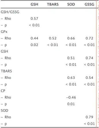
Results from the correlation analysis are summarized in table 4. In RA patients, the GSH/GSSG ratio was positively related to the GSH concentration (r = 0.57; p < 0.01). The GSH and GSSG concentrations were positively related to each other (r = 0.74; p < 0.01). The SOD activity was negatively related to the protein carbonyl content (r = –0.46; p = 0.01), and was posi-tively related to the TBARS levels (r = 0.63; p < 0.01) and the GSSG concentration (r = 0.79; p < 0.01). The GPx activity was positively correlated to the TBARS levels (r = 0.52; p < 0.01) and GSSG concentration (r = 0.72; p < 0.01).
Multiple linear regression models were generated to assess the effect of the covariates age, BMI, TBARS
No part of this publication may be reproduced or photocopying without the prior written permission
of the publisher
.
Table 3. Clinical characteristics and oxidative stress indicators in blood from rheumatoid arthritis patients with active compared to inactive disease
Active disease (n = 10)
median (25th-75th percentile) median (25Inactive disease (n = 19)th-75th percentile) p
Age, years 48.0 (43.5-70) 48.5 (31.2-54.2) 0.27
Duration of illness, years 7.0 (3-15) 2.0 (1-13.5) 0.06
TBARS 0.08 (0.06-0.11) 0.09 (0.07-0.12) 0.43
Protein carbonyls 18.7 (7.3-25.8) 10.8 (6.7-17.9) 0.19
SOD 227.6 (163.7-320.9) 304.0 (164.3-513.6) 0.46
GPx 49.4 (40.7-58.3) 54.3 (42.45-75.7) 0.51
GSH 21.9 (13.3-44.1) 47.2 (17.2-91.4) 0.08
GSSG 3.3 (2.5-5.5) 3.6 (1.8-6.2) 0.77
GSH/GSSG 6.6 (5.3-7.9) 13.0 (9.2-14.6) 0.00
SOD/GPx 4.3 (3.2-4.7) 5.8 (3.7-6.4) 0.15
Active disease: DAS28 ≥ 2.9; TBARS: thiobarbituric acid reactive substances levels (nmol/g Hb); protein carbonyls concentration (nmol/g Hb); SOD: superoxide dismutase activity (U/g Hb); GPx: glutathione peroxidase activity (U/g Hb); GSH: reduced glutathione concentration (nmol/g Hb); GSSG: oxidized glutathione concentration (nmol/g Hb); p = statistical significance by Mann-Whitney U test.
Table 4. Bivariate correlation between oxidative stress indi-cators in blood from rheumatoid arthritis patients
GSH TBARS SOD GSSG
GSH/GSSG
– Rho 0.57
– p < 0.01 GPx
– Rho 0.44 0.52 0.66 0.72
– p 0.02 < 0.01 < 0.01 < 0.01 GSH
– Rho 0.51 0.74
– p < 0.01 < 0.01
TBARS
– Rho 0.63 0.54
– p < 0.01 < 0.01
CP
– Rho –0.46
– p 0.01
SOD
– Rho 0.79
– p < 0.01
TBARS: thiobarbituric acid reactive substances levels (nmol/g Hb); protein carbonyls concentration (nmol/g Hb); SOD: superoxide dismutase activity (U/g Hb); GPx: glutathione peroxidase activity (U/g Hb); GSH: reduced glutathione concentration (nmol/g Hb); GSSG: oxidized glutathione concentration (nmol/g Hb); rho: Spearman’s coefficient; p = statistical significance.
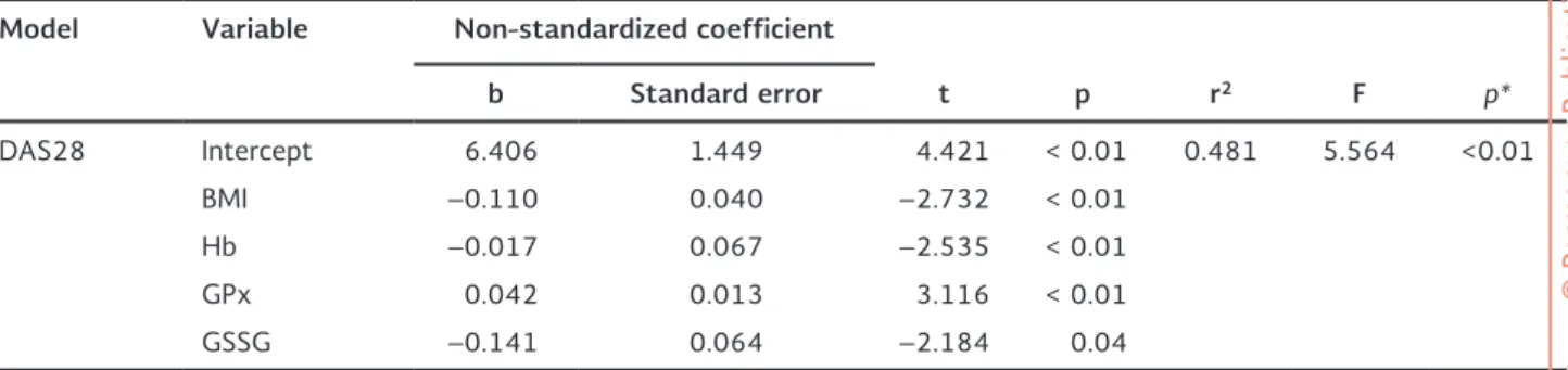
levels, protein carbonyl content, and activities of SOD and GPx on the DAS28 index. Table 5 shows the final model for the correlation of BMI, Hb concentration, GPx activity, and GSSG concentration with the DAS28 index in RA patients (r2 = 0.481; p < 0.01). The
con-stant variance and the normal distribution of the re-siduals did not suggest any trend, confirming the suf-ficiency of the adjusted model (Fig. 1).
DISCUSSION
Despite the divergent opinions on the role of oxidative stress in the genesis and perpetuation of damage ob-served in RA, there is evidence that it may participate in the pathogenesis of the disease30. Oxidative damage
in synovial fluid, with structural changes in hyaluronic acid, cartilage, and collagen, as well as increased lipid peroxidation, protein carbonyl content, and DNA al-terations, have been reported2.
In this study, higher oxidative damage, as assessed by TBARS and protein carbonyl levels, was found in RA patients compared to healthy controls. The GSH con-centration, GPx activity, and the activity of SOD were also higher in RA patients than in healthy individuals; however, the GSH/GSSG ratio was higher and the SOD/ GPx ratio was lower in RA patients than in controls. There were no significant differences in the markers of
No part of this publication may be reproduced or photocopying without the prior written permission
of the publisher
.
Table 5. Lineal multivariate model adjusted for the index of disease activity (DAS28) in rheumatoid arthritis patients
Model Variable Non-standardized coefficient
b Standard error t p r2 F p*
DAS28 Intercept 6.406 1.449 4.421 < 0.01 0.481 5.564 <0.01
BMI –0.110 0.040 –2.732 < 0.01
Hb –0.017 0.067 –2.535 < 0.01
GPx 0.042 0.013 3.116 < 0.01
GSSG –0.141 0.064 –2.184 0.04
DAS28: index of disease activity; BMI: body mass index; Hb: hemoglobin concentration (g/dl); GPx: glutathione peroxidase activity (U/g Hb); GSSG: oxidized glutathione concentration (nmol/g Hb).
2
1
0
–1
Residual
–2
1.0 2.0 3.0
Fitted value
4.0 5.0 6.0
2
1
0
–1
Residual
–2
–2 –1 0
Normal scores
1 2 3
Figure 1. Distribution of the residual values in the lineal model adjusted by the values of DAS28 in individuals with rheumathoid arthritis (RA).
oxidative stress quantified in this study between patients with active compared to inactive RA, which suggests that oxidative stress is characteristic of the morbid process per se, and that it continues even when the patient has no clinical data on disease progression.
The GSH/GSSG ratio and the antioxidant enzyme ac-tivities found in this study are not consistent with the reports of Feijóo, et al.31, who found that in patients
with chronic articular inflammatory disease, these parameters were lower than in control subjects; the authors found differences in oxidative stress between patients with active and inactive disease. Several research groups have found both high and low levels of the antioxidant enzyme activities in RA patients32,33.
This divergence between reports is frequent and may be explained by the variability and complexity of the regulating mechanism of oxidative stress in humans, which is associated with genetic2, epigenetic34, age35,
gender36, and dietary37 factors. Thus, it is appropriate
to continue this type of study to find a unifying set of markers that could be used in clinical practice38,39.
The higher GSH/GSSG ratio in RA patients compared with controls, apparently due to higher levels of GSH with no differences in the concentration of GSSG, could be associated to the higher glutathione reduc-tase (GR) activity in RA. Glutathione reducreduc-tase is a flavoenzyme dependent on NADPH that catalyzes the reduction of GSSH to GSH. This possibility is sup-ported in the study by Feijóo, et al.31, who found that
myeloperoxidase levels are elevated in patients with chronic inflammatory disease, especially those with active disease, and that high myeloperoxidase levels are related to an increase in oxidative damage and the inflammatory response, for myeloperoxidase and GR seem to show a similar activity pattern based on the availability of NADPH. On the other hand, in marine mammals, which have the ability to tolerate the oxi-dative stress that presumably occurs in the isch-emia-reperfusion periods associated to diving apnea, higher GR activity was observed in comparison with non-diving mammals, and this is suggested as one of the adaptive mechanisms of protection against ROS-induced damage40.
No part of this publication may be reproduced or photocopying without the prior written permission
of the publisher
.
In our study, lower SOD/GPx ratios and higher oxida-tive damage were found in RA patients. This, according to the study of Sánchez-Rodríguez, et al.41, suggests
a deficiency in the extracellular antioxidant defense system. Similar observations in construct model were reported in patients with chronic articular inflamma-tory disease in the study by Feijóo, et al.31.
In the present study, the bivariate correlation model was positive between the GSH/GSSG ratio and the GSH concentration. The multiple linear regression analysis showed a negative association between an inflamma-tory activity of the disease and the GSSG content. These data support the possibility of a higher GR ac-tivity in RA. The only antioxidant enzyme that showed a positive correlation with both lipid peroxidation lev-els and the antioxidant protection mechanisms was GPx, suggesting that GPx activity is involved in the primary mechanisms against oxidative stress in RA patients. Both GPx and CAT use H2O2 as substrate; however, CAT acts in the presence of high concentra-tions of the substrate while GPx acts at low concen-trations, which suggests an inverse correlation be-tween these enzymes and, further, that in RA patients the H2O2 concentration may be lower than in other chronic inflammatory diseases, with oxidative dam-age being mediated possibly by HO•42.
Multiple linear regression models to assess the effect of the co-variables of age, BMI, Hb concentration, TBARS levels, protein carbonyl content, activity of SOD and GPx, and GSH and GSSG concentrations on the DAS28 index showed that the most adequate construct was the one including BMI, Hb concentration, GPx activity, and GSSG concentration for the subjects with active or inactive RA (r2 = 0.481; p < 0.01). The
activity of the disease in the model was related to lower BMI and lower Hb content, both described as factors associated to wasting and chronic inflamma-tion characteristic of chronic diseases such as arterial hypertension, diabetes, and RA43, diseases in which
the endothelial damage appears to be the common factor. Vascular endothelium participates in processes such as oxidative stress, inflammation, immune re-sponse, thrombosis, vascular remodeling, and apop-tosis, depending on the precise equilibrium for car-diovascular health44. A unifying theory was recently
postulated to suggest that oxidative stress is the most important pathophysiological mechanism that conditions endothelial damage through at least three
activation mechanisms, such as the NAD(P)/NAD(P) H oxidase system, xanthine oxidase, and endothelial nitric oxide synthase45.
Several laboratory analyses are available to assess oxidative stress. These have been classified into five general procedures that quantify: (i) activity of anti-oxidant enzymes, (ii) concentration of low molecular weight antioxidants, (iii) balance between pro-oxidants and antioxidants, (iv) concentration of oxidants, and (v) concentration of products of oxidative damage16.
However, according to Sánchez-Rodríguez, et al.31,
these measurements have been used as isolated mark-ers and in a static way, interpreting oxidative stress as an increase in oxidized molecules or the decrease in intra– and/or extracellular antioxidants, without taking into account that oxidative stress integrates the effect of the exposure to oxidants coupled to the antioxidant protective mechanisms in vivo in a dy-namic manner. This suggests that there may be indi-viduals with high levels of oxidant molecules but an efficient antioxidant response, as well as subjects with-out elevated oxidant concentrations but with a defi-cient antioxidant response. Thus, if the pro-oxidant and antioxidant systems are evaluated independently, there may be errors in interpretation of results and limitations to their clinical application. Jones16
sug-gests that the increase in pro-oxidants with the con-sequent damage to macromolecules is not the only form of oxidative stress as sustenance of the disease process, but is a more complex situation which includes a severe disorder in the signaling and homeostatic con-trol of metabolic pathways and organs, affecting spe-cific processes such as cell cycle, apoptosis, immune response, and cell membrane functions.
In this study, SOD and GPx activities were higher in RA patients than in healthy individuals; however, these were not high enough to reduce oxidative damage to lipids and proteins. There were no significant differ-ences between patients with active and inactive RA, suggesting an ongoing oxidative stress process in RA patients. Discrepancy with some of the previous studies may be due to differences in study design and popula-tion, which could lead to differences in genetic polymor-phisms, epigenetics, diet, sex, or age. Oxidative stress is a dynamic and complex phenomenon, which re-quires further research and the design of tools for its assessment and clinical application in diagnosis and prognosis of disorders including rheumatoid arthritis.
No part of this publication may be reproduced or photocopying without the prior written permission
of the publisher
.
ACKNOWLEDGMENTS
This study was supported by Centro de Investigaciones Biológicas del Noroeste (CIBNOR) projects PC2.0, PC0.10. The authors thank the personnel and students at the Lab-oratory of Oxidative Stress, CIBNOR, particularly N.O. Ol-guín-Monroy and O. Lugo-Lugo, for technical assistance in sample processing.
REFERENCES
1. Harris ED. Rheumatoid arthritis. Pathophysiology and implica-tions for therapy. N Engl J Med. 1990;322:1277-89.
2. Hitchon CA, El-Gabalawy HS. Oxidation in rheumatoid arthritis. Arthritis Res Ther. 2004;6:265-78.
3. Freire M, Graña J, Galdo F, et al. Guías clínicas: Artritis Reuma-toide. Fisterra. 2004;4:1-6.
4. SER. GUIPCAR: Guía de práctica clínica para el manejo de la artritis reumatoide en España: Sociedad Española de Reumatología 2001. 5. Babior BM. Phagocytes and oxidative stress. Am J Med. 2000;
109:33-44.
6. Forman HJ, Fukuto JM, Torres M. Redox signaling: thiol chemestry defines which reactive oxygen and nitrogen species can act as second messengers. Am J Physiol Cell Physiol. 2004;287:C246-56. 7. McCord JM. The evolution of free radicals and oxidative stress.
Am J Med. 2000;108:652-9.
8. Henrotin YE, Bruckner P, Pujol JP. The role of reactive oxygen species in homeostasis and degradation of cartilage. Osteoar-thritis Cartilage. 2003;11:747-55.
9. Das UN. Free radicals: biology and relevance to disease. J Assoc Physicians India. 1990;38:495-8.
10. Halliwell B. Oxygen radicals, nitric oxide and human inflamma-tory joint disease. Ann Rheum Dis. 1995;54:505-10.
11. Vasanthi P, Nalini G, Rajasekhar G. Status of oxidative stress in rheumatoid arthritis. Int J Rheum Dis. 2009;12:29-33. 12. Kosower NS, Kosower EM. The glutathione status of cells. Int
Rev Cytol. 1978;54:109-60.
13. Pastore A, Piemonte F, Locatelli M, et al. Determination of blood total, reduced, and oxidized glutathione in pediatric subjects. Clin Chem. 2001;47:1467-9.
14. Jones DP. Redox potential of GSH/GSSH couple:assay and bio-logical significance. Methods Enzymol. 2002;348:93-112. 15. Park EM, Ramnath N, Yang GY, et al. High SOD and low GPX
activities in RBC predict susceptibility of lung cancer patients to radiation pneumonitis. Free Radic Biol Med. 2007; 42:280-7. 16. Jones DP. Redefining oxidative stress. Antioxid Redox Signal.
2006;8:1865-79.
17. Henrotin YE, Bruckner P, Pujol JP. The role of reactive oxygen species in homeostasis and degradatioin of cartilage. Osteoar-thritis Cartilage. 2003;11:747-55.
18. De Leo ME, Tranghese A, Passantino M, et al. Manganese su-peroxide dismutase, glutathione peroxidase, and total radical trapping antioxidant capacity in active rheumatoid arthritis. J Rheumatol. 2002;29:2245-6.
19. Karakoc M, Altindag O, Keles H, Soran N, Selek S. Serum oxida-tive-antioxidative status in patients with ankylosing spondilitis. Rheumatol Int. 2008;27:1131-4.
20. Arnet FC. The american rheumatism association 1987 revised criteria for classification of rheumatoid arthritis. Arth Rheum. 1988;31:315-24.
21. Olsson A, Skogh T, Winger G. Comorbidity and lifestyle, repro-ductive factors, and enviromental exposures associated with rheumatoide arthritis. Ann Rheum Dis. 2001;10:934-9. 22. Smolen JS, Breedveld FC, Ebrel G, et al. Vality and reliability of
the twenty-eight-joint count for assesment of rheumatoid ar-thritis activity. Arar-thritis Rheum. 1995;38:38-43.
23. NOM-253-SSA1-2012. Para la disposición de sangre humana y sus componentes con fines terapéuticos. Norma Oficial Mexi-cana. 2012:1-20.
24. Susuki. Measurment of Mn-Sod and Cu, Zn-Sod. In Experimental protocols for reactive oxygen and nitrogen species. Gutteridge J, Taniguchi N (Ed). Oxford, UK. Oxford University Press. 2000; 91-5. 25. Flohé L, Günzler WA. Assays of glutathione peroxidase. Methods
Enzymol. 1984;105:114-20.
26. Vázquez-Medina JP, Zenteno-Savín T, Elsner R. Glutathione pro-tection aginst dive-associated ischemia/reperfusion in ringed seal tissues. J Exp Marine Biol Ecology. 2007;345:110-8. 27. Zenteno-Savin T, Clayton-Hernández E, Elsner R. Diving seals:
are they a model for coping with oxidative stress? Comp Bio-chem Physiol C Toxicol Pharmacol. 2002;133:527-36. 28. Levine RL, Williams JA, Satdtman ER, Shaeter E. Carbonyl assays
for determination of oxydatively modified proteins. Methods Enzymol. 1994;233:346-57.
29. Gaxiola-Robles R, Bitzer-Quintero OK, Méndez-Rodríguez LC, et al. Lipid peroxidation and the response of the antioxidant de-fense system in the obese type 2 diabetic compared with non-obese tyupe 2 diabetic. Nutri Hosp. 2013;28:1905-11. 30. Heliovaara M, Knekt P, Aho K, Aran RK, Alfthan G, Aromaa A.
Serum antioxidants and risk of rheumatoid arthritis. Ann Rheum Dis. 1994;53:51-3.
31. Feijóo M, Túnez I, Ruiz A, Tasset I, Muñoz L, Collantes E. [Oxida-tive stress biomarkers as indicator of chronic inflammatory joint diseases stage]. Reumatol Clín. 2010;6:91-4.
32. Ozturk HS, Cimen MY, Cimen OB, Kacmaz M, Durak I. Oxidant/ antioxidant status of plasma samples from patients with rheu-matoid arthritis. Rheumatol Int. 1999;19:35-7.
33. Taysi S, Polat F, Gul M, Sari RA, Bakan E. Lipid peroxidation, some extracellular antioxidants, and antioxidant enzymes in serum of patients with rheumatoid arthritis. Rheumatol Int. 2002;21:200-4.
34. Sánchez-Pernaute O. [Epigenetic therapies, a step beyond bio-logics for rheumatoid arthritis]. Reumatol Clin. 2010;6:306-10. 35. Samiec PS, Drews-Botsch C, Flagg EW, et al. Glutathione in
human plasma: decline in association with aging, age-related macular degeneration, and diabetes. Free Radic Biol Med. 1998; 24:699-704.
36. Borrás-Blasco C. Importancia del estrés oxidativo en la diferen-cia de longevidad entre machos y hembras. Tesis doctoral. Fac-ultad de Medicina y Odontología. Universidad de Valencia. Es-paña. 2003.
37. Lonn E, Bosch J, Yusuf S, et al. Effects of long-term vitamin E supplementation on cardiovascular events and cancer: a ran-domized controlled trial. JAMA. 2005;293:1338-47.
38. Pérez Gastell PL, Pérez de Alejo JL. Métodos para medir el daño oxidativo. Rev Cubana Med Milit. 2000;29:192-8.
39. Kalpakcioglu B, Senel K. The interrelation of glutathione reduc-tase, catlase, glutathione peroxidase, superoxide dismureduc-tase, and glucose-6-phosphate in the pathogenesis of rheumatoid arthritis. Clin Rheumatol. 2008;27:141-5.
40. Vázquez-Medina JP, Zenteno-Savín T, Elsner R. Antioxidant en-zymes in ringed seal tissues: potential protection against dive-associated ischemia/reperfusion. Comp Biochem Physiol C Toxi-col PharmaToxi-col. 2006;142:198-204.
41. Sánchez-Rodríguez M, Santiago-Osorio E, Vargas L, Mndoza-Núñez V. Propuesta de un constructo para evluar integralmente el estrés oxidativo. Bioquimia. 2004;9:81-90.
42. Cisnero Prego E, Pupo Balboa J, Céspedes Miranda E. Enzimas que participan como barreras fisológicas para eliminar los radi-cales libres: III. Glutatión peroxidasa. Rev Cubana Invest Biomed. 1997;16:10-5.
43. Pasceri VA. A tale of two diseases: atherosclerosis and rheuma-toid arthritis. Circulation. 1999;100:2124-6.
44. Gopaul NK, Manraj MD, Habe A, et al. Oxidative stress could precede endothelial dysfunction and insulin resistance in Indian Mauritians with impaired glucose meatbolism. Diabetologia. 2001;44:706-12.
45. Ceballos-Reyes G, Ramírez-Sánchez I, Calzada-Mendoza C, Oli-vares-Corichi IM. Disfunción endotelial y estrés oxidativo. Endo-crinol Nutr. 2006;14:233-6.
No part of this publication may be reproduced or photocopying without the prior written permission
of the publisher
.