Artículo original
Gallium Scan after the Second and Fourth Chemotherapy Cycle is
Predictive in Aggressive Non-Hodgkin’s Lymphomas
José F Tomás,*,***Joseph Dang,* Barbara Pro,* Maria A. Rodriguez,* Peter McLaughlin,
Jorge Romaguera, Anas Younes,* Fredrick Hagemeister,* Fernando Cabanillas, Donald Podoloff,
Luis Fayad
RESUMEN
Objetivo: evaluar la utilidad del tamizaje con galio como predictor de supervivencia y duración de la respuesta en pacientes con linfoma no Hodgkin agresivo.
Pacientes y método: participaron 190 pacientes con linfoma no-Hodgkin, entre 1992 y 1996, en dos diferentes protocolos de tratamiento con atraciclina. 116 pacientes (61%) tuvieron puntuación Internacional de Pronóstico (IPI) baja y de bajo riesgo intermedio, 74 (39%) la tuvieron alta intermedia y de alto riesgo. Antes del inicio de la quimioterapia todos los pacientes tenían resultados positivos de galio (Ga67), y después de dos (n = 139), cuatro (n = 113) y seis (n = 24) ciclos de quimioterapia se efectuaron exploraciones complementarias. Los pacientes también recibieron radiación en los sitios conocidos con tumor voluminoso.
Resultados: los pacientes en quienes el galio siguió siendo positivo después de dos ciclos de quimioterapia tuvieron una mediana de supervivencia global de sólo 23 meses, comparados con una mediana de 107 meses en quienes las exploraciones de galio fueron nega-tivas (p = .00434). La positividad al Ga67 después de dos ciclos de quimioterapia se asoció con supervivencia libre de enfermedad de 6.6 meses, frente a 106 meses para quienes tuvieron Ga-67 negativo (p =. 00279). Después de cuatro ciclos de quimioterapia, el Ga67 positivo persistente se correlacionó con una media más corta (14.6 meses frente a 102.5 meses, respectivamente, p =. 00092) y supervivencia libre de enfermedad con media más corta (6.2 meses frente a 90 meses, respectivamente, p =. 00006). Mediante análisis de regresión de Cox, el resultado de la gammagrafía con galio es independiente del Índice Pronóstico Internacional (IPI) como un predictor de pobres resultados en el linfoma no Hodgkin agresivo.
Conclusiones: la positividad persistente del tamizaje con galio después de dos, cuatro, o seis ciclos de quimioterapia identifica a grupos de pacientes con gran tendencia a la progresión temprana de la enfermedad y pobre supervivencia global. El efecto parece ser indepen-diente del IPI. El objetivo de esta prueba es identificar la resistencia intrínseca al tratamiento en los pacientes con la enfermedad para que puedan beneficiarse de un esquema más agresivo e innovador, como la intensificación temprana.
Palabras clave: galio, agresivo, linfoma, factores pronósticos.
ABSTRACT
Purpose: To evaluate utility of gallium scans as a predictor of survival and response duration in patients with aggressive NHL.
Patients and Methods: From 1992 to 1996, 190 patients with aggressive non-Hodgkin’s lymphoma (NHL) were enrolled in two different anthracyclin-containing treatment protocols. One hundred-sixteen of these patients (61%) had an International Prognostic Score (IPI), of low, and low-intermediate risk; 74 (39%) had high-intermediate and high-risk. All patients had positive gallium scans (Ga67) prior to initiation of chemotherapy, and additional scans were performed after cycles two (n=139), four (n=113), and six (n=24) of chemotherapy. Patients also received radiation to known sites of bulky disease.
Results: Patients whose gallium scan remained positive after two courses of chemotherapy had a median overall survival (OS) of only 23 months compared to a median OS of 107 months for those whose gallium scans turned negative (p=.00434). Similarly, Ga67 positivity after two cycles of chemotherapy was associated with a disease-free survival (DFS) of 6.6 months compared to 106 months for those who had Ga-67 negativity (p=.00279). After four courses of chemotherapy, persistent Ga67- positivity also correlated with a shorter median OS (14.6 months vs. 102.5 months respectively, p=.00092) and a shorter median DFS (6.2 months vs. 90 months respectively, p=.00006). By Cox regression analysis, the result of the gallium scan was independent of the International Prognostic Index (IPI) as a predictor of poor outcomes in aggressive NHL.
Conclusions: Persistent gallium scan positivity after two, four, or six cycles of chemotherapy identifies a group of patients with great tendency for early disease progression and poor overall survival. The effect appears to be independent of IPI. This test targets patients with disease which has intrinsic resistance to their treatment and who may benefit from more aggressive and innovative therapies such as early intensification.
From the Departments of Lymphoma and Myeloma1 ,Nuclear Medicine_ at the University of Texas. MD Anderson Cancer Center and Department of Hematology at MD Anderson In-ternational España.
Correspondence to: José F Tomás, MD, PhD. Adjunt Proffesor. Department of Lymphoma and Myeloma. The University of Texas. Department of Hematology
MD Anderson International España, Madrid. E-mail [email protected]
A
ggressive NHLs constitute a heterogeneous group of diseases that afflict individuals of all ages. In spite of notable improvements and better outco-me for patients diagnosed from aggressive NHL in the last decade a significant number of them will ultimately die because of lymphoma. The identification of clinical, biological or, recently, genetic features, that identify pa-tients who will not response or relapse after a conventional first-line therapy is very important since it will contribute to a more efficient use of the available drugs and treatment strategies for aggressive NHL, and contribute to a more individualized therapy. Several investigators have defined pretreatment prognostic features that identify patients likely to have short responses to initial chemotherapy and poor overall prognosis. These include, among others, the IPI,_ b2 microglobulin (b2M),2 serum interleukin-6,3 Ki-67,4 and the M D Anderson Tumor Score.5Although not a pretreatment factor, the quality of response to initial treatment is one of the most important prognostic features determining the ultimate outcome of patients with aggressive lymphomas. Those who do not achieve a CR with initial therapy have much lower survival rates than those who do. Various investigators have suggested that patients who achieve early remission during treatment will have longer disease-free survival results.6-11 However, despite achieving CR, many patients
will still relapse, usually within the first two years of initial treatment.12,13 Some of those who develop relapse will be
eligible for potentially curative high-dose chemotherapy with stem cell rescue. If clinicians could successfully identify patients who will not achieve CR or who will likely respond initially to treatment but have a very high relapse-risk before medical signs of disease reoccurrence develop then aggressive strategies involving salvage therapy treatments could be employed in the hopes of improving prognoses.
Relatively common findings in the treatment of this disease are large tumor masses that slowly shrink with therapy. Distinguishing active tumor from necrotic tissue can be difficult in the evaluation of post treatment residual masses.14,15,16 Image-guided core biopsy has been useful
in this situation, but may not be completely reliable when biopsies are negative for disease.17 While computed
tomography (CT) scans are invaluable for determining the extent of disease at diagnosis, gallium imaging, with or without the use of single photon emission computed tomography (SPECT), and Positron emission tomography (PET) may have a greater specificity for assessing the presence of viable tumor in residual masses of patients with Hodgkin’s Lymphoma (HL) and NHL.18-27
Multiple investigators have studied gallium imaging during and after chemotherapy treatment and suggested that persistent Ga-67 positivity is a significant adverse prognostic factor.28-33 Janicek et al reported the significance
of persistent Ga-67 positive scans in 30 patients with poor prognosis aggressive NHL and tumor masses ³ 10 cm.28
Another retrospective analysis of 75 patients with HL and NHL, Zinzani et al demonstrated gallium scan usefulness in evaluation of residual mediastinal masses at the completion of therapy.29 Others have studied whether Ga-67 scanning
early during therapy can predict treatment outcomes.30,31 In
these earlier studies, conclusions regarding the predictive utility of gallium scanning has been limited by small patient numbers and scant information on the pretreatment prog-nostic factor profile of patients studied. In this analysis, we report on the utility of gallium scans after the second, fourth and sixth cycle of frontline chemotherapy in 190 patients with good and poor prognosis aggressive histology NHL.
PATIENT AND METHODS
Eligibility Criteria, Treatment, and Accrual Data
Newly diagnosed patients with aggressive NHL were eva-luated at M D Anderson from June 1992 through December 1996 and enrolled on clinical protocols 92-054 and 93-003. These studies were designed to treat patients with intensive chemotherapy. Because of the similarities of the two trials and the concurrent time of patient recruitment, data from these two studies are pooled for analysis purposes.
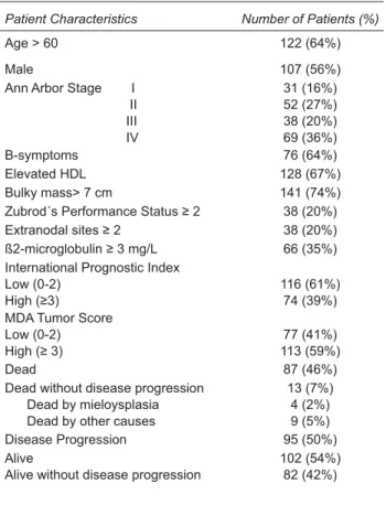
Table I. Patient Characteristics (n=190)
Patient Characteristics Number of Patients (%)
Age > 60 122 (64%)
Male 107 (56%)
Ann Arbor Stage I II III IV
31 (16%) 52 (27%) 38 (20%) 69 (36%)
B-symptoms 76 (64%)
Elevated HDL 128 (67%)
Bulky mass> 7 cm 141 (74%)
Zubrod´s Performance Status ≥ 2 38 (20%)
Extranodal sites ≥ 2 38 (20%)
ß2-microglobulin ≥ 3 mg/L 66 (35%)
International Prognostic Index Low (0-2)
High (≥3)
116 (61%) 74 (39%) MDA Tumor Score
Low (0-2) High (≥ 3)
77 (41%) 113 (59%)
Dead 87 (46%)
Dead without disease progression Dead by mieloysplasia Dead by other causes
13 (7%) 4 (2%) 9 (5%)
Disease Progression 95 (50%)
Alive
Alive without disease progression
102 (54%) 82 (42%)
registered for treatment under protocol 92-054. Patients with an M D Anderson tumor score ³ 3 were eligible. They were treated with alternating triple therapy (ATT) as previously reported.35Seventy-seven patients with similar aggressive histology NHL with a tumor score <3 were treated in protocol 93-003. A tumor score of < 3 is associated with a good prognosis. Patients received CHOP-based chemotherapy for six cycles. Only 190 of the 223 patients from both protocols had positive baseline gallium scans and are subjects of this analysis. All patients received one or more additional gallium scans after their baseline pretreatment scan.
Patient Characteristics (Table 1)
Biopsy slides were reviewed at MD Anderson by one or more experienced hemato-pathologists. Information on Ann Arbor stage, LDH, serum b2 microglobulin, serum albumin, Zubrod’s performance status, size of the largest tumor, number of extranodal areas, number of treatment cycles, treatment response, and baseline gallium scans results were collected prospectively. For this report, bulky disease was defined by a mass³ 7 cm.
Gallium Scan Technique
Gallium scans were performed with the intravenous injection of 8 - 10 mCi of Ga-67 citrate with subsequent imaging performed 48-120 hours later. Scans were done before treatment, and 3 weeks after the second or more cycles of chemotherapy. Each patient received 8-10 mi-llicuries of gallium-67 citrate as a bolus injection. Using a dual detector gamma camera, with a medium energy collimator, total body images were performed at a scan speed of 10 cm/min at a minimum of 48 hours after Ga-67 injection. The 93KeV, 184 KeV, and 296 KeV peaks were used with 20% and 10% windows. The image typically had 2 to 4 m counts. SPECT imaging was performed at 48 hours and up to 72 hours, depending upon patient’s clinical status. Images were displayed in the transaxial, saggital and coronal views, and results interpretation was performed from hard copy transparencies and interactive console display with volume-rendered three-dimensional images. Interpretations were performed with full knowledge of clinical, laboratory, and other imaging findings.
Criteria for Response
A CR was defined as no detectable evidence of disease by physical examination, x-rays, and CT scans. Bone marrow was confirmed to be free of lymphoma by repeat biopsy if involved by disease prior to the start of treatment. Uncon-firmed CR (CRu) was defined as the presence of minimal residual abnormalities on radiographic imaging (a mass <25% of the original volume calculated from the product of two diameters) with no palpable disease on physical examination. Partial response (PR) was defined as a ³ 50% reduction in the product of two diameters of tumor measured by radiological criteria or physical examination. Those who met the above criteria but still had histological evidence of lymphoma by biopsy were designated as ha-ving achieved PR. Progressive disease (PD) was defined as any tumor reduction less than 50%, significant tumor growth between courses despite an initial ³ 50% reduction, or lymphoma relapse in a patient achieving CR. Clinical responses lasting less than at least two months were de-signated PD.
Additional Biopsies
fine needle aspirations (FNA), seven underwent surgical biopsies, and 10 had both FNA and surgical biopsies.
Statistical Methods
Disease-free survival was calculated with the start of therapy to the time of first relapse, disease progression, or toxic death. Disease progression was not dependent on the status of the gallium scan. Thus, patients with sig-nificant tumor reduction and persisting Ga-67 positivity were not considered to have disease progression unless there was clear clinical or radiological evidence. Overall survival was calculated from beginning of therapy until death. Survival analysis was performed using the Kaplan and Meier method.36 The positive predictive value (PV+)
was calculated using the following equation: PV+= TP/ (TP+FP), where TP=true-positive and FP=false positive. A test was considered TP when results of the test were positive, and patient had clear evidence of disease relapse or a positive FNA or biopsy after treatment. A test was con-sidered FP when the test was positive without clear disease relapse and with a negative biopsy. Negative predictive value (PV-) was calculated using the following equation: PV-= TN/(TN+FN), where TN=true-negative results and FN=false negative results. A test was considered to be TN when the test results and biopsy (if available) were negative, and no progressive disease occurred. A test was considered to be false negative when results were nega-tive and biopsy (if available) was posinega-tive; or if patient developed progressive disease.
Statistical differences observed were assessed using the log-rank test. All p values were two-sided. Overall follow-up duration was calculated from the beginning of treatment to the last day of follow-up evaluation or death.
RESULTS (Table 1)
Of the 190 patients with positive baseline gallium scans, 139 had a gallium scan performed after the second cycle of chemotherapy, and 43 of these remained positive. Fifty-three of the 139 patients did not have additional gallium scans performed afterwards due to disease progression or other reasons including patient or physician preference. One hundred and thirteen patients had gallium scans after the fourth course of chemotherapy resulting in 23 posi-tive scans. Eighty-seven of the 113 patients had gallium scans after both the second and fourth treatment courses. Twenty-four patients did not have scans after the second or
fourth course but had gallium scans after the sixth course of chemotherapy due to physician or patient preference. Only three patients had gallium scans that were initially negative and later positive during repeat testing. All three had disease progression quickly after this observation with DFS of 3.5, 3.9, and 6.6 months. For the purpose of this study, the final gallium scan is defined by the status of the last scan done after course two, four or six of treatment. All patients with positive scans after the second course had an additional scan after course four or six unless disease progression developed. Ten of 43 patients with positive scans after the second course eventually had negative scans following additional courses of treatment.
Responses to Therapy
Eighty-one patients (72%) treated with the ATT regimen achieved CR (44 CR, and 37 CRu). Twenty-six (23%) achieved PR, and six (5%) patients progressed during therapy. Sixty-four (83%) patients treated with CHOP-Bleomycin/OPEN regimen achieved CR (33 CR, and 31 CRu). Ten (13%) achieved PR, and three (4%) progressed on therapy. Of the 95 patients who relapsed or have not respon-ded, 24 patients had autologous bone marrow transplants; eight had allogeneic transplants; and one had both.
Overall Survival and Disease-free survival Results (Tables 1-3)
With a median follow up duration of 88 months for the surviving patients, 87 (46%) patients died. Seventy-four deaths (39%) were due to lymphoma; 13 (7%) died of other causes, including four (2%) of myelodysplasia.
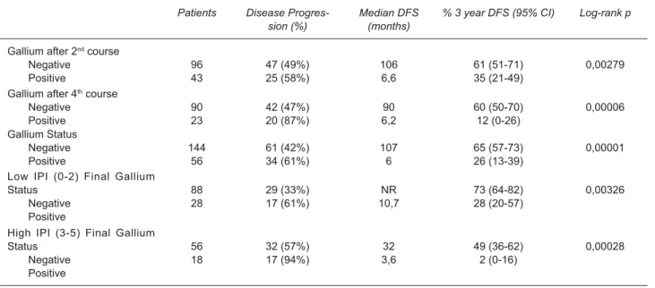
Table II. Disease-free Survival
Patients Disease
Progres-sion (%)
Median DFS (months)
% 3 year DFS (95% CI) Log-rank p
Gallium after 2nd course
Negative Positive 96 43 47 (49%) 25 (58%) 106 6,6 61 (51-71) 35 (21-49) 0,00279
Gallium after 4th course
Negative Positive 90 23 42 (47%) 20 (87%) 90 6,2 60 (50-70) 12 (0-26) 0,00006 Gallium Status Negative Positive 144 56 61 (42%) 34 (61%) 107 6 65 (57-73) 26 (13-39) 0,00001
Low IPI (0-2) Final Gallium Status Negative Positive 88 28 29 (33%) 17 (61%) NR 10,7 73 (64-82) 28 (20-57) 0,00326
High IPI (3-5) Final Gallium Status Negative Positive 56 18 32 (57%) 17 (94%) 32 3,6 49 (36-62) 2 (0-16) 0,00028
NR: Not Reached
Table III. SURVIVAL
Patients Dead (%) Median Survival
(months)
% 3 year Survival (95% CI)
Log-rank p
Gallium after 2nd course
Negative Positive 96 43 41 (43%) 26 (60%) 111 18 71 (63-79) 38 (24-52) 0,00017
Gallium after 4th course
Negative Positive 90 23 42 (47%) 16 (70%) 102 14,6 66 (56-76) 26 (7-45) 0,00092 Gallium Status Negative Positive 144 56 61 (42%) 27 (48%) 111 18 71 (63-79) 38 (24-52) 0,00017
Low IPI (0-2) Final Gallium Status Negative Positive 88 28 27 (31%) 12 (43%) 116 44,7 82 (78-86) 52 (3-71) 0,02650
High IPI (3-5) Final Gallium Status Negative Positive 56 18 34 (61%) 15 (83%) 51 6,8 51 (38-64) 19 (2-26) 0,00114
the Ga-67 negative group vs. 6.2 months and 12% for the Ga-67 positive group (log-rank p= 0.00006) (Figure 3). The median OS and 3-year OS was 102 months and 66% for the negative group vs. 14.6 months and 26% for the positive group (log-rank p=00092) (Figure 4).
Final gallium scan analysis revealed the following. Of 144 patients who had negative final scans, 61
IPI and Gallium Analysis
Patients were analyzed according to low IPI (0-2) or high IPI (3-5) status. In the low IPI group, 28 of 116 patients had positive final gallium scans resulting in a median DFS
Figure 1. Kaplan-Meier Estimates of Disease-free Survival for Gallium Scans After Cycle Two
Figure 2. Kaplan-Meier Estimates of Overall Survival for Gallium Scans After Cycle Two
Figure 3. Kaplan-Meier Estimates for Disease-free Survival for Gallium Scans After Cycle Four
Figure 4. Kaplan-Meier Estimates for Overall Survival for Gallium Scans After Cycle Four
Figure 5. Kaplan-Meier Estimates of Disease-free Survival for the Last Gallium Scan
Figure 6. Kaplan-Meier Estimates of Overall Survival for the Last Gallium Scan
OS and 3-yr OS was 116 months and 82% vs. 44.7 mon-ths, and 52% for the negative and positive Ga-67 groups respectively (log-rank p= 0.02650) (Table 3).
In the high IPI group, 18 of 74 patients had persistently positive final gallium scans resulting in a median DFS of 32 months for the negative Ga-67 group vs. 3.6 months for the Ga-67 positive group (Table 2). The 3-yr DFS was 49% for the negative Ga-67 group vs. 2% for the positive Ga-67 group (log-rank p= 0.00028). Median OS and 3-yr OS was 50.8 months and 51% for the negative Ga-67 group vs. 6.8 months and 19% for the positive Ga-67 group (log-rank p= 0.00114) (Table 3).
A Cox regression analysis showed that gallium scans performed after the second cycle of chemotherapy were independent of the IPI status (high vs low) for predicting failure-free survival (p= 0.001396 and p= 0.000102 res-pectively). This independence from the IPI status was also seen in scans after the fourth chemotherapy (p= 0.00002 and p= 0.021463) and in the final gallium scan (p= 0.000001 and p= 0.000005).
Predictive Values
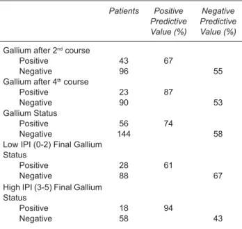
The positive predictive value of disease progression of a gallium scan after the second and fourth course of
treatment was 67% and 87% (Table 5). Fifty-six patients had persistently positive gallium scans on their last evalua-tion resulting in a 74% overall positive predictive value. Negative predictive value of the gallium scan after the second and fourth course of treatment was 55% and 53% respectively. One hundred forty-four patients had negative gallium scans at their last evaluation resulting in a 58% overall negative predictive value. Further analysis of pa-tients according to IPI status reveals a positive predictive value of gallium scans of 61% for low IPI and 94% for high IPI patients. The negative predictive value is 67% and 43% for low and high IPI patients respectively.
Landmark Analysis
In contrast, the positive Ga67 group had a median DFS of 40 months and 3-year DFS of 51%. This difference did not meet statistical significance (p=0.087).
DISCUSSION
Gallium scintigraphy has been frequently used at the end of the therapy in NHL and HD to evaluate treatment response, particularly in the setting of residual masses. It offers superior sensitivity to detect residual disease than CT imaging.28,29,30 Post treatment Ga67 positivity is an
adverse prognostic factor for disease-free survival and overall survival.29 Our study, the largest mid-cycle and
post-treatment gallium scanning analysis to date, conclu-des that persistent Ga67 positivity after cycle two, four, or six of frontline treatment in patients with aggressive NHL with either favorable or poor prognosis is a hallmark for early disease progression. These patients may benefit
Table IV. Predictive values
Patients Positive Predictive Value (%)
Negative Predictive Value (%)
Gallium after 2nd course
Positive Negative
43 96
67
55 Gallium after 4th course
Positive Negative
23 90
87
53 Gallium Status
Positive Negative
56 144
74
58 Low IPI (0-2) Final Gallium
Status Positive Negative
28 88
61
67 High IPI (3-5) Final Gallium
Status Positive Negative
18 58
94
43
Table V. Landmark Analysis Disease-free Survival
Patients Dead (%) Median Survival
(Months)
3-year Survival (95% CI)
Log-rank p value
Gallium Status Negative Positive
125 24
41 (33%) 12 (50%)
NR 40
74 (66-82) 51 (31-71)
0,087
NR: Not Reached
from an early switch to different therapies and should be strongly considered for novel treatments.
Ten patients who initially had positive Ga-67 scans after cycle two developed negative Ga-67 scans after cycle four. Of these patients, seven eventually had PD after achieving an initial PR while three continued to be disease-free after achieving CR. Only three patients whose gallium scans were initially negative after cycle two developed positive scans upon later testing, and all relapsed shortly afterwards. Based on this small subset of patients, it is difficult to determine when the optimal time to maximize the usefulness of gallium scintigraphy to assess response to a regimen would be. Nevertheless, scans done after treatment cycles two and four were able to delineate with great significance a group of patients likely to have disease progression.
The landmark analysis censoring patients with early PD prior to six months shows that these gallium tests do not reliably predict which patients will have late relapse (beyond six months). It is very likely that the sensitivity of the gallium scan limits its ability to predict these later events. Patients who relapse late probably have a smaller tumor volume that escapes detection of the gallium scan than those who relapse early. Newer, more advanced te-chnologies may provide needed sensitivity to predict later relapses in these patients.
Other tests such as magnetic resonance imaging 37-9 and positron emission tomography (PET) may also be useful in predicting response and disease-free survival.40
Accumulating evidence shows that PET offers greater specificity and sensitivity over gallium scintigraphy, due to a poor spatial resolution and low sensitivity at the abdominal level of the gallium scan.40-47 Use of PET
of gallium scintigraphy to predict late relapses. However, its cost effectiveness and availability in different clinical settings needs further exploration, and we are currently comparing both procedures in a prospective study for DLBCL at our institution.
The gallium scan status was independent of the M D Anderson Tumor Score and the IPI by Cox regression analysis. As expected by Bayesian statistics, the positive predictive value of a gallium scan done in patients with a worse overall prognosis predicted by a high IPI score was better than in patients with a more favorable pretreatment prognosis. Likewise, the prognostic utility of a positive gallium scan after the fourth chemotherapy course was bet-ter than a positive scan afbet-ter the second treatment course. Patients who are at extremely high risk for early disease progression may possibly be selected through combining multiple prognostic factors such as MD Anderson Tumor Score, IPI, and gallium scan status in the midst of their current chemotherapy treatment.
In conclusion, although gallium scans are been progres-sively substituted by FDG-PET studies in the management of aggressive NHL, in our experience, the largest series ever analyzed, we have shown that gallium scans done during mid-cycle or at the end of therapy can predict which patients with aggressive NHL have poor prognosis and are likely to have disease progression. Of note was that this results were independent from the most common risk-factor clinical classification employed, the IPI score. In this patients, a change to different, more intense chemo-therapy regimens and new treatment strategies is justified. Whether a similar strategy can be adopted with the use of new metabolic imaging techniques need to be clarified, but emerging data from recent series have demonstrated that PET imaging performed in the mid-course of therapy had a predictive value for clinical outcome.45
REFERENCES
1. Shipp MA, Harington DP, Anderson JR, et al. A predictive model for aggressive NHL: The International Non-Hodgkin’s Lympho-ma Prognostic Factors Project. N Engl J Med 1993;329:987-994.
2. Swan F, Velasquez WS, Tucker S, et al. A new serologic system for the large-cell lymphomas based on initial b2-microglobulin and lactate dehydrogenase levels. J Clin Oncol 1989;7:1518-1527.
3. Seymour JF, Talpaz M, Cabanillas F, et al. Serum Interleukin-6 levels correlate with prognosis in diffuse large-cell lymphoma. J Clin Oncol 1995;13:575-582.
4. Miller TP, Grogan TM, Dahlberg S, et al. Prognostic significan-ce of the Ki-67-associated proliferative antigen in aggressive non-Hodgkin’s lymphomas: A prospective Southwest Oncology Group Trial. Blood 1994;83:1460-1466.
5. Rodriguez J, Cabanillas F, McLaughlin F, et al. A proposal for a simple staging system for intermediate grade lymphoma and immunoblastic lymphoma based on the “tumor score”. Annals Oncol 1992;3:711-717.
6. Coiffier B, Bryon PA, Berger F, et al. Intensive and sequential combination chemotherapy for aggressive malignant lympho-mas (Protocol LNH-80). J Clin Oncol 1986;4:147-153. 7. Armitage JO, Weisenburger DD, Hutchins M, et al.
Chemothera-py for diffuse large-cell lymphoma -Rapidly responding patients have more durable remission. J Clin Oncol 1986;4:160-164. 8. Amadori S, Guglielmi C, Anselmo AP, et al. Treatment of diffuse
aggressive NHL with intensive multidrug regimen included high dose cytosine arabinoside (F-MACHOP). Semin Oncol 1985;12:218-222.
9. Vitolo U, Bertini M, Tarella C, Bertoncelli MC, et al. MACOP -B treatment for advanced stage diffuse large cell lympho-mas: A multicentre Italian study. Eur J Cancer Clin Oncol 1989;25:1441-1449.
10. Guglielmi C, Amadori S, Ruco LP, et al. Combination chemo-therapy for the treatment of diffuse aggressive lymphomas: F-MACHOP update. Semin Oncol 1987;14:104-109. 11. Anderson JR, Ginsberg S, Gottlieb AJ. Chemotherapy of
diffuse large cell lymphoma -Rapidly responding patients have more durable remissions. J Clin Oncol 1986;4:1420-1421. (Letter)
12. Gordon LI, Harrington D, Andersen J, et al. Comparison of a second-generation combination chemotherapeutic regimen (m-BACOD) with standard regimen (CHOP) for advanced diffu-se non-Hodgkin’s lymphoma. N Engl J Med 1992;327:1342-1349.
13. Fisher RI, Gaynor ER, Dahlberg S, et al. Comparison of a standard regimen (CHOP) with three intensive chemotherapy regimens for advanced non-Hodgkin’s lymphoma. N Engl J Med 1993;328:1002-1006.
14. Radford JA, Cowan RA, Flanagan M, et al. The significance of residual mediastinal abnormality on the chest radiograph following treatment for Hodgkin’s disease. J Clin Oncol 1988;6:940-946.
15. Surbone A, Longo D, DeVita V, et al. Residual abdominal masses in aggressive non-Hodgkin’s lymphoma after combi-nation chemotherapy: significance and management. J Clin Oncol 1988;6:1832-1837.
16. Canellos GP. Residual mass in lymphoma may not be residual disease (Editorial). J Clin Oncol 1988;6:931-932.
17. Pappa VI, Hussain HK, Reznek RH, et al. Role of image-guided core-needle biopsy in the management of patients with lymphoma. J Clin Oncol 1996;14:2427-2430.
18. Anderson KC, Leonard RCF, Canellos GP, et al. High-dose Gallium imaging in lymphoma. Am J Med 1983;75:327-331. 19. Israel O, Front D, Lam M, et al. Gallium 67 imaging in
monito-ring lymphoma response to treatment. Cancer 1988;61:2439-2443.
21. Holman BL, Tumeh SS. Single-photon emission computed tomography (SPECT). Applications and potential. JAMA 1990;263:561-564.
22. Weeks JC, Yeap BY, Canellos GP, et al. Value of follow-up procedures in patients with large-cell lymphoma who achieved complete remission. J Clin Oncol 1991;9:1193-1203. 23. Front D, Ben-Haim S, Israel O, Epelbaum R, et al.
Predicti-ve value of Ga-67 scintigraphy after treatment. Radiology 1992;182:359-363.
24. Front D, Bar-Shalom R, Epelbaum R, et al. Early detection of lymphoma recurrence with Gallium-67 scintigraphy. J Nucl Med 1993;34:2101-2104.
25. Front D, Israel O. The role of Gallium-67 in evaluating the results of therapy of lymphoma patients. Semin Nucl Med 1995;25:60-71.
26. Even-Sapir E, Bar Shalom R, Israel O, et al. Single-photon emission computed tomography quantification of gallium citrate uptake for the differentiation of lymphoma from benign hilar uptake. J Clin Oncol 1995;13:942-946.
27. Vose JM, Bierman PJ, Anderson JR, et al. Single-photon emission computed tomography gallium imaging versus computed tomography: Predictive values in patients under-going high-dose chemotherapy and autologous stem-cell transplantation for non-Hodgkin’s lymphoma. J Clin Oncol 1996;14:2473-2479.
28. Janicek M, Kaplan W, Neuberg D, et al. Early restaging gallium predicts outcome in poor-prognosis patients with aggressive non-Hodgkin’s Lymphoma treated with high-dose CHOP chemotherapy. J Clin Oncol 1997;15:1631-1637.
29. Zinzani PL, Magagnoli M, Franchi R, et al. Diagnostic role of gallium scanning in the management of lymphoma with me-diastinal involvement. Hematological 1999;84(7): 604-607. 30. Front D, Bar-Shalom R, Mor M, et al. Aggressive non-Hodgkin
lymphoma: early prediction of outcome with 67Ga scintigraphy. Radiology 2000;214(1):253-257.
31. Israel O, Mor M, Epelbaum R, et al. Clinical pretreatment risk factors and Ga-67 scintigraphy early during treatment for prediction of outcome of patients with aggressive non-Hodgkin lymphoma. Cancer 2002;94(4):873-878.
32. Gasparini M, Bombardieri E, Castellani M, et al. Gallium-67 scintigraphy evaluation of therapy in non-Hodgkin’s lymphoma. J Nucl Med 1998;39(9):1586-1590.
33. Tuli MN, Al-Shemmari SH, Ameen RM, et al. The use of gallium-67 scintigraphy to monitor tumor response rates and predict long-term clinical outcome in patients with lymphoma. Clin Lymphoma 2004;5(1):56-61.
34. Rodriguez J, Cabanillas F, McLaughlin P, et al. A proposal for a simple staging system for intermediate grade lymphoma and immunoblastic lymphoma based on the ‘tumor score’. Ann Oncol 1992;3(9):711-717.
35. Cabanillas F, Rodriguez-Diaz Pavon J, Hagemeister FB, McLaughlin P, et al. Alternating triple therapy for the treatment of intermediate grade and immunoblastic lymphoma. Ann Oncol 1998;9(5):511-518.
36. Kaplan E, Meier P. Non-parametric estimation for incomplete observations. J Am Stat Assoc 1958;53:457-481.
37. Brice P, Rain JD, Frija J. Value of imaging for the diagnosis of residual mediastinal mass in malignant lymphomas. Nouv Rev Fr Hematol 1991;33:531-532.
38. Hill M, Cunningham D, MacVicar D, et al. Role of magnetic re-sonance imaging in predicting relapse in residual masses after treatment of lymphoma. J Clin Oncol 1993;11:2273-2278. 39. Devizzi L, Maffioli L, Bonfante V, et al. Comparison of
ga-llium scan. Computed tomography, and magnetic resonance in patients with mediastinal Hodgkin’s disease. Ann Oncol 1997;8(Suppl 1):S53-S56.
40. Rˆmer W, Hanauske A-R, Ziegler S, et al. Positron emission tomography in non-Hodgkin’s lymphoma: Assessment of chemotherapy with fluorodeoxyglucose. Blood 1998;91:4464-4471.
41. Kostakoglu L, Leonard JP, Kuji I, et al. Comparison of fluo-rine-18 fluorodeoxyglucose position emission tomography and Ga-67 scintigraphy in evaluation of lymphoma. Cancer 2002;94:879-888.
42. Wirth A, Seymour JF, Hicks RJ, et al. Fluorine-18 fluoro-deoxyglucose positron emission tomography, gallium-67 scintigraphy, and conventional staging for Hodgkin’s disease and non-Hodgkin’s lymphoma. Am J Med 2002;112:262-268.
43. Zijlstra JM, Hoekstra OS, Raijmakers PG, et al. 18FDG positron emisino tomography versus 67Ga scintigraphy as prognostic test during chemotherapy for non-Hodgkin’s lym-phoma. Br J Haematol 2003;123:454-462.
44. Filmont JE, Vranjesevic D, Quon A, et al. Conventional ima-ging and 2-deoxy-2-[18F] fluoro-D-glucose positron emission tomography for predicting the clinical outcome of previously treated non-Hodgkin’s lymphoma patients. Mol Imaging Biol 2003;5:232-239.
45. Haioun C, Itti E, Rahmouni A, et al. [18F]fluor-2-deoxy-D-glucose positron emission tomografie (FDG-PET) in aggres-sive lymphoma: an early prognostic tool for predicting patient outcome. Blood 2005;106:1376-1381.
46. Torizuka T, Nakamura F, Kanno T, et al. Early therapy moni-toring with FDG-PET in aggressive non-Hodgkin’s lymphoma and Hodgkin’s lymphoma. Eur J Nucl Med Imaging 2003;31(1): 22-28.