hAGT inhibitors as chemotherapy enhancers
Maria Tintoré Gazulla
Aquesta tesi doctoral
està subjecta a la llicència
Reconeixement 3.0. Espanya de Creative
Commons.
Esta tesis doctoral está sujeta a la licencia
Reconocimiento 3.0.
España de Creative
Commons
.
UNIVERSITAT DE BARCELONA
FACULTAT DE FARMÀCIA
“hAGT inhibitors as chemotherapy enhancers”
Maria Tintoré Gazulla
UNIVERSITAT DE BARCELONA
FACULTAT DE FARMÀCIA
Programa de Doctorat en Biotecnologia
hAGT inhibitors as chemotherapy enhancers
Memòria presentada per Maria Tintoré Gazulla per a optar al títol de
doctor per la Universitat de Barcelona
Dr. Carme Fàbrega Claveria Dr. Ramón Eritja Casadellà
Dr. Glòria Rosell Pellisé
Directora
Director
Tutora
The work described in this thesis has been performed in the Institute for Advanced
Chemistry of Catalonia (CSIC) and in the Institute for Research in Biomedicine of Barcelona,
IRB Barcelona. Chapter 5 is the result of a short stay of 4 months in the University of Milan.
Durant aquest temps al laboratori he tingut la sort de conèixer molta gent maca.
Voldria agrair especialment als meus companys del laboratori de Química dels Àcids
Nucleics tot el que he après, tant científica com personalment. Gràcies al Ramon per
donar-me l’oportunitat d’entrar en aquest magnífic grup. A la Cardonar-me, que em va ensenyar gairebé
a utilitzar una pipeta i de qui he après que el més important en la recerca són la paciència i
el rigor. A l’Anna, que m’ha cuidat com una mare. Al Santi, per la seva paciència amb els
meus múltiples dubtes sobre síntesi química; a la Sònia, sempre disposada a donar un cop
de mà amb els HPLC, els MALDIs o el que faci falta; i a l’Adele, per la seva disponibilitat i
ajuda amb els cultius cel·lulars. Al Rubén per la seva ajuda amb els experiments de masses
i per tots els bons moments i consells. Al Nacho, que m’aguanta amb alegria les invasions de
papers a la seva taula i els robatoris de calculadores i bolis. I a la resta de persones que han
passat pel lab durant aquest temps, de qui he pogut aprendre moltes coses.
Vull agrair molt a la Stefi la seva carinyosa acollida durant la meva estada al seu
laboratori a Milà. I a la Laura, el Leo, la Gigliola, la Sabrina i l’Angela.
També vull mencionar els meus companys estudiants de doctorat, en especial a
l’Albert, per tots els berenars en què hem convertit un moment de desànim per experiments
que no sortien en unes bones rialles. Al Michael i l’Andrey pels dinars i ping-pongs
compartits. I als meus companys i amics del màster, en especial a la Laura per la seva ajuda
durant l’escriptura de la tesi i al Jesús per donar-me un cop de mà amb l’estadística.
Moltes gràcies als meus amics de farmàcia pel seu suport, amb ells vaig començar
l’aventura de la ciència. Agraeixo molt al Germán la seva ajuda amb el disseny de la portada
de la tesi. I gràcies també al Carlos Roca, la Laia i la resta d’amics per la seva curiositat i
interès, em fa molta il·lusió que amics que treballen en camps tan allunyats tinguin tantes
ganes de saber el què faig.
INDEX
Introduction... 1
Objectives... ...29
1.
Potential inhibitors of hAGT’s DNA repair activity: study of the hAGT-compound complex
formation by ESI-MS and toxicity in cell culture………...33
1.1.
Study of the hAGT-compound complex formation by ESI-MS and toxicity in cell
culture………...35
1.2.
Appendix 1. Receptor-based virtual screening and biological characterization of new
inhibitors
of
Human
Apurinic/Apyrimidinic
Endonuclease
Enzyme
(Ape1)………...59
2.
Development of a novel fluorescence assay based on the use of the Thrombin Binding
Aptamer
for
the
detection
of
O
6-alkylguanine–DNA-alkyltransferase
activity………75
2.1.
Development of a novel fluorescence assay based on the use of the Thrombin Binding
Aptamer
for
the
detection
of
O
6-alkylguanine–DNA
Alkyltransferase
activity……….….79
2.2.
Supplementary information………...89
2.3.
Appendix 2: Thrombin Binding Aptamer, more than a simple aptamer: chemically
modified derivatives and biomedical applications...93
3.
DNA Origami as DNA repair nanosensor at the single-molecule level……….…………...109
3.1.
DNA Origami as DNA repair nanosensor at the single-molecule level……..………..113
3.2.
Supplementary information………...117
3.3.
Appendix 3: DNA nanoarchitectures: steps towards biological applications………..141
4.
A fluorescence biosensor for hAGT activity………...163
4.1.
A fluorescence biosensor for hAGT activity………...167
4.2.
Supplementary information……….179
4.3.
Appendix 4:
In vitro
assay to evaluate potential inhibitors of hAGT by a new fluorescence
method: preliminary results……….203
5.
Molecular biosensing using gold-coated superparamagnetic nanoparticles functionalized
with DNA aptamers……….209
General discussion………..221
Conclusions……….237
Summary……….……..…241
ABBREVIATIONS
3
H-MNU: Tritiated 1-methyl-1-nitrosourea
3’-Dabsyl CPG:
1-(4,4’-Dimethoxytrityloxy-3-[O-
(N-4'-sulfonyl-4-(dimethylamino)-azobenzene)-3-aminopropyl]-propyl-2-O-succinoyl-long chain
alkylamino-CPG
3'-Dabcyl CPG:
1-(4,4'-Dimethoxytrityloxy)-3-[O-(N-4'-carboxy-4-(dimethylamino)-
azobenzene)-3-aminopropyl)]-propyl-2-O-succinoyl-long
chain alkylamino-CPG
6’-FAM:
6-[(3’,6’-dipivaloylfluoresceinyl)-carboxamido]-hexyl-1-O-[(2-cyanoethyl)-(
N,N
-diisopropyl)]-phosphoramidite
Ac: acetyl
Ac
2O: acetic anhydride
ACN: acetonitrile
AcOEt: ethyl acetate
AFM: atomic force microscopy
anh: anhydrous
Ape1: human apurinic/apyrimidinic
endonuclease
Ar: aromatic
Au(OOCCH
3): gold acetate
AuNPs: gold nanoparticles
AuSPIONs: gold-coated superparamagnetic iron
oxide nanoparticles
BCNU: 1,3-Bis(2-chloroethyl)-1-nitrosourea or
carmustine
BME: 2-mercaptoethanol
Bz: benzyl
Bzl: benzoyl
Cpd: compound
CPG: controlled pore glass
Dabcyl: 4-(4-(dimethylamino)phenyl-azobenzoic
acid
Dabsyl:
N
-4’-sulfonyl-4-(dimethylamino)-azobencene)-3-aminopropyl
DCM: dichloromethane
DEAD: diethyl
azodicarboxylate (IUPAC
diethyl diazenedicarboxylate)
DIEA:
N,N
-Diisopropylethylamine
DMF:
N,N
-dimethylformamide
DMSO: dimethylsulfoxide
DMT: 4,4’-dimethoxytrityl
DMT-Cl: 4,4’-dimethoxytrityl chloride
DTT: dithiothreitol
EDTA: ethylenediaminetetraacetic acid
ESI: electrospray ionization
EtTFA: ethyl trifluoroacetate
FAM/F: fluoresceine
F
G: O
6-fluorescein-benzylguanosine
FITC: fluorescein isothiocyanate
FL: full length
FRET: fluorescence resonance energy transfer
GST: gluthatione-S-transferase
hAGT: human O
6-alkylguanine-DNA
alkyltransferase
HBA: hydrogen bond acceptors
HBD: hydrogen bond donors
HPLC: high performance liquid chromatography
IC
50: inhibitory concentration 50%
IPTG: isopropyl-β-D-1-thiogalactopyranoside
LCAA: long chain amino alkyl
LD
50: lethal dose 50%
MALDI: matrix-assisted laser
diserption/ionization
MeOH: methanol
MB: molecular beacon
MNU: 1-methyl-1-nitrosourea
MTT:(3-(4,5-dimethylthiazol-2-yl)-2,5-diphenyltetrazolium bromide)
NMR: nuclear magnetic resonance.
NPs: nanoparticles
OD: optical density
O
6-MeG: O
6-methylguanine
Q
T:
(N-4'-carboxy-4-(dimethylamino)-
azobenzene)-aminohexyl-3-acrylamido]-2'-deoxyuridine
RP-HPLC: reverse phase high pressure liquid
chromatography
rt: room temperature
SPIONs: superparamagnetic iron oxide
nanoparticles
TBA: thrombin-binding aptamer
cTBA: complementary TBA
TCA: trichloroacetic acid
TEA: triethylamine
TCEP·HCl: tris(2-carboxyethyl)phosphine
hydrochloride
TEAAc: triethylammonium acetate
TEM: transmission electron microscopy
TFA: trifluoroacetic acid
THAP: 2’,4’,6’-trihydroxyacetophenone
monohydrate
THF: tetrahydrofurane
Thr: α-thrombin
TLC: thin-layer chromatography
Tm: melting temperature
TMAOH: tetramethylammonium hydroxide
TOF: time of flight
Tris: tris(hydroxymethyl)aminomethane
UV: ultraviolet
Deoxyribonucleic acid (DNA)
The DNA double helix was first described by Watson and Crick in 1953.
[1]Since then, the
scientific advances based in DNA have been prodigious and far beyond expectations in
many fields, reaching areas as diverse as medicine, basic biology, genetics, forensics,
archeology among others, and culminating in the sequencing of the human genome.
[2]Deoxyribonucleic acid is a self-assembling biopolymer that encodes the genetic
information used in the development and functioning of all known living organisms and
many viruses. DNA, together with proteins and carbohydrates, compose the three major
macromolecules essential for all forms of life. DNA forms double helices directed by
canonical Watson-Crick base pairing and is stabilized by hydrogen bonds,
π
-
π
stacking and
hydrophobic interactions.
[1]The two DNA strands are composed of simpler units called
nucleotides. Each nucleotide is composed of a nitrogen-containing nucleobase—either
guanine (G), adenine (A), thymine (T), or cytosine (C)—as well as a monosaccharide sugar
called deoxyribose and a phosphate group. The four nucleobases are classified in
pyrimidines and purines, depending on their structure: pyrimidines (T and C) consist of a
single heterocycle of six atoms and purines (A and G) are composed by a pyrimidine ring
fused to an imidazole ring (figure 1). Nucleotides are joined to one another by covalent
bonds between the sugar of one nucleotide and the phosphate of the next, resulting in a
chain formed by an alternating sugar-phosphate backbone. According to base pairing rules
(A with T and C with G), hydrogen bonds bind the nitrogenous bases of the two separate
polynucleotide strands to make double-stranded DNA. This double helix is further
stabilized by
π
-
π
stacking and hydrophobic interactions.
DNA structures
DNA exists in many possible conformations that include A-DNA, B-DNA, and Z-DNA
forms (figure 2), although only B-DNA and Z-DNA have been directly observed in
functional organisms.
[4]The conformation adopted by DNA depends on its hydration level,
its sequence, the amount and direction of supercoiling, the chemical modifications of its
bases, the type and concentration of metal ions, as well as the presence of polyamines in
solution.
[5]The more compact A form of DNA has 11 base pairs per turn and exhibits a
large tilt of the base pairs with respect to the helix axis. In addition, the A form has a
central whole (figure 2, left). This helical form is adopted by RNA–DNA and RNA–RNA
helices.
[6]The B-form of DNA, usually found in cells, consists of well-defined structures
that are repeated along the strands: the helical turn measures ̴
3.4 nm,
the helical
[image:17.595.122.456.510.728.2]nature, is right-handed and possesses a well-defined structure. Z-DNA is conferred as a zig-zag and is a form of higher energy than B-DNA.
The remarkable flexibility of nucleic acids allows them to form a great variety of
structures, in addition to the double helix described above, as for example triplexes,
i-motifs
and G-quadruplexes. This flexibility is caused mainly by the high degree of freedom
of the deoxyribose ring, and secondly, by the rotation of the glycosidic bond and the
phosphate backbone.
Triplex forming oligonucleotides (TFOs)
[10]bind in the major groove of duplex DNA
with high specificity and affinity. A DNA triplex is formed when pyrimidine or purine bases
occupy the major groove of the DNA double helix forming Hoogsteen pairs with purines of
the Watson-Crick base-pairs. Intermolecular triplexes are formed between triplex forming
oligonucleotides (TFO) and target sequences on duplex DNA
[11](figure 3).
[12]TFO should
have the identical sequence of the complementary strand of the associating strand in the
DNA duplex, while the direction of the TFO relies on the type of DNA triplex. Depending on
which bases (pyrimidine or purine) of the TFO interact with the purine base of the duplex,
there are two kinds of DNA triplexes: parallel and anti-parallel, respectively.
[12]Because of
these characteristics, TFOs have been proposed as homing devices for genetic
manipulation in vivo to alter gene expression and mediate genome modification.
[13]Figure 3. Parallel triplex which consists of T × A·T and C+ × G·C triads (light green). Triplex forming oligonucleotide (TFO), bound at the major groove of the DNA duplex, is coloured in pink. Adapted from ref. [12].
particular C-rich sequences, i-motifs can fold close to neutral pH.
[15]The i-motif may be
formed by a single strand containing four cytidine repeats, by association of two strands
containing two cytidine repeats or by four strands containing a single cytidine stretch. In
vivo
, intramolecular i-motifs can be found in C-rich sequences of centromeric and
[image:19.595.204.394.174.319.2]telomeric sequences in the chromosome.
Figure 4. Representation of an i-motif quadruplex structure, with its hemiprotonated C:C+ base pair in cyan. Adapted from ref. [12].
G-quadruplexes and TBA
Figure 5. A. Structure of a G-quadruplex structure with the square and planar arrangement of four guanines forming a G-quartet. Adapted from ref. [12] B. Schematic representation of intermolecular quadruplexes, tetra and bimolecular. C. Schematic representation of intramolecular quadruplexes, antiparallel and parallel respectively. Adapted from ref. [19][19]
Modifications in the base composition of the tetrads are poorly tolerated by these
structures. As an example, inosine
[20]and O
6-methylguanosine
[21], (figure 6) both
nucleosides containing non-natural bases, can form a smaller number of hydrogen bonds,
provoking the loss of the quadruplex conformation.
[22]Figure 6. Chemical structure of methylguanosine and inosine.
oligonucleotide aptamers that possess a G-quadruplex structure, as the G-quadruplex
aptamers that inhibit the HIV integrase.
[26]In particular, the
α
-thrombin binding aptamers (TBA) are well characterized
chair-like, antiparallel quadruplex structures that bind specifically to
α
-thrombin at nanomolar
concentrations and therefore have interesting anticoagulant properties.
[27]α
-thrombin is
able to interact with two different TBAs in two binding sites of the protein.
[28]These two
sequences are known to bind specifically and cooperatively to two specific and almost
opposite epitopes of
α
-thrombin when folded into their quadruplex structure.
[28b]TBA1 is
a 15mer nucleotide composed of two G-tetrads that are connected by three edge-wise
loops, forming a well-characterized intramolecular chair-like, antiparallel quadruplex
which binds
α
-thrombin in its primarily fibrinogen exosite.
[28a]In contrast, TBA2, a 29mer
nucleotide that forms a combined quadruplex/duplex structure, interacts with
α
-thrombin
in its heparin-binding exosite, placed in the opposite side of the protein
[28b](figure 7).
Figure 7. α-thrombin interaction with the α-thrombin binding aptamers. The fibrinogen exosite (I) of α -thrombin interacts with the 15mer aptamer while the heparine exosite (II) is recognized by the longer aptamer (29mer).
DNA stability, damage and repair
rays and gamma radiation,
[33]and human-made mutagenic chemicals,
[34]specially
aromatic compounds that act as intercalating agents.
[35]Some viruses can also cause
mutations in DNA during their replication cycle inside the host cell.
[36]In contrast,
endogenous damage is caused by spontaneous mutations and replication errors,
[37]or by
reactive oxygen species produced from normal metabolic byproducts.
[38]There are several types of damage to DNA due to exogenous factors and
endogenous cellular processes:
[39]oxidation of bases, as for example
8-oxo-7,8-dihydroguanine (8-oxoG);
[40]alkylation of bases
[41](usually methylation), such as
formation of 7-N-methylguanine,
[42]3-N-methyladenine
[43]and 6-O-methylguanine;
[44]or
hydrolysis of bases, such as deamination,
[45]depurination,
[46]and depyrimidination.
[47]Other damage includes bulky adduct formation, mismatch of bases due to errors in DNA
replication and crosslinks of different types.
[48]Figure 8. Scheme of the different types of damage that DNA can suffer with their according DNA repair pathways. Adapted from ref. [52]
[image:23.595.86.512.70.392.2]Figure 9. Schematic representation of the enzymes involved in the base excision repair pathway.
Other DNA repair mechanisms are based on a single reaction modulated by a
single enzyme. For example, the DNA repair O
6-alkylguanine DNA alkyltransferase (AGT or
MGMT) is in charge of repairing alkylating damage, removing alkyl adducts from the O
6position of guanines. The human AGT (hAGT), the most thoroughly characterized AGT
protein, repairs alkylated DNA by flipping the damaged base out of the helix, and the alkyl
group is transferred from the point of lesion to the active site Cys145 residue to be
repaired.
[57]Once alkylated, this protein is degraded by the ubiquitin pathway.
[58]hAGT is
normally present in all cells, but its overexpression can be triggered in tumoral cells that
are being attacked by alkylating chemotherapeutic agents.
DNA alkylating agents as chemotherapy for cancer
methylmethane sulfonate (MMS) or temozolamide (TMZ) in ovarian,
[63]breast,
[63b, 64]colon
[65]and HT1080 fibrosarcoma cancer cells.
[66]Another subset of alkylating agents, which includes nitrosoureas and
temozolamide, have a preference for alkylating guanine at the O
6position, which is the
most important in terms of mutagenesis and carcinogenesis.
[42, 44, 67]In particular,
1,3-bis-(2-chloroethyl)-1-nitrosourea (BCNU or carmustine) attacks initially at the O
6guanine
position, causing its rearrangement in a cyclic intermediate due to the attack at the N
1position of guanine, giving rise to N
1,O
6-ethanoguanine.
[68]Finally, a cross-link with the
opposite cytosine is formed (figure 10) and, as a consequence, DNA replication is blocked,
producing G2/M arrest.
[69]N H N
N NH2 NH O
N H N
N NH2 N O N O N H Cl Cl N O BCNU Cl N H N
N NH2 N O DNA DNA DNA N N O H2N
DNA
N H N
N NH2 N O DNA N N O DNA HN
Figure 10. Mechanism of action of 1,3-bis-(2-chloroethyl)-1-nitrosourea, which alkylates the O6 position of guanines, causing a crosslink with the opposite cytosine.
The DNA-repair O
6-alkylguanine DNA alkyltransferase (hAGT) is in charge of
removing alkyl adducts from the O
6position of guanines, blocking their cytotoxic effects
and playing an important role as a chemotherapy resistance mechanism (figure 11).
[70]It is well established that tumor cells frequently express higher levels of this
protein, which appears to be predictive of poor response to chemotherapeutic drugs. This
effect has been observed in a large number of cancers, ranging from colon cancer, lung
tumors, breast cancer, pancreatic tumors, non-Hodgkin’s lymphoma, myeloma and
glioblastoma multiforme, among others.
[71]Additionally, methylation of hAGT promoter
and consequently hAGT complete depletion has been associated with longer survival in
patients with gliomas under radiation-chemotherapy combining treatment.
[72]Therefore,
pharmacological inhibition of hAGT has the potential to enhance cytotoxicity of a diverse
range of anticancer agents.
[73]Adducts formed at the O
6position of the guanine are of
major importance in both the initiation of mutations and in the cytotoxic effects of these
agents. For all these reasons, hAGT is considered relevant as a prognosis marker of cancer
and represents a potential therapeutic target.
[70c]Several research groups have focussed
their research on the identification of small molecules capable of inhibiting hAGT activity
and enhancing the cytotoxic effect of the alkylating agents in tumour cells.
[74]DNA Nanotechnology
At the end of the XX century, Ned Seeman set the bases for the use of DNA as a scaffold
for nanoscale building material.
[78]The remarkable specificity of the molecular recognition
between complementary nucleotides in the DNA base pairing has made it an attractive
molecule for scientists and engineers interested in micro- and nano-fabrication. Its
predictability, rigidity, and precise structural control, as well as the creation of algorithms
for de novo design of new self-assembled structures,
[79]make it a useful building material
to develop different kinds of nanotechnological platforms. Compared to other
self-assembling molecules, DNA nanostructures offer programmable interactions and surface
features for the precise positioning of other nanoparticles and biomolecules.
[80]Seeman’s original goal was the creation of regular 3D lattices of DNA which could be
used as scaffolding for the rapid, orderly binding of biological macromolecules to speed
the formation of suitable crystals for 3D protein structure elucidation in x-ray diffraction
studies.
[78]This concept gave rise to the tile-based assembly method, used to synthesize
two-dimensional periodic lattices
[81]and three-dimensional architectures as for example a
cube in solution
[82]and different polyhedra in solid phase.
[83]Some examples of 3D DNA
constructs are represented in figure 12.
[84]Figure 12. A. Schematic representation of a tridimensional cube, containing twelve equal-length double-helical edges arranged around eight vertices. Adapted from ref. [82] B. Double-double-helical representation of an ideal truncated octahedron. Adapted from ref. [83b] C. Schematic representation of a DNA tetrahedron. Adapted from ref. [83c] D. Reconstituted 3D image of an octahedron. Secondary structures of the octahedron consisted of different DNA motifs for 3D formation. Adapted from ref. [83a] E. Scheme of the polyhedral structures: a tetrahedron, a dodecahedron and a bucky ball were assembled from three-point star building blocks. Adapted from reference [83d].
kilobase, the M13 phage genome, is folded with the help of hundreds of short ‘staples’ to
create a rational desired two-dimensional shape (figure 13). Since then, various DNA
motifs have been designed in 2D and 3D, and extensive studies are currently ongoing to
apply these nanostructures to a large amount of biomedical, computational and molecular
motor purposes.
Figure 13. Scheme of the formation of a DNA origami by the annealing of a viral circular scaffold and the complementary staple strands. Adapted from ref [87].
DNA origami is a versatile tool for the self-assembly of other molecular species
[86]and
constitutes an excellent platform to create a variety of new nanoscale devices
[87]with great
biological potential and applications.
[88]As examples, Yao and co-workers have used the
DNA origami as an addressable support for label-free detection of RNA hybridization
[89]and later, Seeman and co-workers have developed a nanosensor to detect single
nucleotide polymorphism (SNP).
[90]Both strategies represent an innovative way to use the
DNA origami methodology to create a nanosensor for biomedical applications at the single
molecule level using atomic force microscopy (AFM) (figure 14A). In addition, DNA
origami has been applied for the study of DNA repair proteins, as represented in figure
14B, where the methyl transfer reaction of EcoRI is visualized in a frame-like DNA origami.
In this particular case, Sugiyama and collaborators studied by AFM the regulation of DNA
methylation using different tensions of double strands. They introduced two different
double helical tensions (tense and relaxed) into an origami frame, to control the methyl
transfer reaction of EcoRI and examine the structural effect of this methylation.
[91]Endo et
al.
further evolved this idea to create a versatile nanochip for direct analysis of DNA
base-excision repair. In this case, the studied enzymes were 8-oxoguanine glycosylase (hOgg1)
and t4 pyrimidine dimer glycosylase (PDG).
[92]The exact positioning and displacement of
the enzymes in the reaction can be monitored and analyzed.
the design of DNA-based channel that can punch pores into the lipidic cell membrane
which are able to discriminate single DNA molecules (figure 14D).
[94]This could be of great
interest for the delivery of oligonucleotide-based drugs for gene therapy, which do not
enter the cell easily due to their chemical features.
Figure 14. A. Detection of single molecule polymorphisms via atomic force microscopy over an origami surface. Adapted from ref. [90]. B. Methyl transfer reaction of EcoRI and analysis of the structural effect of this methylation using a same frame-like design of DNA origami. Adapted from ref. [91] C. DNA-origami based channel that can punch pores into the lipidic cell membrane, with ability to discriminate single DNA molecules. Adapted from ref. [93]. D. Logic-gated DNA nano-pill for the selective delivery of molecular payloads to the cell. Adapted from ref. [94].
DNA Nanoparticles
In recent years, a great variety of chemical methods has been developed to
synthesize functionalized nanoparticles for biomedical applications such as drug delivery,
cancer therapy, diagnostics, tissue engineering and molecular biology, and the
structure-function relationship of these structure-functionalized nanoparticles has been extensively
examined.
[95]Nanoparticles are particles between 1 and 100 nanometres in size. In
nanotechnology, a particle is defined as a small object that behaves as a whole unit with
respect to its transport and properties. Particles are classified according to their
composition and diameter. Their properties allow the functionalization with diverse
biomolecules, including DNA, by different chemical means.
Figure 15. Schematic representation of the colorimetric change suffered by gold colloid upon aggregation and its UV spectra displacement. Adapted from ref [103].[101]
In contrast, super paramagnetic iron oxide nanoparticles possess different
interesting features for nanomedicine. SPION are one of the most promising agents in
diagnostics, due to their advantages as MRI contrast agents.
[102]Under an applied magnetic
field, SPION shorten the spin-spin relaxation time (T2) of the proton, which results in
darkening of MR images. A schematic representation of this process is shown in figure
16.
[103]Figure 16. Super paramagnetic iron oxide nanoparticles as negative contrast agents. Under an applied magnetic field, SPION shorten the spin-spin relaxation time (T2) of the proton, which results in darkening of MR images. Adapted from ref. [103]
Gold and iron-based magnetic nanoparticles seem to be one of the most promising
nanoparticles for biomedical applications for their unique properties. The combination of
them through a gold coating over the magnetic core provides the benefits from both
nanoparticles, adding the magnetic properties to the robust chemistry provided by the
thiol functionalization of the gold coating (figure 17).
[107]As a result, an increasing number
of laboratories is working on the synthesis and applications of this type of gold-coated
nanoparticles.
[105c, 105d, 108]Figure 17. Steps in the synthesis and functionalization of gold-coated magnetic nanoparticles. Adapted from ref [107].
As mentioned before,
DNA functionalized nanoparticles are suitable for several
applications, as drug delivery or the development of sensors for the detection of
biomolecules. Nanoparticles provide the benefits from controlled drug delivery and
cell-specific targeting, compared to the traditional forms of drug administration. A drug is
transported to the place of action, hence, its influence on vital tissues and undesirable side
effects can be minimized. Accumulation of therapeutic compounds in the target site
increases and, consequently, the required doses of drugs are lower.
[109]However, for
nanoformulations used in drug delivery the focus in most researches is mainly on the
reduction of toxicity of the incorporated drug, whereas the possible toxicity of the carrier
used is not considered. Their toxicity due to bioaccumulation still needs to be studied and
minimized, as the kind of hazards they may cause are beyond that produced by the
chemicals or biomolecules with which they are functionalized.
References:
[1]
J. D. Watson, F. H. Crick, Nature 1953, 171, 737-738.
[2]
a) F. Sanger, G. M. Air, B. G. Barrell, N. L. Brown, A. R. Coulson, C. A. Fiddes, C. A.
Hutchison, P. M. Slocombe, M. Smith, Nature 1977, 265, 687-695; b) K. B. Mullis, F.
A. Faloona, Methods Enzymol. 1987, 155, 335-350; c) F. W. Studier, Proc Natl Acad
Sci U S A 1989
, 86, 6917-6921; d) A. Martin-Gallardo, W. R. McCombie, J. D.
Gocayne, M. G. FitzGerald, S. Wallace, B. M. Lee, J. Lamerdin, S. Trapp, J. M. Kelley, L.
I. Liu, et al., Nat Genet 1992, 1, 34-39; e) X. C. Huang, M. A. Quesada, R. A. Mathies,
Anal. Chem. 1992
, 64, 2149-2154.
[3]
P. Shing Ho and Megan Carter (2011). DNA Structure: Alphabet Soup for the
Cellular Soul, DNA Replication-Current Advances, Dr Herve Seligmann (Ed.), ISBN:
978-953-307-593-8,
InTech,
DOI:
10.5772/18536.
Available
from:
http://www.intechopen.com/books/dna-replication-current-advances/dna-structure-alphabet-soup-for-the-cellular-soul
[4]
A. Ghosh, M. Bansal, Acta Crystallogr D Biol Crystallogr 2003, 59, 620-626.
[5]
H. S. Basu, B. G. Feuerstein, D. A. Zarling, R. H. Shafer, L. J. Marton, J Biomol Struct
Dyn 1988
, 6, 299-309.
[6]
Saenger W. Principles of nucleic acids structure, Springer-Verlag, New York, 1984
[7]
F. M. Pohl, T. M. Jovin, J Mol Biol 1972, 67, 375-396.
[8]
M. de Rosa, D. de Sanctis, A. L. Rosario, M. Archer, A. Rich, A. Athanasiadis, M. A.
Carrondo, Proc Natl Acad Sci U S A 2010, 107, 9088-9092.
[9]
H. Zhang, H. Yu, J. Ren, X. Qu, Biophys J 2006, 90, 3203-3207.
[10] G. Felsenfeld, A. Rich, Biochim Biophys Acta 1957, 26, 457-468.
[11] M. D. Frank-Kamenetskii, S. M. Mirkin, Annu Rev Biochem 1995, 64, 65-95.
[12] J. Choi, T. Majima, Chem Soc Rev 2011, 40, 5893-5909.
[13] a) H. E. Moser, P. B. Dervan, Science 1987, 238, 645-650; b) M. Grigoriev, D.
Praseuth, A. L. Guieysse, P. Robin, N. T. Thuong, C. Helene, A. Harel-Bellan, Proc
Natl Acad Sci U S A 1993
, 90, 3501-3505.
[14] a) K. Gehring, J. L. Leroy, M. Gueron, Nature 1993, 363, 561-565; b) H. A. Day, P.
Pavlou, Z. A. Waller, Bioorg Med Chem 2014, 22, 4407-4418; c) S. A. Benabou, A.;
Eritja, R.; González, C.; Gargallo, R., RSC Advanced 2012.
Anet, J. Feigon, Biochemistry 1996, 35, 15383-15390; d) S. Burge, G. N. Parkinson,
P. Hazel, A. K. Todd, S. Neidle, Nucleic Acids Res 2006, 34, 5402-5415.
[17] S. W. Blume, V. Guarcello, W. Zacharias, D. M. Miller, Nucleic Acids Res 1997, 25,
617-625.
[18] S. Neidle, Curr Opin Struct Biol 2009, 19, 239-250.
[19] G. Song, J. Ren, Chem Commun (Camb) 2010, 46, 7283-7294.
[20] A. G. Petrovic, P. L. Polavarapu, J Phys Chem B 2008, 112, 2255-2260.
[21] C. S. Mekmaysy, L. Petraccone, N. C. Garbett, P. A. Ragazzon, R. Gray, J. O. Trent, J. B.
Chaires, J Am Chem Soc 2008, 130, 6710-6711.
[22] M. Trajkovski, P. Sket, J. Plavec, Org Biomol Chem 2009, 7, 4677-4684.
[23] a) L. Oganesian, T. M. Bryan, Bioessays 2007, 29, 155-165; b) M. L. Duquette, P.
Handa, J. A. Vincent, A. F. Taylor, N. Maizels, Genes Dev 2004, 18, 1618-1629.
[24] J. R. Williamson, M. K. Raghuraman, T. R. Cech, Cell 1989, 59, 871-880.
[25] S. Rankin, A. P. Reszka, J. Huppert, M. Zloh, G. N. Parkinson, A. K. Todd, S. Ladame, S.
Balasubramanian, S. Neidle, J Am Chem Soc 2005, 127, 10584-10589.
[26] J. R. Wyatt, T. A. Vickers, J. L. Roberson, R. W. Buckheit, Jr., T. Klimkait, E. DeBaets,
P. W. Davis, B. Rayner, J. L. Imbach, D. J. Ecker, Proc Natl Acad Sci U S A 1994, 91,
1356-1360.
[27] A. Avino, C. Fabrega, M. Tintore, R. Eritja, Curr Pharm Des 2012, 18, 2036-2047.
[28] a) L. C. Bock, L. C. Griffin, J. A. Latham, E. H. Vermaas, J. J. Toole, Nature 1992, 355,
564-566; b) D. M. Tasset, M. F. Kubik, W. Steiner, J Mol Biol 1997, 272, 688-698.
[29] T. Lindahl, Nature 1993, 362, 709-715.
[30] B. A. Lodish H, Matsudaira P, Kaiser CA, Krieger M, Scott MP, Zipursky SL, Darnell J.
Molecular Biology of the Cell
, New York, NY., 2004.
[31] a) R. De Bont, N. van Larebeke, Mutagenesis 2004, 19, 169-185; b) S. Burney, J. L.
Caulfield, J. C. Niles, J. S. Wishnok, S. R. Tannenbaum, Mutat Res 1999, 424, 37-49.
[32] a) S. E. Mancebo, S. Q. Wang, Rev Environ Health 2014, 29, 265-273; b) A.
Kammeyer, R. M. Luiten, Ageing Res Rev 2015, 21, 16-29.
[33] a) D. J. Shah, R. K. Sachs, D. J. Wilson, Br J Radiol 2012, 85, e1166-1173; b) A. I.
Gaziev, Radiats Biol Radioecol 2013, 53, 117-136.
[34] D. O. Carpenter, S. Bushkin-Bedient, J Adolesc Health 2013, 52, S21-29.
[35] a) M. D. Faddeeva, T. N. Beliaeva, Tsitologiia 1991, 33, 3-31; b) L. B. Hendry, V. B.
Mahesh, E. D. Bransome, Jr., D. E. Ewing, Mutat Res 2007, 623, 53-71.
[37] a) G. R. Stuart, Y. Oda, J. G. de Boer, B. W. Glickman, Genetics 2000, 154, 1291-1300;
b) B. A. Kunz, K. Ramachandran, E. J. Vonarx, Genetics 1998, 148, 1491-1505.
[38] C. Nathan, A. Cunningham-Bussel, Nat Rev Immunol 2013, 13, 349-361.
[39] K. S. Gates, Chem Res Toxicol 2009, 22, 1747-1760.
[40] P. Fortini, B. Pascucci, E. Parlanti, M. D'Errico, V. Simonelli, E. Dogliotti, Mutat Res
2003
, 531, 127-139.
[41] F. Drablos, E. Feyzi, P. A. Aas, C. B. Vaagbo, B. Kavli, M. S. Bratlie, J. Pena-Diaz, M.
Otterlei, G. Slupphaug, H. E. Krokan, DNA Repair (Amst) 2004, 3, 1389-1407.
[42] R. Saffhill, G. P. Margison, P. J. O'Connor, Biochim Biophys Acta 1985, 823, 111-145.
[43] D. E. Shuker, E. Bailey, A. Parry, J. Lamb, P. B. Farmer, Carcinogenesis 1987, 8,
959-962.
[44] B. Singer, Cancer Res 1986, 46, 4879-4885.
[45] a) R. Shapiro, M. Danzig, Biochemistry 1972, 11, 23-29; b) R. Shapiro, R. S. Klein,
Biochemistry 1966
, 5, 2358-2362; c) L. A. Frederico, T. A. Kunkel, B. R. Shaw,
Biochemistry 1990
, 29, 2532-2537; d) T. Lindahl, B. Nyberg, Biochemistry 1974, 13,
3405-3410; e) P. Karran, T. Lindahl, Biochemistry 1980, 19, 6005-6011.
[46] a) P. Auerbach, R. A. Bennett, E. A. Bailey, H. E. Krokan, B. Demple, Proc Natl Acad
Sci U S A 2005
, 102, 17711-17716; b) S. Boiteux, M. Guillet, DNA Repair (Amst)
2004
, 3, 1-12.
[47] A. B. Robertson, A. Klungland, T. Rognes, I. Leiros, Cell Mol Life Sci 2009, 66,
981-993.
[48] a) P. A. Muniandy, J. Liu, A. Majumdar, S. T. Liu, M. M. Seidman, Crit Rev Biochem
Mol Biol 2010
, 45, 23-49; b) N. R. Jena, J Biosci 2012, 37, 503-517; c) D. M. Noll, T.
M. Mason, P. S. Miller, Chem Rev 2006, 106, 277-301; d) Y. Huang, L. Li, Transl
Cancer Res 2013
, 2, 144-154; e) B. Said, M. K. Ross, T. Salib, R. C. Shank,
Carcinogenesis 1995
, 16, 3057-3062.
[49] a) H. Fung, B. Demple, Mol Cell 2005, 17, 463-470; b) L. A. Loeb, B. D. Preston, Annu
Rev Genet 1986
, 20, 201-230.
[50] B. Demple, Curr Biol 1995, 5, 719-721.
[51] J. Smith, L. M. Tho, N. Xu, D. A. Gillespie, Adv Cancer Res 2010, 108, 73-112.
[52] http://jonlieffmd.com/blog/the-many-ways-neurons-repair-their-own-dna.
[53] S. S. Wallace, DNA Repair (Amst) 2014, 19, 14-26.
d) D. M. Wilson, 3rd, M. Takeshita, A. P. Grollman, B. Demple, J Biol Chem 1995,
270
, 16002-16007.
[55] A. R. Evans, M. Limp-Foster, M. R. Kelley, Mutat Res 2000, 461, 83-108.
[56] a) B. Demple, T. Herman, D. S. Chen, Proc Natl Acad Sci U S A 1991, 88,
11450-11454; b) D. S. Chen, T. Herman, B. Demple, Nucleic Acids Res 1991, 19, 5907-5914;
c) J. H. Hoeijmakers, Nature 2001, 411, 366-374.
[57] D. S. Daniels, T. T. Woo, K. X. Luu, D. M. Noll, N. D. Clarke, A. E. Pegg, J. A. Tainer, Nat
Struct Mol Biol 2004
, 11, 714-720.
[58] K. S. Srivenugopal, X. H. Yuan, H. S. Friedman, F. Ali-Osman, Biochemistry 1996, 35,
1328-1334.
[59] M. R. Middleton, G. P. Margison, Lancet Oncol 2003, 4, 37-44.
[60] D. B. Longley, P. G. Johnston, J Pathol 2005, 205, 275-292.
[61] J. P. Belzile, S. A. Choudhury, D. Cournoyer, T. Y. Chow, Curr Gene Ther 2006, 6,
111-123.
[62] A. M. Reed, M. L. Fishel, M. R. Kelley, Future Oncol 2009, 5, 713-726.
[63] a) M. L. Fishel, Y. He, M. L. Smith, M. R. Kelley, Clin Cancer Res 2007, 13, 260-267; b)
M. L. Fishel, M. R. Kelley, Mol Aspects Med 2007, 28, 375-395.
[64] M. Luo, M. R. Kelley, Anticancer Res 2004, 24, 2127-2134.
[65] P. Taverna, L. Liu, H. S. Hwang, A. J. Hanson, T. J. Kinsella, S. L. Gerson, Mutat Res
2001
, 485, 269-281.
[66] S. Madhusudan, F. Smart, P. Shrimpton, J. L. Parsons, L. Gardiner, S. Houlbrook, D. C.
Talbot, T. Hammonds, P. A. Freemont, M. J. Sternberg, G. L. Dianov, I. D. Hickson,
Nucleic Acids Res 2005
, 33, 4711-4724.
[67] a) A. Sabharwal, M. R. Middleton, Curr Opin Pharmacol 2006, 6, 355-363; b) A. E.
Pegg, Cancer Invest 1984, 2, 223-231; c) M. Belanich, M. Pastor, T. Randall, D.
Guerra, J. Kibitel, L. Alas, B. Li, M. Citron, P. Wasserman, A. White, H. Eyre, K.
Jaeckle, S. Schulman, D. Rector, M. Prados, S. Coons, W. Shapiro, D. Yarosh, Cancer
Res 1996
, 56, 783-788; d) R. S. Foote, S. Mitra, B. C. Pal, Biochem Biophys Res
Commun 1980
, 97, 654-659; e) A. E. Pegg, Cancer Res 1990, 50, 6119-6129.
[68] W. P. Tong, M. C. Kirk, D. B. Ludlum, Cancer Res 1982, 42, 3102-3105.
[69] L. Yan, J. R. Donze, L. Liu, Oncogene 2005, 24, 2175-2183.
[70] a) T. P. Brent, P. J. Houghton, J. A. Houghton, Proc Natl Acad Sci U S A 1985, 82,
2985-2989; b) G. Tagliabue, L. Citti, G. Massazza, G. Damia, R. Giavazzi, M. D'Incalci,
[71] a) S. L. Gerson, J Clin Oncol 2002, 20, 2388-2399; b) G. P. Margison, A. C. Povey, B.
Kaina, M. F. Santibanez Koref, Carcinogenesis 2003, 24, 625-635; c) S. L. Gerson,
Nat Rev Cancer 2004
, 4, 296-307.
[72] a) M. E. Hegi, A. C. Diserens, T. Gorlia, M. F. Hamou, N. de Tribolet, M. Weller, J. M.
Kros, J. A. Hainfellner, W. Mason, L. Mariani, J. E. Bromberg, P. Hau, R. O.
Mirimanoff, J. G. Cairncross, R. C. Janzer, R. Stupp, N Engl J Med 2005, 352,
997-1003; b) M. Esteller, J. Garcia-Foncillas, E. Andion, S. N. Goodman, O. F. Hidalgo, V.
Vanaclocha, S. B. Baylin, J. G. Herman, N Engl J Med 2000, 343, 1350-1354.
[73] a) A. E. Pegg, K. Swenn, M. Y. Chae, M. E. Dolan, R. C. Moschel, Biochem Pharmacol
1995
, 50, 1141-1148; b) E. Alvino, R. Pepponi, E. Pagani, P. M. Lacal, C. Nunziata, E.
Bonmassar, S. D'Atri, J Pharmacol Exp Ther 1999, 291, 1292-1300.
[74] A. E. Pegg, Chem Res Toxicol 2011, 24, 618-639.
[75] B. Kaina, G. P. Margison, M. Christmann, Cell Mol Life Sci 2010, 67, 3663-3681.
[76] A. E. Pegg, S. Kanugula, S. Edara, G. T. Pauly, R. C. Moschel, K. Goodtzova, J Biol Chem
1998
, 273, 10863-10867.
[77] O. Khan, M. R. Middleton, Expert Opin Investig Drugs 2007, 16, 1573-1584.
[78] N. C. Seeman, J Theor Biol 1982, 99, 237-247.
[79] P. W. Rothemund, N. Papadakis, E. Winfree, PLoS Biol 2004, 2, e424.
[80] J. Fu, M. Liu, Y. Liu, H. Yan, Acc. Chem. Res. 2012, 45, 1215-1226.
[81] E. Winfree, F. Liu, L. A. Wenzler, N. C. Seeman, Nature 1998, 394, 539-544.
[82] J. H. Chen, N. C. Seeman, Nature 1991, 350, 631-633.
[83] a) W. M. Shih, J. D. Quispe, G. F. Joyce, Nature 2004, 427, 618-621; b) Y. S. Zhang,
N.C., J. Am. Chem. Soc 1994, 116, 1661; c) R. P. Goodman, I. A. Schaap, C. F. Tardin, C.
M. Erben, R. M. Berry, C. F. Schmidt, A. J. Turberfield, Science 2005, 310,
1661-1665; d) Y. He, T. Ye, M. Su, C. Zhang, A. E. Ribbe, W. Jiang, C. Mao, Nature 2008,
452
, 198-201.
[84] M. Tintore, R. Eritja, C. Fabrega, Chembiochem 2014, 15, 1374-1390.
[85] P. W. Rothemund, Nature 2006, 440, 297-302.
[86] a) R. Chhabra, J. Sharma, Y. Ke, Y. Liu, S. Rinker, S. Lindsay, H. Yan, J Am Chem Soc
2007
, 129, 10304-10305; b) H. T. Maune, S. P. Han, R. D. Barish, M. Bockrath, W. A.
Iii, P. W. Rothemund, E. Winfree, Nat Nanotechnol 2010, 5, 61-66; c) S. Pal, Z. Deng,
B. Ding, H. Yan, Y. Liu, Angew Chem Int Ed Engl 2010, 49, 2700-2704; d) B. Sacca, R.
Meyer, M. Erkelenz, K. Kiko, A. Arndt, H. Schroeder, K. S. Rabe, C. M. Niemeyer,
Nanotechnol 2012
, 7, 169-173; c) N. V. Voigt, T. Torring, A. Rotaru, M. F. Jacobsen, J.
B. Ravnsbaek, R. Subramani, W. Mamdouh, J. Kjems, A. Mokhir, F. Besenbacher, K.
V. Gothelf, Nat Nanotechnol 2010, 5, 200-203.
[88] A. V. Pinheiro, D. Han, W. M. Shih, H. Yan, Nat Nanotechnol 2011, 6, 763-772.
[89] Y. Ke, S. Lindsay, Y. Chang, Y. Liu, H. Yan, Science 2008, 319, 180-183.
[90] H. K. Subramanian, B. Chakraborty, R. Sha, N. C. Seeman, Nano Lett 2011, 11,
910-913.
[91] M. Endo, Y. Katsuda, K. Hidaka, H. Sugiyama, Angew Chem Int Ed Engl 2010, 49,
9412-9416.
[92] M. Endo, Y. Katsuda, K. Hidaka, H. Sugiyama, Angew. Chem. Int. Ed. Engl. 2010, 49,
9412-9416.
[93] S. M. Douglas, I. Bachelet, G. M. Church, Science 2012, 335, 831-834.
[94] M. Langecker, V. Arnaut, T. G. Martin, J. List, S. Renner, M. Mayer, H. Dietz, F. C.
Simmel, Science 2012, 338, 932-936.
[95] a) R. Subbiah, M. Veerapandian, K. S. Yun, Curr Med Chem 2010, 17, 4559-4577; b)
S. Hassan, A. V. Singh, J Nanosci Nanotechnol 2014, 14, 402-414; c) A. E. Nel, L.
Madler, D. Velegol, T. Xia, E. M. Hoek, P. Somasundaran, F. Klaessig, V. Castranova,
M. Thompson, Nat Mater 2009, 8, 543-557.
[96] a) S. K. Ghosh, T. Pal, Chem Rev 2007, 107, 4797-4862; b) M. C. Daniel, D. Astruc,
Chem Rev 2004
, 104, 293-346; c) R. Baron, B. Willner, I. Willner, Chem Commun
(Camb) 2007
, 323-332; d) E. Katz, I. Willner, Angew Chem Int Ed Engl 2004, 43,
6042-6108; e) N. L. Rosi, C. A. Mirkin, Chem Rev 2005, 105, 1547-1562; f) J. J.
Storhoff, C. A. Mirkin, Chem Rev 1999, 99, 1849-1862; g) W. Zhao, M. A. Brook, Y. Li,
ChemBioChem 2008
, 9, 2363-2371.
[97] S. Link, M. A. El-Sayed, Annu Rev Phys Chem 2003, 54, 331-366.
[98] S. Zeng, D. Baillargeat, H. P. Ho, K. T. Yong, Chem Soc Rev 2014, 43, 3426-3452.
[99] a) J. H. Teichroeb, J. A. Forrest, V. Ngai, L. W. Jones, Eur Phys J E Soft Matter 2006,
21
, 19-24; b) M. Stobiecka, Biosens Bioelectron 2014, 55, 379-385; c) C. Ge, L. Yu, Z.
Fang, L. Zeng, Anal Chem 2013, 85, 9343-9349; d) H. Dong, Z. Zhu, H. Ju, F. Yan,
Biosens Bioelectron 2012
, 33, 228-232; e) C. T. Ng, S. T. Dheen, W. C. Yip, C. N. Ong,
[100] R. Elghanian, J. J. Storhoff, R. C. Mucic, R. L. Letsinger, C. A. Mirkin, Science 1997,
277
, 1078-1081.
[101] http://bionems.cloud.ntu.edu.tw/research1.php
[102] R. Weissleder, Science 2006, 312, 1168-1171.
[103] http://bme240.eng.uci.edu/students/08s/ykim30/03.htm
[104] a) Y. X. Wang, S. M. Hussain, G. P. Krestin, Eur Radiol 2001, 11, 2319-2331; b) R.
Weissleder, G. Elizondo, J. Wittenberg, C. A. Rabito, H. H. Bengele, L. Josephson,
Radiology 1990
, 175, 489-493.
[105] a) A. Z. Wang, V. Bagalkot, C. C. Vasilliou, F. Gu, F. Alexis, L. Zhang, M. Shaikh, K.
Yuet, M. J. Cima, R. Langer, P. W. Kantoff, N. H. Bander, S. Jon, O. C. Farokhzad,
ChemMedChem 2008
, 3, 1311-1315; b) G. K. Kouassi, J. Irudayaraj, J
Nanobiotechnology 2006
, 4, 8; c) G. K. Kouassi, J. Irudayaraj, Anal Chem 2006, 78,
3234-3241; d) R. Hao, R. Xing, Z. Xu, Y. Hou, S. Gao, S. Sun, Adv Mater 2010, 22,
2729-2742; e) M. V. Yigit, D. Mazumdar, Y. Lu, Bioconjug Chem 2008, 19, 412-417.
[106] W. Wu, Q. He, C. Jiang, Nanoscale Res Lett 2008, 3, 397-415.
[107] L. Y. Zhou, J.; Wei, J., J. Mater. Chem. 2011, 21, 2823-2840.
[108] a) I. Robinson, D. Tung le, S. Maenosono, C. Walti, N. T. Thanh, Nanoscale 2010, 2,
2624-2630; b) L. Wang, J. Luo, Q. Fan, M. Suzuki, I. S. Suzuki, M. H. Engelhard, Y. Lin,
N. Kim, J. Q. Wang, C. J. Zhong, J Phys Chem B 2005, 109, 21593-21601.
OBJECTIVES:
The long term objective of this thesis is the search for inhibitors of hAGT repair activity to
be used as adjuvants in chemotherapy treatment. In order to achieve this goal, in this
thesis we have evaluated potential inhibitors of hAGT and we have developed several
analytical methods to find a robust technique for the in vitro evaluation of hAGT activity
that will help us to characterize hAGT inhibitors. The specific objectives of this thesis are:
A.
Primary screening of the compounds with potential capacity to inhibit hAGT.
1.
Evaluation of the complex formed by hAGT and its potential inhibitors by ESI-MS.
2.
Evaluation of the toxicity and efficacy of the candidate compounds in cell culture.
B.
Development of methods for the detection of hAGT repair activity in vitro:
1.
Methods based on the conformational change of a G-quadruplex:
1.1.
Fluorescence assay based on the conformational change of a G-quadruplex
oligonucleotide.
1.2.
Design of a supramolecular platform that allows the detection of hAGT
activity through the direct monitoring of the conformational change of a
pair of G-quadruplexes using Atomic Force Microscopy.
2.
New method to detect hAGT activity by transfer of fluorescence from the substrate
oligonucleotide to the active site of the protein.
In this first chapter, we evaluate the potency of small molecules which can be
inhibitors of the DNA repair activity of the protein hAGT. A virtual screening was realized
in collaboration with the Bioinformatics Unit of the CMBSO. A library of 4 million
compounds was screened for shape complementarity to the active site of the protein,
assuming that the best candidates could be good hits for hAGT inhibition due to their
ability to enter the active site and occupy the space where the alkylated guanine of the
DNA needs to enter in order to be repaired, as well as for their interaction with the amino
acids implied in the repair. A set of 15 compounds with potential inhibitory activity
against hAGT was selected. From these 15 compounds, only 10 were commercially
available. These 10 compounds were studied by ESI-MS to pre-establish their ability to
form a complex with hAGT
in vitro
, and subsequently, their toxicity was assessed in cell
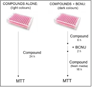
culture by MTT and colony formation assays.
Potential inhibitors of hAGT’s DNA repair activity: study of the
hAGT-compound complex formation by ESI-MS and its toxicity in cell culture.
Table of contents:
1.
Introduction: inhibition of hAGT
2.
hAGT: structure, overexpression and purification
3.
Mass Spectrometry study of the complex formation between hAGT and
its potential inhibitors
4.
Toxicity in cell culture
4.1.
MTT assays
4.2.
Colony formation assays
5.
Conclusions
1.
Introduction: inhibition of hAGT.
The human O
6-alkylguanine-DNA-alkyltransferase (hAGT)
[1]is a DNA repair
protein that removes alkyl mutations from the O
6position of guanines, restoring the DNA
which has been damaged by alkylating agents. This group of chemotherapeutic anticancer
drugs, alkylating agents, produce their cytotoxic effects by introducing alkyl groups which
generate adducts and crosslinks at multiple sites in DNA.
[2]For this reason, hAGT
represents a relevant pharmacological target in the fight against chemotherapy resistance
and patients’ survival.
[3]Chemotherapy based on alkylating agents, together with the
inhibition of hAGT, may result in a higher efficiency of the treatment, due to the blocking
in the repair of the alkylated-DNA by hAGT that leads to apoptosis of cancer cells.
[3]hAGT is a suicidal enzyme which repairs alkyl damage from the O
6position of
guanines, and in minor rate, from the O
4position of thymidines. It has 207 amino acids, a
molecular weight of 21645.9 Da and a theoretical isoelectric point of 8.28. Its
crystallographic structure was solved by Daniels and coworkers in 2000.
[1b]hAGT is a
monomer and has 2 domains, the N- and the C-terminal, where the active site cysteine is
found (Figure 1).
Figure 1. hAGT structure, described by Daniels et. al. The two domains are clearly seen. The violet sphere corresponds to a zinc atom present in the N-terminal domain. PBD code 1EH8.
Figure 2. hAGT (green) bound to DNA (red), with Arginine 128 (yellow, left) entering the double helix and pushing outside the alkylated guanine (blue), which interacts with the active site, Cysteine 145 (yellow, right). PDB code 1T39.
Compound Chemical structure xlogP HBD HBA Charge Molecular Weight
1 1.78 3 13 0 453.492
2 3.68 3 11 0 471.572
3 2.93 3 13 0 467.519
4 1.91 4 14 0 483.518
Compound Chemical structure xlogP HBD* HBA** Charge Molecular Weight
6 2.05 3 13 0 467.519
7 1.15 3 14 -1 478.430
8 1.79 3 15 -1 516.454
9 1.96 3 12 -1 383.348
[image:52.595.77.518.68.598.2]10 1.90 1 2 0 148.205
Table 1. Chemical structure and properties of the compounds tested in this thesis as potential hits of hAGT activity, found by a virtual screening. *HBD: hydrogen bond donors. **HBA: hydrogen bond acceptors.
2.
hAGT: structure, overexpression and purification
Recombinant hAGT full length (FL) (Scheme 1A) was used for the MS study of the
complex formation with the potential inhibitors, but here we also describe in full detail the
overexpression and purification of a mutant version of hAGT which will be used in the
following chapters of this thesis. This inactive mutant of hAGT (hAGT C145S) had a shorter
sequence, where 30 amino acids were removed from its C-terminal end without affecting
its folding, as represented in scheme 1B. Its active site cysteine was replaced by a serine,
causing the loss of its repair activity but maintaining its capacity to recognize and bind
alkylguanine-DNA.
[4]This hAGT mutant was designed in order to use it as a negative
control for the different
in vitro
assays.
Scheme 1. Amino acid sequence of the hAGT proteins. 1A. hAGT FL with the active site, cysteine 145, in red.
1B. hAGT ΔC177 C145S, the shorter version of hAGT FL with a mutation at the active site (serine 145) in red.
The two types of hAGT were overexpressed and purified following the protocol
described by Ruiz
et al
.
[5]This protocol implied the introduction of a hystidine tag with
Figure 3. Purification of hAGT full length with Hys-tag. 3A. Chromatogram of the nickel HiTrap-column purification. 3B. Chromatogram of the size exclusion column, where a single pure peak can be observed. 3C. PAGE of the purification process.
In the case of the hAGT C145S, a dyalisis at 4ºC with prescission protease (PrePro)
was carried out after the nickel HiTrap column. PrePro is a protease which cuts a specific
sequence introduced between the hystidine tag and the N-terminal of hAGT, removing the
Hys tag. Finally, a Size exclusion column was used to further purify the protein and to
slowly exchange the buffer to the storing buffer.
Figure 4. Purification of hAGT C145S with Hys-tag. 4A. Nickel HiTrap purification chromatogram. 4B. Size exclusion purification chromatogram. In this case, two peaks are observed: the first and smaller one corresponds to a portion of hAGT which is aggregated and the second one corresponds to the well folded hAGT C145S. 4C. PAGE of the purification process. The top gel shows the sample preparation prior to the nickel column, the nickel column and the GST column. The bottom gel corresponds to the analysis of the fractions collected from the size exclusion column, showing the pure hAGT C145S.