Aluminium Tolerance Mechanisms in Brachiaria sp.
96
0
0
Texto completo
(2)
(3) Acknowledgements. This Ph.D Thesis was granted by a fellowship PIF 2008–2012 from Autonomous University of Barcelona and by a research stay award to Catalina Arroyave (travel fund fellowships for research outside Cataluña) from the Autonomous University of Barcelona, Spain 2010-2011 at the College of Agriculture Food and Natural Resources, Division of Plant Science, University of Missouri–Columbia. USA. Experimental work was financed by MICIN through the research project BFU201014873/subprogram BFI Technical assistance from the Microscopy Service of UAB is gratefully acknowledged. Especially thank to Alejandro Sánchez Chardi. Thanks to Sílvia Bronsoms a member of ProteoRed-ISCIII network..
(4) Agradecimientos. Con el más sincero agradecimiento a Dra Charlotte Poschenrieder Wiens y a la Dra Roser Tolrà Pérez por todo su gran apoyo en la realización de este trabajo. Al Dr Juan Barceló Coll y su familia por todo su gran apoyo, no sólo en el trabajo sino además por todo su respaldo emocional. Por toda su colaboración a todos los miembros de la unidad de Fisiología Vegetal. A Rosa Padilla, Than Thuy y Jesús Goday por toda su contribución en la realización de este trabajo..
(5) Index ABBREVIATIONS USED OBJECTIVES............................................................................................................................1 INTRODUCTION .................................................................................................................... 2 General Introduction................................................................................................. 3 1. Brachiaria . ............................................................................................................ 3 2. Aluminium: speciation. ......................................................................................... 4 3. Physiological mechanisms of aluminium toxicity. ................................................ 5 3.1. Phytotoxic Al species ......................................................................................... 5 3.2. Mechanisms of Al resistance ............................................................................. 6 3.2.1. Mucilage and border cells ................................................................. 6 3.2.3. Rhizosphere pH .................................................................................. 6 3.2.2. Detoxification of Al by organic acids ................................................. 7 4. References ............................................................................................................ 9 Chapter 1. Evaluation of Aluminium Tolerance in Brachiaria Species ............................. 13 1.1. Introduction . ................................................................................................... 14 1.1.1. Influence of Al on growth and mineral nutrition of Brachiaria species. ....... 14 1.2. Material and methods ..................................................................................... 15 1.2.1. Plant materials and growth conditions. ........................................................ 15 1.2.1.1. Cultivating plants in low-ionic-strength nutrient solutions Discussion ................................................................................................... 15 1.2.2. Mineral nutrition and growth analysis ......................................................... 16 1.2.2.1. Growth and mineral nutrition ................................................................... 16 1.2.3. Statistical analysis ........................................................................................ 17 1.2.3.1. Statistical analysis ......................................................................... 17 1.3. Results. ............................................................................................................. 17 1.3.1. Effect of Al on root growth .......................................................................... 17 1.3.2. Mineral nutrition .......................................................................................... 19.
(6) 1.3.2.1. Effects of aluminium exposure on nutrient uptake by Brachiaria species. ....................................................................................................... 19 1.4. Discussion ........................................................................................................ 24 1.5. References chapter 1........................................................................................ 28. Chapter 2. Toxic Effects and Distribution of Aluminium in the Root Apex of Brachiaria Species. ................................................................................................................................ 32 2.1. Introduction . .................................................................................................... 33 2.2. Material and methods . .................................................................................... 33 2.2.1. Plasma membrane integrity. ............................................................ 33 2.2.2. Hematoxylin staining of roots ......................................................... 34 2.2.3. Aluminium detection with morin staining .......................................34 2.2.4. Fixation, embedding and sectioning for microscopy ....................... 34 2.2.5. Aluminium detection with lumogallion-DAPI stain ......................... 34 2.3. Results. ............................................................................................................. 35 2.3.1. Comparison of Al-resistance between three Brachiaria species. ..... 35 2.3.2. Localization of aluminium by lumogallion-Al, and morin-Al complex fluorescence in roots of B. decumbens and B. brizantha .......................... 36 2.4. Discussion. ........................................................................................................ 36 2.4.1. The integrity of the plasma membrane ........................................... 36 2.4.2. Differential aluminium tolerance between two Brachiaria species. 39 2.5. References chapter 2........................................................................................ 41 Chapter 3. Changes in Root-Morphology of Brachiaria Species (SEM and TEM observations) ....................................................................................................................... 44 3.1. Introduction. ..................................................................................................... 45 3.1.1 Changes in root-morphology of Brachiaria Species .......................... 45 3.2. Materials and methods ....................................................................................45 3.2.1 Transmission electron microscopy (TEM) ......................................... 45 3.2.2 Scanning electron microscopy coupled with spectrometer of energy dispersive X-ray microanalysis (SEM-EDX). ................................................ 45 3.3. Results . ............................................................................................................ 46 3.3.1. Effect of Al on root of Brachiaria species structural changes........... 46.
(7) 3.4. Discussion......................................................................................................... 51 3.5. References chapter 3 ....................................................................................... 52. Chapter 4. A role for Phenolic Substances in Al Tolerance of Brachiaria ....................... 54 4. Introduction. ....................................................................................................... 55 4.1. Phenolic substances in roots............................................................................ 55 4.2. Materials and methods .................................................................................... 57 4.2.1. Determination of capacity for Fe III complexation in root tips (cyclic hydroxamates content in Brachiaria species) ............................................ 57 4.2.2. Analysis of phenolics substances in roots ........................................ 57 4.2.2.1. Phenolics soluble. ............................................................. 57 4.2.2.2. Phenolics from cell wall in Brachiaria decumbens. .......... 58 4.2.3. Autofluorescence ............................................................................ 58 4.3 Results. .............................................................................................................. 59 4.3.1. Iron complexation capacity .............................................................. 59 4.3.2. Phenolics substances. ...................................................................... 59 4.3.2.1. Phenolics soluble in B. decumbens and B. brizantha. ...... 59 4.3.2.2. Phenolics from cell wall in B. decumbens ....................... 60 4.4. Discussion......................................................................................................... 63 4.5. References chapter 4 ....................................................................................... 66 Chapter 5. DIGE Analysis of Root Proteome in Brachiaria decumbens ............................ 69 5.1. Introduction . ................................................................................................... 70 5.1. Proteomic analysis of Brachiaria decumbens roots under aluminium stress .. 70 5.2. Materials and methods .................................................................................... 70 5.2.1. Harvest. ............................................................................................ 70 5.2.2. Protein extraction. ........................................................................... 71 5.2.3. 2D DIGE analysis. .............................................................................. 71 5.2.3.1. Sample preparation and CyDye labelling ......................... 71 5.2.3.2. Isoelectric focusing. .......................................................... 73 5.2.3.3. In-gel digestion with trypsin. ............................................ 74.
(8) 5.2.3.4. Protein identification by MALDI-TOF mass spectrometry. ....................................................................................................... 74 5.2.3.5. Database Search ............................................................... 74 5.3. Results. ............................................................................................................. 75 5.3.1. Protein affected by Al stress............................................................. 75 5.4. Discussion. ........................................................................................................ 78 5.5. References chapter 5........................................................................................ 81. GENERAL CONCLUSIONS.................................................................................................. 84 General Conclusions . .............................................................................................. 85.
(9) Index of abbreviations used. Al Aluminium Al3+ Aluminium ion ALMT Al activated malate transporter CHAPS 3-[(3-Cholamidopropyl) dimethylammonio]-1propanesulfonate DAHP 3-deoxy-d-arabino-heptulosonate 7-phosphate DAPI 4'-6-Diamidino-2-phenylindole DFQA 1,3-di-O-trans-feruloylquinic acid DIGE Difference gel electrophoresis DIMBOA 2,4-dihydroxy-7-methoxy-l,4 - benzoxazin-3-one 2D DIGE Two-dimensional difference gel electrophoresis DTT Dithiothreitol GAX glucoarabinxilans IEF Isoelectric focusing ICP-OES Inductively coupled plasma atomic emission spectroscopy IPG Immobilized pH gradient. MALDI-TOF MS Matrix-assisted laser desorption ionization-time of flight mass spectrometry MATE Mitogen activated toxin extrusion MES 4-Morpholineethanesulfonic acid MeOH methanol Morin 2',3',4',5,7-pentahydroxyflavanone NAD nicotinamide adenine dinucleotide NADH nicotinamide adenine dinucleotide reduce form NCBI Biotechnology Information non-redundant protein database Nrat1 Nramp aluminium transporter 1 Lumogallion (3-[2,4 dihydroxyphenylazo]-2-hydroxy-5-chlorobenzene sulfonic acid) OA organic acid.
(10) OsO 4 osmium tetroxide PMF Peptide mass fingerprinting R Receptor ROS Reactive oxygen species SEM Scanning electron microscopy TFA trifluoroacetic acid TCA trichloroacetic acid TEM Transmission electron microscopy XET xyloglucan endotransglucosylase.
(11) OBJECTIVES Aluminium toxicity is among the main abiotic stress factors limiting crop productivity in acidic environments, which comprise up to 50% of the world's arable lands. The general objective of this Thesis is to elucidate the mechanisms underlying differences in aluminium tolerance, by physiological, biochemical and proteomic approaches in Brachiaria species, B. decumbens, B. Brizantha, and B. ruziziensis. First, we aim to get a general overview of the species differences in tolerance to the low nutrient supply and high Al3+ availability that typically occur in acid mineral soils of the tropics. For this purpose root elongation and mineral nutrition of Brachiaria species growing in low ionic strength nutrient solutions is studied. The possible relevance of differences in Al location in root tips, the Alinduced alterations in root architecture, and the production of phenolics compounds as potential Al binding substances is a second step for typifying species differences in Al tolerance. Once characterized the most Al tolerant species, the proteomic study is focused on this species. Proteomics is considered to bridge the gap between gene sequence and phenotype. In the case of Brachiaria genomic information is scarce. Under these circumstances the proteomic approach may help not only to better understand the metabolic events underlying Al-induced phenotype changes leading to the expression of Al tolerance in Brachiaria, but also to focus future investigations for identifying the responsible genes.. 1.
(12) INTRODUCTION. 2.
(13) General Introduction 1. Brachiaria Brachiaria (Poaceae), a genus of forage grasses of African origin, comprises about 100 species. Currently, some of them are gaining considerable relevance as fodder crops in tropical regions worldwide. The economic interest in these grasses is greatest in tropical America. Cultivation of Brachiaria is of increasing importance as a pasture for livestock production, especially in South American savannas with 40±70 million hectares in Brazil alone. In this region extensive adoption of Brachiaria cultivation over the past three decades has had a revolutionary impact on the productivity of vast areas of previously underused or marginal soils (Boddey et al., 1996; Wenzl et al., 2000; Kochian et al., 2004) The reasons for this fast expansion are good nutritional quality combined with withstanding to heavy grazing, high seed production, good feeding quality, and high resistance to multiple environmental stress factors (Miles et al., 1998). Acidic soils limit crop production in half the world's potentially arable land, mostly in developing countries in Africa, Asia and South America. Aluminium toxicity is considered among the most relevant abiotic stress factors limiting crop production on acid mineral soils in the tropics. In soils with a pH below 5 and especially if pH values fall below 4.5, toxic forms of Al are solubilised into the soil solution. Plant available Al ions inhibit root growth and functioning, and thus reducing yields of crops, such as wheat (Triticum aestivum L.), corn (Zea mays L.), or soybean (Glycine max (L.) Merr.). Besides Al toxicity, acid soils are characterized by having excess H+ and Mn2+, as well as deficiency of Ca2+, Mg2+, and PO 4 3− (Kochian et al., 2004; Panda and Matsumoto, 2007). Certain Brachiaria species are performing well under the same or similar harsh conditions. The condition of being plants well-adapted to tropical climate with C4-type photosynthesis makes Brachiaria even more attractive and further breeding efforts. Brachiaria species are a main basis for animal nutrition. Those plants are commercially exploited in tropical regions because they are best adapted to both the low-fertility and the aluminium toxicity typically found in these zones. Brachiaria brizantha (A. Rich) (palisadegrass) besides Al-tolerance also has resistance to spittlebug. Brachiaria ruziziensis (ruzigrass) is less adapted to acid soil conditions, but has better nutritional quality than other species of Brachiaria. Brachiaria decumbens (signalgrass) is one of the most widely sown forage grasses 3.
(14) in the tropics with 26.4 million ha in Brazil alone. Unlike most food crops, it is directly derived from a wild apomictic germplasm accession that deserves special attention because of its extreme Al tolerance and good adaptation to infertile acid soils (Wenzl et al., 2001, 2002). 2. Aluminium: speciation Aluminium is a major constituent of the mineral fraction of most soils. Aluminium usually is tightly bound in the silicate lattice; so this amphoterous element is sparingly soluble and mostly unavailable to plants. However, in soils with acid or highly alkaline pH the solubility of Al is enhanced. Aluminium has the electron configuration [Ne]3s23p1. At acid pH the formation and stability of the Al aqua complex Al(H 2 O) 6 3+ is favoured, This trivalent ionic compound usually written as Al3+ seems to be the most phytotoxic Al species. Part of the difficulty of studying Al-related processes in plants can be attributed to the complex chemistry of Al. Depending on the conditions; Al can undergo hydrolysis reactions to several different mononuclear hydroxides or polymerization reactions to polycations (Casey, 2006). The most important variables in the aqueous chemistry of aluminium are the pH and soluble organic matter. At low pH Al hydrolyzes in solution yielding the aluminium hexahydrate cation [Al(OH 2 )6]3+ (aq) This trivalent cationic Al species simply written as Al3+, is the predominant form of the monomeric aluminium ion in the strongly acidic range (pH ≤ 4.), whereas hydroxyl species such as [AlOH]2+ and [Al(OH)2]+ form as the pH increases to values around 6. In the near neutral pH range (5 < pH < 8) aluminium trihydroxide (Al(OH) 3(s) ) or gibbsite, occurs, whereas Al(OH) 4 -, or aluminates, dominate in alkaline conditions. Many of the monomeric Al cations bind to various organic and inorganic ligands such as PO 4 3-, SO 4 2 -, F-, organic acids, proteins, and lipids (Poschenrieder et al., 2008; Casey, 2006; Delhaize and Ryan, 1995) The phytotoxic Al3+ is the hardest Lewis acid among other trivalent metal cations such as La3+, Cr3+ or Ga3+. Hard Lewis acids are characterized by a low covalent and a high ionic index. Despite the fact that Al is the most abundant metal on earth, so far there is no recognized biological function for this element. This may be attributed to different factors such as slow ligand exchange rates, hampered membrane transport, and its interference with Mg2+ binding sites at phosphate esters (DNA/RNA, ATP, etc.) (Merkx and Averill, 1999; Poschenrieder and Barceló, 2004) 4.
(15) 3. Physiological mechanisms of aluminium toxicity 3.1 Phytotoxic Al species The aluminium hexahydrate [Al(OH 2 ) 6 ]3+ , which is solubilised into the soil solution under low pH, is considered the main toxic species for plants. It has six coordination sites and as class A metal preferentially binds to oxygen donor ligands. Multiple molecular targets in plant cells can act as potential ligands for Al3+ and both the toxicity mechanisms and the means for detoxification are based on the availability of potential binding sites (Barceló and Poschenrieder, 2002). The target site of Al toxicity is the root apex. The primary symptom of Al-toxicity in plants is the inhibition of root growth. This process has been studied at the tissue and cellular levels. At tissue level, the distal transition zone has been identified as the most Al sensitive root zone (Horst et al., 2010). This region corresponds to the zone where cells leaving the meristem (cell division zone) are about to enter the elongation zone (cell expansion zone). Aluminium causes fast inhibition of both root tip cell division and cell elongation. Moreover, Al toxicity changes the entire root architecture by fast induction of lateral root development (Doncheva et al.,2005; Amenós et al., 2009). In sensitive species the distal transition accumulates more Al than the mature root tissues. Multiples studies have clearly shown that both apoplast and symplast are sites of aluminium location in plants (Vázquez et al., 1999; Zheng and Yang, 2005; Horst et al., 2010) Cell walls seem primary sites for Al binding and toxicity. In Al-sensitive plants Al causes changes in the amount of cell wall polysaccharides. It has been proposed that Al3+ is attracted to the negatively charged carboxyl groups of unmethylated pectins. Both a low degree of methylesterification of cell wall pectins and an Alinduced accumulation of β-1,3-glucan (callose) synthesis in the apoplast have been found to be related to Al sensitivity in different crop plants (Eticha et al., 2005 and Yang et al., 2008; Panda and Matsumoto, 2007). Also Yang et al., (2008) found in resistant rice cultivars a relation between higher degree of methylesterification of cell wall polysaccharides and less Al-binding sites. Thus the degree of methylesterification of cell wall pectins could be a mechanism of Al resistance. The plasma membrane is a further target for Al toxicity. Aluminium may damage the plasma membrane by inducing reactive oxygen species causing lipid 5.
(16) peroxidation and oxidative stress (Liu et al., 2008). Wheat roots treated with Al showed increased H+ efflux near the root apex and root cap. Aluminium interfered with calmodulin-stimulated ATPase activity and proton transport in plasmamembrane (Miyasaka et al., 1989). Furthermore, Al3+ affects the mechanisms controlling the organization of microtubule cytoskeleton; dividing root-tip cells and the nucleus could be targets for injury (Frantzios et al., 2000; Kochian et al., 2004; Doncheva et al., 2005) 3.2 Mechanisms of Al resistance 3.2.1 Mucilage and border cells Border cells have been described as a possible mechanism for Al-tolerance. The root border cells (RBCs) exude mucilage that may help to protect plants by inhibiting Al uptake into the root meristem. Studies in pea (Pisum sativum) also suggest a possible role of cell-wall pectins in the root border cells for immobilizing Al (Miyasaka et al., 2001). Comparative studies in soybean cultivars Zhechun (Altolerant cultivar) and Huachun (Al-sensitive cultivar) revealed enhanced inhibition of root growth by Al when RBCs were removed from the root apex (Cai et al., 2011.) Aluminium induced a thicker mucilage layer around detached border cells in two snapbean (Phaseolus vulgaris) cultivars, avoiding Al uptake into the roots (Yu et al., 2009) 3.2.2 Rhizosphere pH The most important factor for root Induced changes in rhizosphere pH is the uptake of nutrients (cation-anion balance). The rhizosphere pH, in turn, has a marked influence on the availability to plants of essential nutrients and toxic elements present in the soil solution. Increase of rhizosphere pH has been proposed as an important mechanism to avoid aluminium toxicity. This would be one of the most effective external Al resistance mechanisms of plants (Marschner, 2012). Several studies were directed toward the analysis of the possible correlation between changes in the rhizosphere pH and plant Al tolerance. In two varieties on the wheat (Triticum aestivum L.) under Al stress, the Al-tolerant variety showed a stronger capacity of enhancing rhizosphere pH than the Alsensitive variety. Similar results are now available for other species (wheat, sorghum, and Arabidopsis thaliana mutant talr-104) that can change the rhizosphere pH increased to tolerate Al toxicity (Degenhardt et al., 1998; Yang et al., 6.
(17) 2011). Al-sensitive mutants (als3 and als5) exhibited lower rhizosphere pH values (Bose et al., 2010). Contrastingly, cultivation of Al hypertolerant plants, such as tea causes decrease of soil pH rather than an increase (Alekseeva et al., 2011).. 3.2.3 Detoxification of Al by organic acids. Fig1. Hypothetical models for the aluminium (Al)-stimulated secretion of organic acid anions (OA) 3+ from plant roots. Pattern I-type responses: Al activates a constitutively expressed anion efflux 3+ channel that facilitates exudation of organic acids (OA) from root tips upon Al exposure. Al 3+ resistance genotypes show greater expression than Al -sensitive ones. Activation of organic anion (OA) efflux by Al interacting directly with the pre-existing proteins in the plasma membrane (members 3+ of the ALMT and MATE families of proteins in wheat). In the Pattern II response, Al first interacts with the cell, possibly involving a specific receptor (R) on the plasma membrane; this induces the activation of genes coding for the transport proteins. Organic acid anions form stable, non toxic complex with Al(Ma et al., 2001; Delhaize et al., 2007). Plants produce a range of mono-, di- and tri- carboxylates. Most of these organic acids, such as citrate, malate, and oxalate, form strong complexes with Al3+ as indicated by the high stability constants (Poschenrieder et al., 2008). Many plant species can increase the efflux of these organic acids at the root level as a mechanism for Al tolerance. The kind of organic acids secreted, as well as secretion patterns differ among plant species (fig. 1 taken from Ma et al., 2001). A fast Al–induced efflux of malate, following pattern 1, seems responsible for Al 7.
(18) tolerance in wheat (Triticum aestivum L.). In Al3+-resistant Arabidopsis thaliana (Delhaize et al., 1993; Sasaki et al., 2004; Delhaize et al., 2007) exposure to Al nearly instantaneously activated a concentration-dependent citrate release. Citrate exudation from root tips has also been observed in Al-tolerant maize (Zea mays) and barley (Hordeum vulgare L.) (Piñeros et al., 2002; Furukawa et al., 2007). Aluminium-resistant buckwheat secretes oxalic acid from roots in response to aluminium stress (Ma et al., 1997), while radish (Raphanus sativus) and rye (Secale cereale) release two organic acids malate and citrate (Ma et al., 2001) Recently there is molecular and genetic evidence showing that other properties of the root may contribute to Al resistance. In recent investigations Ma et al., 2009 were able to discover two genes STAR1 and STAR2 that are implicated in Al resistance in rice (Oryza sativa L.). STAR1 interacts with STAR2 to form a complex that functions as a unique ATP binding cassette ABC transporter. The complex works as a UDP-GLc efflux transporter, which is required for detoxification of aluminium. Recently a study localized an aluminium transporter (Nrat1) in plasma membrane of rice. This transporter is a member of the Nramp family (Xia et al., 2010). This finding is highly relevant because identification of Al- transporters helps to a better understanding of the mechanisms that are involved in Altolerance or Al-toxicity. Despite the fact that much progress has been made in our understanding of Al toxicity and tolerance mechanisms in major crop plants with a relatively low tolerance level, such as wheat or bean, the mechanisms underlying Al hyperresistance as observed in Brachiaria are still not established. In this investigation we have compared the Al resistance in three species of Brachiaria, Brachiaria decumbens, Brachiaria brizantha and Brachiaria ruziziensis. B. decumbens and B, brizantha, both species have a high tolerance to aluminium toxicity. However, B. ruziziensis is sensitive to high concentrations of aluminium. Previous investigations into the mechanisms of Al hyperresistance in Brachiaria evidenced that they are neither based on external detoxification of Al by organic acid exudation nor on apical root-induced alkalinisation (Wenzl et al., 2001).. 8.
(19) References Amenós, M., Corrales, I., Poschenrieder, C., Illés, P., Baluska, F., Barceló, J. 2009. Different Effects of Aluminum on Actin Cytoskeleton and Brefeldin A-sensitive Vesicle Recycling in Root Apex Cells of Two Maize Varieties Differing in Root Elongation Rate and Al Tolerance. Plant Cell Physiol 50: 528-540 Barceló, J., Poschenrieder, C. (2002) Fast root growth responses, root exudates, and internal detoxification as clues to the mechanisms of aluminium resistance and tolerance: a review. Environ Exp Bot 48: 75-92 Boddey, R., Rao, I. and Thomas R. 1996. Nutrient cycling and environmental impact of Brachiaria pastures. In: Miles J. Maass B and do Velle C. (eds). Brachiaria: Biology, Agronomy and improvement. Cali: CIAT and Brasilia: EMBRAPA. 72-86 Bose, J., Babourina, O., Shabala, S., Rengel, Z. 2010. Aluminium-induced ion transport in Arabidopsis: the relationship between Al tolerance and root ion flux. J Exp Bot 61: 11 3163–3175 Casey, W. 2006. Large Aqueous Aluminum Hydroxide Molecules. Chem Rev 106: 1 1–16 Cai, M., Wang, F., Li, R., Zhang, S., Wang, S., Xu, D. Response and tolerance of root border cells to aluminum toxicity in soybean seedlings. J Inorg Biochem 105: 966971 Degenhardt, J., Larsen, P., Howell, S., Kochian, L. 1998. Aluminum Resistance in the Arabidopsis Mutant alr-104 Is Caused by an Aluminum-Induced Increase in Rhizosphere pH. Plant Physiol 117: 19–27 Delhaize, E., Gruber, B., Ryan PR. 2007. The roles of organic anion permeases in aluminium resistance and mineral nutrition. FEBS Letters 581:2255–2262 Delhaize, E., Ryan PR. 1995. Aluminum Toxicity and Tolerance in Plants. Plant Physiol 107: 31 5-321 Doncheva, S., Amenós, M., Poschenrieder, C., Barceló, J. 2005. Root cell patterning: a primary target for aluminium toxicity in maize. J Exp Bot 56: 414 1213–1220. 9.
(20) Furukawa, J. Yamaji, N. Wang, H. Mitani, N. Murata, Y. Sato, K. Katsuhara, M. Takeda, K. Ma, J. 2007. An aluminum-activated citrate transporter in barley. Plant Cell Physiol 48:1081–1091 Frantzios, G., Galatis, B., Apostolackos, P. 2000. Aluminium effects on microtubule organization in dividing root-tip cells of Triticum turgidum. I. Mitotic cells. New Phytol 145: 211-224 Horst, W., Wang, Y., Eticha, D. 2010. The role of the root apoplast in aluminiuminduced inhibition of root elongation and in aluminium resistance of plants: a review. Ann Bot 106: 1 185-197 Huang, C., Yamaji, N., Mitani, N., Yano, N., Nagamura, Y., Ma, J. 2009. A BacterialType ABC Transporter Is Involved in Aluminum Tolerance in Rice W. Plant Cell 21: 655–667 Kochian, L., Hoekenga, O., Piñeros, M. 2004. How do Crop Plants Tolerate Acid Soils? Mechanisms of Aluminum Tolerance and Phosphorous Efficiency. Annu Rev Plant Biol 55:459-493 Ma, J. 2007. Syndrome of aluminum toxicity and diversity of aluminum resistance in higher plants. Int Rev Cytol 264: 225–252 Ma, J., Ryan, P., Delhaize, E. 2001. Aluminium tolerance in plants and the complexing role of organic acids. Trends Plant Sci 6: 6 273-278 Ma, J., Zheng, S., Hiradate, S., Matsumoto, H. 1997b. Detoxifying aluminum with buckwheat. Nature 390: 569–570 Marschner P. 2012. Third edition Marschner’s Mineral Nutrition of Higher Plants. Elsevier Ltd. ISBN-978-0-12-384905-2. 353Merkx and Averill. 1999. Probing the Role of the Trivalent Metal in Phosphate Ester. J Am Chem Soc 121: 6683-6689 Miles J.W. Maass, L., Do Valle, C., Kumble, V (Eds.). 1998. Brachiaria: Biología, Agronomía y Mejoramineto. Centro Inter de Agricult Trop Publication number 154, Cali Colombia 1998 Miyasaka, S,. Hawes, M. 2001. Possible Role of Root Border Cells in Detection and Avoidance of Aluminum Toxicity. Plant Physiol 125: 1978–1987. 10.
(21) Miyasaka,S,. Kochian, L., Shaff, J. Foy, C. 1989. Mechanisms of Aluminum Tolerance in Wheat. Plant Physiol 91: 1188-1196 Liu, Q., Yang, J., He, L., Li, Y. Zheng, S. 2008. Effect of Aluminum on Cell Wall, Plasma Membrane, Antioxidants and Root Elongation in Triticale. Biol Plant 52:1 87-92 Panda and Matsumoto. 2007. Molecular physiology of aluminum toxicity and tolerance in plants. Bot Rev 73: 4 326-347 Piñeros, M., Magalhaes J., Carvalho, V., Kochian, L. 2002. The Physiology and Biophysics of an Aluminum Tolerance Mechanism Based on Root Citrate Exudation in Maize. Plant Physiol 129: 1194–1206 Poschenrieder, C., Gunsé, B., Corrales, I., Barceló, J. 2008. A glance into aluminum toxicity and resistance in plants. Sci Total Environ 400: 356-368 Ryan,P., Tyerman, S., Sasaki,T., Furuichi,T., Yamamoto, Y., Zhang, W., Delhaize, E. 2011. The identification of aluminium-resistance genes provides opportunities for enhancing crop production on acid soils. J Exp Bot 62: 1 9–20 Sasaki, T., Yamamoto, Y., Ezaki, B., Katsuhara, M., Ahn, S., Ryan, P., Delhaize, E., Matsumoto, H.2004. A wheat gene encoding an aluminium-activated malate transporter. Plant J 37, 645–653 Tice, K., Parker, D., DeMason, D. 1992 Operationally Defined Apoplastic and Symplastic Aluminum Fractions in Root Tips of Aluminum-intoxicated Wheat. Plant Physiol 100: 309-318 Vázquez, M., Poschenrieder, Ch., Corrales, I., Barceló, J. 1999. Change in apoplastic aluminum during the initial growth response to aluminum by roots of a tolerant maize variety. Plant Physiol 119: 435-444 Wenzl, P., Chaves, A., Buitrago, M., Patiño, M., Meyer, J., Rao, I. 2002. Aluminium Stress Stimulates the Accumulation of organic acids in Root Apice of Brachiaria species. J Plant Nutr Soil Sci 165: 582-588 Wenzl, P., Chávez, L., Mayer, J., Rao, I. and Nair, M. 2000. Roots of nutrientdeprived Brachiaria species accumulate 1,3-di-0-trans-feruloylquinic acid. Phytoche 55: 389-395 11.
(22) Wenzl, P., Patiño, G., Chaves, A. 2001.The high level of aluminum resistance in signalgrass is not associated with known mechanisms of external aluminum detoxification in root apices. Plant Physiol 125: 3 1473-1484 Xia, J., Yamaji, N., Kasai, T., Ma, J. 2010. Plasma membrane-localized transporter for aluminum in rice. PNAS 107: 43 18381–18385 Yang, Y., Wang, Q., Geng, J., Guo1, Z., Zhao, Z. 2011. Rhizosphere pH difference regulated by plasma membrane H+-ATPase is related to differential Al-tolerance of two wheat cultivars. Plant Soil Environ 57: 5 201–206 Yang, J., Li, Y., Zhang, Y., Zhang, S., Wu, Y., Wu, P., Zheng, S. 2008. Cell Wall Polysaccharides Are Specifically Involved in the Exclusion of Aluminum from the Rice Root Apex. Plant Physiol 146: 602–611 Yu, M., Shen, R., Liu, J., Chen, R., Xu, M., Yang, Y., Xiao, H., Wang, H., Wang, H., Wang, C. 2009. The role of root border cells in aluminum resistance of pea (Pisum sativum) grown in mist culture. J Plant Nutr Soil Sci 172: 528–534 Zheng and Yang. 2005. Target sites of aluminum phytotoxicity. Biol Plant 49: 3 321-331. 12.
(23) Chapter 1. Evaluation of Aluminium Tolerance in Brachiaria Species. 13.
(24) Chapter 1 1. Introduction 1.1. Influence of Al on growth and mineral nutrition of Brachiaria species The acquisition of water and nutrients from soil are the main functions of plant roots. The ability of species to develop on soils with low nutrient availability depends on both nutrient uptake efficiency and nutrient use efficiency. High uptake efficiency implies powerful mechanisms for mobilization of essential nutrients from the rhizosphere and high affinity uptake and transport mechanisms. Nutrient use efficiency, in turn, depends on low internal requirements for optimal growth (Rengel et al., 2009; Hinsinger et al., 2011) In the case of acid soils, the extremely low ionic strength of the soil solution is associated with deficiency of essential nutrients and complex other stress factors including aluminium (Al) and manganese (Mn) toxicities. At lower pH, H+ toxicity may also affect plant growth disturbing the plant nutrient balance, especially affecting tissue concentrations of Ca, Mg, K and B (Poschenrieder et al., 1995). Aluminium exposure can inhibit root elongation and specific nutrient absorption , decreasing uptake of Ca, Mg, K, P and B. Aluminium-induced alteration of Ca2+ and K+ homeostasis have frequently been observed. For example, Piñeros and Kochian (2001) found that Al3+ interact with plasma-membrane channel proteins, blocking the uptake of K+ and Ca2+. Other experiments suggest that the Al-induced callose synthesis depends on Al-induced disturbance of Ca homeostasis (Llugany et al., 1994; Massot et al., 1999; Rengel and Zhang, 2003). Furthermore, Mg2+ is influenced by acidity and presence of Al3+, since its uptake by plants decrease with the pH in the solutions. Whenever an increase in the aluminium concentration is observed, magnesium availability is affected (Kamprath and Foy, 1985). Maize varieties exposed to acid solutions containing Al showed alterations of nutrient uptake. This was due to both H+ toxicity and shortterm exposure to Al. Aluminium induced changes in the B uptake rate and decreased vacuolar K+, and phosphate concentrations (Poschenrieder et al., 1995; Garzón et al., 2011) High acid soil tolerance is a common feature of Brachiaria species such as Brachiaria decumbens and Brachiaria brizantha. These species must have high tolerance to Al3+ and H+ toxicity. However, the absolute importance of different 14.
(25) soil nutrients influencing growth and productivity of Al and proton tolerant plants also depends on their physiological adaptation to the low availability of essential nutrient in the acid soils (Wenzl et al., 2001, 2003) The aim of this study was to investigate the influence of different high Al concentrations in low ionic strength nutrient solution on growth and mineral nutrition of three Brachiaria species in order to further clarify the role of the ability to maintain nutrient homeostasis in acid soil tolerance of Brachiaria. For this purpose the hydroponic solution used here was based on the modified solution from Wenzl et al., 2001, which was designed for being a real approximation to the chemical soil properties that limit forage productivity on acid mineral soils. This is a highly relevant aspect because practically useful conclusions only can be drawn if experimental conditions simulate natural environments and closely resemble those that would be observed under natural conditions.. 1.2. Materials and methods 1.2.1. Plant materials and growth conditions 1.2.1.1. Cultivating plants in low-ionic-strength nutrient solutions Seeds of different Brachiaria species, signalgrass B. decumbens (Stapf), palisade grass B. brizantha (Hochst. Ex A. Rich.) Stapf, and Congo grass B. ruziziensis (R.Germ. and C.M.Evrard) Unipasto Marangatu, Brasil and Agrosemillas, Medellín, Colombia) were germinated on floating treys on distilled water. Emerged seedlings were transferred to continuously aerate nutrient solution (plastic vessels of 5 L capacity; 10 plants per beaker). The solution contained low nutrient concentrations. The composition was (in μM): 106 (NH 4 ) 2 SO 4 ; 100 KNO 3 ; 20 K 2 SO 4 ; 200 Ca(NO 3 ) 2 ; 1 NaH 2 PO 4 .H 2 O; 169 CaCl 2 ; 120 MgSO 4 .7H 2 O; 10 Fe-EDTA; 1 ZnSO 4 .7H 2 O; 0.2 CuSO 4 .5H 2 O; 1 MnSO 4 .H2O; 0.004 (NH 4 ) 2 SO 4 ; 20 SiCl 4 ; 0,001 (NH 4 ) 6 MoO 24 .4H 2 O; 6 H 3 BO 3 . Nutrient solution's pH was checked daily and adjusted to pH 4.2±0.1. The solutions were renewed every two days. After 15 days in control nutrient solution, plants were transferred to treatment solutions supplemented or not (controls) with 200 μM Al as AlCl 3 . The activity of Al3+ in the solution was 31.7 μM (GEOCHEM-EZ, Cornell University, USA). Plants were grown in an environmental-controlled growth chamber (Conviron® SH10) under the 15.
(26) following conditions: 12/12 h photoperiod, photon fluence rate 600 μEm−2 s−1; day/night temperature 27/25 °C, and relative humidity 60%. 1.2.2. Mineral nutrition and growth analysis 1.2.2.1. Growth and Mineral nutrition Length of the main root was measured before Al supply and every 24h until 96h after start of the treatments (time samples 24, 48, 72 and 96h). Nutrition assays were performed in triplicate (plastic vessels of 6 L capacity; 21 plants per beaker). Plants pre-cultured for 15 days in control nutrient solution were used for the 72h Al uptake and mineral nutrition experiments. For this purpose plants were divided into 4 groups. One group was maintained in control nutrient solution for the following 72h (control), the others were transferred to solutions containing 200 μM Al either immediately (total Al exposure time 72h) or after a further 24h or 48h period in control solution. So these last two groups received Al for 48 and 24h, respectively. This experimental design allowed using the same control for all Al exposure treatments (all plants were of the same age when harvested). Before sample processing the root samples collected from control and Al treatment plants were desorbed with 1 mM citrate for 30 minutes followed by distilled water for 15 minutes. Root dry weight was measured on 42 plants of each species and treatment after oven drying (105°C). Dried samples were ground to fine powder. The analysis of nutrients was done after digestion of plant material with acid mixture in an open hotblock digestion system (Item No.: SC154- 54-Well HotBlock™, United Kingdom). The method prepares plant tissue for the quantitative determination of the concentration of boron (B), calcium (Ca), copper (Cu), iron (Fe), magnesium (Mg), manganese (Mn), phosphorus (P), potassium (K), zinc (Zn) and aluminium (Al) using 5 ml nitric acid 69 % and 2 ml hydrogen peroxide 30 % (HN0 3 /H 2 0 2 ) and 25 mL miliQ water. Concentrations of these elements were determined in three samples per species and treatment. The concentrations of nutrients were determined by inductively coupled plasma optical emission spectrometry (ICPOES, Thermo Jarrell-Ash, model 61E Polyscan, England).. 16.
(27) 1.2.3 Statistical analysis 1.2.3.1. Statistical analysis The experiment considers nutrient uptake kinetics by determining element concentrations in the Brachiaria species after three different exposure times 24, 48 and 72h under aluminium treatment and plants without aluminium (controls). Three replicates per species and treatment (21 plants for each replicate n = 3) were analysed. Variance-stabilizing transformations were undertaken, where necessary, to conform to the assumptions of ANOVA. Subsequently, data were analysed by ANOVA followed by Tukey HSD to identify significant differences between Brachiaria species for each time. Test of Dunnet HSD was applied in order to compare upon time the nutrient uptake for each species with its control.. 1.3. Results 1.3.1. Effect of Al on root growth. Fig. 2 Elongation of main root of three Brachiaria species exposed to Control (no Al supply) nutrient solutions. Values are means ± SE from n = 4 plants per time sample in the case of B. decumbens and B. brizantha, n=21 for B. ruziziensis. Under control conditions the three species maintained constant root elongation. In B. brizantha root elongation rate was approximately 0.7 cm /day while the mean elongation rate in B. decumbens was around 0.3 cm day−1, and nearby 0.15 cm day−1 for B. ruziziensis (Fig 2). Roots elongation rates in B. decumbens and B. 17.
(28) ruziziensis were about 56 % and 78 %, respectively, lower than the rate observed in B. brizantha. The Brachiaria species where exposed to 200 μM Al (Fig 3). In this case, roots of B. brizantha maintained a constant elongation rate of 0.4 cm day−1. But after 96h exposure to Al the relative root length was 41% that of control plants. Aluminium supply had a time-dependent effect on the root elongation rate in B. decumbens. A strong initial reduction on root elongation of 75% was observed after 24h exposure to Al. However, the root elongation rate recovered upon time. After 96h a reduction of only 17% in comparison to control in the root elongation rate was observed (Fig3). In the case of B. ruziziensis, after 24 h a strong initial reduction of 68 % was observed. No recovering of the root elongation rate occurred during the experimental period of 96h when a reduction rate of 67% was found in the Al treated plants (Fig. 3). Species differences in root elongation rates were statistically significant according to ANOVA (p < 0.05).. Fig. 3 Effect of Al supply on root elongation of different Brachiaria species, B.decumbens, B. brizantha and B. ruziziensis. Elongation root was calculated from root elongation of 10-dold seedlings exposed to Control (no Al supply) or Al-spiked nutrient solutions, pH 4,2, containing 200 µM Al. Data are means ± SE (for B. decumbens and B. brizantha n=4, for B. ruziziensis n=21). Y- Axis (root elongation) is different for each Brachiaria species. 18.
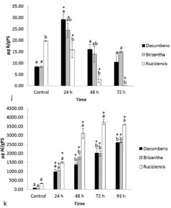
(29) 1.3.2. Mineral nutrition 1.3.2.1. Effects of aluminium exposure on nutrient uptake by Brachiaria species Under control conditions B. decumbens, B. brizantha, and B. ruziziensis showed significant differences among their root concentrations of several essential nutrients. (Fig. 4 b, c, d, e, g, h and i). The root concentrations of essential alkaline and alkaline earth elements were lower in B. decumbens (K, Ca, and Mg) and B. brizantha (K and Ca) than in B. ruziziensis. Excepting Fe, concentrations of essential transition metals were also lower in roots of B. decumbens (Cu, Zn) and B. brizantha (Mn, Zn, Cu) than in those of B. ruziziensis. Root boron concentrations were substantially lower in B. decumbens and B. brizantha than in B. ruziziensis. Under control conditions no statistically significant differences in root P concentrations among the Brachiaria species were observed. Brachiaria brizantha had significantly lower root sulphur concentrations than the other Brachiaria species. Although all species had low Al root concentrations under control conditions, the mean value for B. ruziziensis was slightly higher than in the other species. When exposed to Al the root concentrations of this element steadily increased with exposure time in all species. Roots of B. ruziziensis accumulated considerably higher concentrations than those of B. decumbens and B. brizantha. Aluminium supply had a marked, time and species dependent influence on the root concentrations of mineral nutrients. In B. decumbens root concentrations of P and K exhibited a U shape behaviour in response to Al exposure time with lowest values for short (24 and /or 48h) and recovering after 72h exposure. Root concentrations of P and K of the other Brachiaria species were little affected by Al supply. Aluminium exposure considerably decreased root Mg and Mn concentrations, while Cu and Fe concentrations rather tended to increase, especially Fe in roots of B. ruziziensis and Cu in those of B. brizantha and B. decumbens. The most notable species differences in Al-induced changes were observed for boron. Exposure for 24h to Al caused a 2-3 fold increase of root B concentrations in B. decumbens and B. brizantha, but did not affect the considerably higher B concentration in B. ruziziensis. Longer Al exposure however decreased root B levels in B. ruziziensis below detection limit (< 1 µg/g d.w.), while B concentrations in B. decumbens and B. ruziziensis returned to values after 72h of Al exposure. 19.
(30) 20.
(31) 21.
(32) 22.
(33) Fig. 4 Root nutrient uptake kinetics for Brachiaria species. Kinetics was evaluated at three times 24, 48 and 72h, under aluminium-treatment 200 μM AlCl 3 and control without aluminium. Mean±SE is -1 -1 shown. ANOVA-Tukey HSD a. Concentration of Phosphorus (P: μg g PS ). b. Concentration of -1 -1 -1 -1 potassium (K: μg g PS ).c. Concentration of calcium (Ca: μg g PS ). d. Concentration of -1 -1 -1 -1 magnesium (Mg: μg g PS ) e. Concentration of sulphur (S: μg g PS ). f. Concentration of iron (Fe: -1 -1 -1 -1 -1 -1 μg g PS ). g. Concentration of zinc (Zn: μg g PS ). h. Concentration or copper (Cu: μg g PS ). i. -1 -1 -1 -1 Concentration of manganese (Mn: μg g PS ). j. Concentration of Boron (B: μg g PS ). k. -1 -1 Concentration of aluminium (Al: μg g PS ). Test Dunnet HSD for species B. decumbens, B. brizantha and B. ruziziensis regarding their respective controls (*). 23.
(34) 1.4. Discussion Considerable species differences in root elongation rates and root mineral nutrient concentrations were observed in control plants growing in acid; low ionic strength nutrient solution without Al. Brachiaria ruziziensis exhibited the lowest root elongation rate in control solutions. Moreover, this species had by far the highest root Ca and B concentrations under control conditions. In general, plant species largely differ in their Ca and B requirements. Monocots and more specifically grasses have low requirements. But considerable differences in Ca and B accumulation within grasses have been observed. As an example Agrostis setacea from acid soils accumulate less Ca than A. stolonifera adapted to calcareous soils (Clarkson 1965). Both Ca and B are typically required for cell wall stability (Bolaños et al., 2004; Marschner, 2012) and the higher concentrations of these elements in roots of B. ruziziensis suggest higher Ca and B requirements that may reflect differences in cell wall composition. Those, in turn, may be related to the considerably higher Al sensitivity in this species (see below) in comparison to B. brizantha and B. decumbens. Taken together these results suggest that under our control conditions the root elongation rate of B. ruziziensis may have been limited by the acid pH and the low ionic strength of the nutrient solution. The use of this nutrient solution is justified, however, because it represents a realistic approximation to chemical acid soil properties (Wenzl et al., 2003). The marked growth difference between the three Brachiaria species under simultaneously Al-treatment, low pH and by low ionic strength nutrient solution conditions probably was the result of an interaction between the effects of the three stress factors (Poschenrieder et al., 1995; Wenzl et al., 2003; Schroth et al., 2003). Here only the influence of Al-toxicity was evaluated in detail, but comparative study of the mineral nutrients in the plants revealed clear differences in both the quantitative requirements under control conditions and the ability to maintain nutrient homeostasis under Al supply. According to relative root elongation rates under Al supply, the Al tolerance of Brachiaria species decreased in the order of B. decumbens >B. brizantha > B. ruziziensis. In most crop species root growth is reduced by about 50% when exposed to Al3+ activities between < 1 to 5 μM. The inhibitory effect can be fast and may occur within 30 to 120 minutes after the exposure to Al (Barceló and Poschenrieder 2002, Doncheva et al. 2005; Llugany et al., 2005; Poschenrieder et 24.
(35) al., 2008). In this work, the roots of B. decumbens and B. brizantha continued to elongate in solutions containing 200 μM total Al corresponding to 32 μM Al3+ activity, for the entire period of evaluation. After 96 h Al reduced root elongation in B. brizantha by 41% while in B. decumbens the reduction was only 17%. By contrast, B. ruziziensis suffered a 67% inhibition of elongation after this exposure period (96 h). The response to Al in B. ruziziensis found here is in line with the observation by Wenzl et al., 2001 who reported a 50% inhibition in root elongation for B. ruziziensis growing in solution with 115 µM total Al. Our results clearly reveal species differences in Al tolerance within the genus Brachiaria. Aluminium tolerance in the three species considered here followed the order B. decumbens > B. brizantha > B. ruziziensis. The comparative use of relative root elongation under ion stress conditions as a tolerance index can be influenced by differences among species in root elongation rates under control conditions. A low root elongation rate is a common characteristic in many Al-resistant species or varieties (Amenós et al., 2009). This is also the case in the Brachiaria species considered here. The root elongation rate of B. decumbens is characteristically lower than that of B. brizantha. This lower growth rate can contribute to a certain overestimation of Al resistance, at least in short-term evaluations. However, the observation that B. decumbens exposed to Al recovered a root elongation rate close to control values after a lag time of 48 to 72 hours not only confirms the Al hyperresistance of this species, but also suggest that at least part of the Al tolerance mechanism is Al-inducible. In this sense B. decumbens behaves similar to some Al tolerant maize varieties were also a lag time of several hours was required for full expression of Al tolerance (Kidd et al., 2001). Although B. ruziziensis is the most Al sensitive species among those analysed here, its ability to maintain a low but still significant root elongation at 32 μM Al3+ activity makes it clearly more tolerant than most other fodder grasses. In this sense B. ruziziensis can be classified as a species with low Al sensitivity, while B. brizantha and B. decumbens can be classified as moderately tolerant and hypertolerant, respectively. The higher Al tolerance in B. decumbens and B. brizantha than in B. ruziziensis was clearly related to better Al exclusion from the roots as indicated by the lower root Al concentrations in these species (Fig 4 k). Besides Al tolerance, tolerance to H+, 25.
(36) and Mn toxicity, and Mg, Ca, K, P and Mo deficiency seem important characteristics for performance in acid tropical soils (Kamprath and Foy, 1985; Rao, 2001; Poschenrieder et al., 1995, 2008). Brachiaria species (B. decumbens, B. brizantha and B. ruziziensis) showed differences in their ability to maintain nutrient homeostasis under Al-treatment upon time. Exposure to Al can inhibit the uptake of many cations including Ca2+, Mg2+, K+, and NH+ 4 . Al3+may interact directly with several different plasma-membrane channel proteins blocking the uptake of ions such as K+ and Ca2+ (Kochian et al., 2005). All three Brachiaria species analysed here maintained low but constant root Ca2+ levels under Al supply, while Mg concentrations were substantially decreased in all of them. In rice and bean it has been demonstrated that Mg can alleviate Al toxicity. This, ameliorative effect of Mg might be related to greater citrate efflux (Yang et al., 2007). While reduction of Mg concentrations has frequently been described as a sign of Al toxicity, better Al tolerance in wheat or maize varieties were not accompanied by the ability to avoid this Al-induced decrease of Mg tissue levels (Poschenrieder et al., 1995; Silva et al., 2010). This is in line with the findings in Brachiaria reported here and also by Wenzl et al. (2003) in signalgrass and ruzigrass. Also root Mn concentrations decrease with Al supply in all three species. Nonetheless, concentrations remained higher than root Fe concentrations. The considerably high root Mn concentrations may reflect the high Mn tolerance reported for these Brachiaria species by Rao (2001). Potassium is extremely important for osmotic balances, and therefore for cell extension (Silvia et al., 2010). Root K+ concentrations were hardly affected in B. brizantha and B. ruziziensis. Surprisingly, in the most Al tolerant B. decumbens K+ was significantly reduced by exposure to Al for 24 h, but recovered after 48 to 72h. This decrease is coincident with the transient Al induced inhibition of root elongation in B. decumbens. In many studies it has been proposed that the Al resistance may be associated with an immobilization of Al by P in the root tissues. Zheng et al., 2005 found that the concentration of Al and P in an Al-resistant cultivar was significantly higher than that of a sensitive cultivar of Fagopyrum esculentum. They suggested that immobilization of Al with P within cell walls was involved in the high Al resistance of buckwheat. Similar results have also been found in maize (Vázquez et al., 1999) and Avena sativa (Marienfeld and Stelzer, 1999). Root P concentrations observed 26.
(37) here are not in line with this view. Phosphorus and S concentrations were more affected (in the kinetic) by Al in B. decumbens than in B. brizantha and B. ruziziensis. The levels for all Brachiaria species were below the usual levels reported for grasses (Bergman, 1988). Many soils affected by metal toxicity have lower-than-optimal concentrations of available essential nutrients, low pH, and combinations of toxic metals, thus intensifying the effect of individual toxic metals other than aluminium (Rao, 2001; Wenzl et al., 2003). Furthermore, as Al availability increases under those conditions, the accumulation of other elements such as Cu or Fe also may increase and eventually may reach toxic levels in Al sensitive genotypes (Silvia et al., 2010). In fact, the most Al sensitive B. ruziziensis exhibited a strong Al-induced increase in roots Fe concentrations (Fig. 4 f). Contrastingly, B. ruziziensis maintained Cu concentrations along the Al exposure period while an Al-induced increase of Cu levels in the Al tolerant B. brizantha was found (Fig. 4 h). The most conspicuous differences among species were found for Al–induced changes in root boron levels. While in B. decumbens a sharp but transient increase of B levels was observed after 24h exposure to Al, in the Al sensitive B. ruziziensis Al decreased B concentrations (Fig. 4 j). After 72h Al exposure the B concentrations in roots of B. ruziziensis felt below detection limit (< 1μg g-1 dry weight). Previous investigations revealed better maintenance of B homeostasis in Al tolerant than in Al sensitive maize varieties (Poschenrieder et al., 1995). Bdeficiency exacerbates Al toxicity in both dicots and monocots (Corrales et al., 2008). Boron deficiency has been reported to increase the proportion of unmethylated pectins in root tip cell wall. In primary cell walls (PCW) rhamnogalacturonans II (RG-II) predominantly exist as dimer (dRG-II) that is covalently cross-linked by borate diester. So, cell wall structure and function largely depends on the interaction between borate and this type of pectic polysaccharide. Pectins are required for the formation of the pectin network in cell walls and contribute to the mechanical strength and physical properties of the PCW and are essential to normal plant growth and development (Yapo, 2011). When the pectins are not methylated, the pectic matrix may strongly bind Al to the cell wall. In this sense the degree of pectin methylation has been proposed as a main factor for Al resistance of roots (Horst et al., 2010).. 27.
(38) In accordance to this, the strong Al-induced reduction of root B concentrations in B. ruziziensis may decrease pectin methylation and enhance Al binding, thus causing severe Al toxicity in the form of inhibition of root elongation. In addition, root growth in B. ruziziensis was probably further inhibited due to acute B deficiency induced by Al in combination with poor adaptation to deficiency of other mineral nutrients (Wenzl et al., 2003). Otherwise, the Al resistant B. decumbens and B. brizantha species exhibited a strong but transient increase of root B 24h after Al exposure. In B. decumbens this increase was coincident in time with Al induced inhibition of root elongation and substantial alterations of cell wall structure. If this jump in cell wall B concentrations is related or not to the activation of Al tolerance mechanisms in Brachiaria decumbens clearly deserves further investigations (see also chapter 5).. 1.5. References chapter 1 Amenós M., Corrales, I., Poschenrieder, C., Illes, P., Baluska, F., Barceló J. 2009. Different effects of aluminum on the actin cytoskeleton and brefeldin A-sensitive vesicle recycling in root apex cells of two maize varieties differing in root elongation rate and aluminum tolerance. Plant Cell Physiol 50: 528-540 Bolaños L., Lukaszewski K, Bonilla I, Blevins D., 2004. Why boron? Plant Physiol Biochem 42: 907-912 Clarkson, DT., 1965 Calcium uptake by calcicole and calcifuges species in the genus Agrostis L. J Ecol 53: 427-435 Fukuda, T., Saito, A., Wasaki, J., Shinano, T., Osaki, M. 2007. Metabolic alterations proposed by proteome in rice roots grown under low P and high Al concentration under low pH. Plant Sci 172: 1157–1165 Garzón, T., Gunsé, B., Rodrigo, A., Tomos, D., Barceló, J., Poschenrieder, C. 2011. Aluminium-induced alteration of ion homeostasis in root tip vacuoles of two maize varieties differing in Al tolerance. Plant Sci 180: 709–715 Hinsinger, P., Brauman, A., Devau, N., Gerard, F., Jourdan, C., Laclau, JP., Le Cadre., E., Jaillard, B., Plassard, C. Acquisition of phosphorus and other poorly mobile nutrients by roots. Where do plant nutrition models fail? Plant Soil 348: 29-61 28.
(39) Horst, W., Wang, Y., Eticha, D. 2010. The role of the root apoplast in aluminiuminduced inhibition of root elongation and in aluminium resistance of plants: a review. Ann Bot 106: 1 185-197 Kamprath E. and Foy, C. 1985. Lime fertiliser plant interactions in acid soils. In: O. Engelstad. (Ed.) Fertiliser Plant technology and use. Soil Sci Soc Amer Madison, Wisconsin 91-151 Kidd, P., Llugany, M., Poschenrieder, C., Gunsé, B., Barceló, J. 2001. The Role of exudates in aluminum resistance and silicon–induced amelioration of aluminum toxicity in three varieties of maize (Zea mays L.). J Exp Bot 52: 1339 – 1352 Kochian, L., Piñeros, M., Hoekenga, A. 2005. The physiology, genetics and molecular biology of plant aluminum resistance and toxicity. Plant Soil 274: 175– 195 Llugany, M., Massot, N., Wissemeier, AH., Poschenieder, C., Horst WJ., Barceló, J. 1994. Aluminum tolerance of maize cultivars as assessed by callose production and root elongation. Z Pflzernähr Bodenk 157: 447-451 Marienfeld ,S., Stelzer, R. 1993. X-ray microanalyses in roots of Al-treated Avena sativa plants. J Plant Physiol 141: 569–573 Marschner, P. 2012. MARSCHNER’S Mineral Nutrition of Higher Plants. 3rd edition Academic Press, Amsterdam Massot, N., Llugany, M., Poschenrieder, C., Barceló, J. 1999. Callose production as indicator of aluminium toxicity in bean cultivars. J Plant Nutr 22: 1-10 Piñeros, A., Kochian, L. 2001. A patch-clamp study on the physiology of aluminum toxicity and aluminum tolerance in maize. Identification and characterization of Al (3+)-induced anion channels. Plant Physiol 125: 292–305 Poschenrieder, C., Gunsé , B., Corrales, I., Barceló, J. 2008. A glance into aluminum toxicity and resistance in plants. Sci Total Environ 400: 356–368 Poschenrieder, C., Llugany, M., Barceló, J. 1995. Short-term effect of pH and aluminium on mineral nutrition in maize varieties differing in proton and aluminium tolerance. J Plant Nutr 18: 7, 1495-1507. 29.
(40) Louw-Gaume, A., Rao, M., Gaume, A., Frossard, E. 2010. A comparative study on plant growth and root lasticity responses of two Brachiaria forage grasses grown in nutrient solution at low and high phosphorus supply. Plant Soil 328:155–164 Rao, I. 2001. Adapting tropical forages to low-fertility soils. In: J. A. Gomide, W. R. S. Mattos and S. C. da Silva (Eds.) Proceedings of the XIX International Grassland Congress. Brazilian Society of Animal Husbandry, Piracicaba, Brazil 247-254 Rengel, Z. Damon, PM. 2009. Crops and genotypes differ in efficiency of potassium uptake and use. Physiol Plant 133: 624-636 Rengel, Z., Zhang, WH. 2003. Role of dynamics of intracellular calcium in aluminium-toxicity syndrome. New Phytol 159: 295-314 Schroth, G., Lehmann, J., Barrios, E., 2003. Soil nutrient availability and acidity. In: Schrith, Sinclair (Eds.), Trees, Crops and Soil Fertility. CAB International 93–130 Vázquez, M., Poschenrieder, C., Corrales, I., Barceló J. 1999. Changes in apoplastic aluminum during the initial growth response to aluminum by roots of tolerant maize variety. Plant Physiol 119: 435–444 Wenzl, P., Chaves, A., Patiño, M., Meyer, J., Rao, I. 2001. The High Level of Aluminum Resistance in Signalgrass Is Not Associated with Known Mechanisms of External Aluminum Detoxification in Root Apices. Plant Physiol 125: 1473–1484 Wenzl, P., Chaves, A., Buitrago, M., Patiño, M., Meyer, J., Rao, I. 2002. Aluminium Stress Stimulates the Accumulation of organic acids in Root Apice of Brachiaria species. J Plant Nutr Soil Sci 165: 582-588 Wenzl, P., Mancilla, L., Mayer, J., Albert, R., Rao, I. 2003. Simulating Infertile Acid Soils with Nutrient Solutions: The Effects on Brachiaria Species. Soil Sci Soc Am J 67:1457–1469 Wenzl, P., Arango, A., Chaves, A., Buitrago, M., Patiño, M., Miles, J. Rao, I. 2006. A Greenhouse Method to Screen Brachiariagrass Genotypes for Aluminum Resistance and Root Vigor. Crop Sci 46:968-973 Yapo, B. 2011. Pectin Rhamnogalacturonan II: On the “Small Stem with Four Branches” in the Primary Cell Walls of Plants. Int J of Carb Chem ID 964521. 2011:10.1155- 964521 30.
(41) Yang, J., Jiang, F., Ya, Y., Ping, W., Shao, J. 2007. Magnesium enhances aluminuminduced citrate secretion in rice bean roots (Vigna umbellata) by restoring plasma membrane H+-ATPase activity. Plant Cell Physiol 48: 66–73. 31.
(42) Chapter 2 Toxic Effects and Distribution of Aluminium in the Root Apex of Brachiaria Species. 32.
(43) Chapter 2 2.1. Introduction Root tips have been identified as the primary site of Al accumulation and Al toxicity effects in sensitive plants. Target sites for aluminium attack in plant root tips are located in the apoplasm (cell walls), at the plasma membrane, and in the symplasm (cytosol). Cell walls are primary sites for Al binding. The chemical and mechanical properties of the cell wall can be modifying by aluminium. Cell wall pectins containing negatively charged carboxyl groups may attract the trivalent Al3+, causing cross linking of cell wall components and reduce cell wall extensibility. Al has a higher binding affinity to carboxylic groups than Ca2+, thus resulting in a displacement of cell wall Ca2+ by Al. Al stress often results in the deficiency of calcium. In Al-sensitive wheat it was found that aluminium modifies the metabolism of cell-wall components and thus makes the cell wall thick and rigid, increased the weight-average molecular mass of hemicellulosic polysaccharides. Al3+ modifies Ca2+ homeostasis, inducing perturbations in cellular Ca2+metabolism. Exposure to aluminium may induce alterations of the structure of calcium receptors that regulate the biological activities of a large variety of proteins. The binding of Al ions to the plasma membrane proteins can accelerate the efflux of K+ and inhibit the influx of K+. Aluminium exposure caused rapid depolarization of the plasma membrane and long-term may cause peroxidation of membrane lipids causes’ loss of membrane integrity (Ishikawa and Wagatsuma, 1998; Piñeros et al., 2001; Basset and Matsumoto, 2008). Aluminium affects the hydraulic conductivity of root cortical cells and of the entire root system (Gunsé et al., 1997; Tabuchia and Matsumoto, 2001; Yang et al., 2008) The objectives of the present study were to investigate location of Al in roots of Brachiaria species differing in AI tolerance in relation to toxic effects visualized by membrane damage. 2.2. Materials and methods 2.2.1. Plasma membrane integrity The loss of plasma membrane integrity was evaluated by a spectrophotometric assay using Evans blue stain. Roots tips were stained in 0.25% (w/v) aqueous solution of Evans blue for 15 min, washed three times with distilled water, for 10 min each, according Baker and Mock (1994) and photographed. After staining with 33.
(44) Evans blue, 1 cm root tips of 15 roots per species and treatment were incubated for quantification of membrane damage with 1 mL N, N dimethylformamide. Optical density was measured spectrophotometrically at 600 nm, according to Kikui et al., (2005) 2.2.2. Hematoxylin staining of roots After 24-h exposure to 200 µM Al, seedlings were stained for visual detection of aluminium using the method proposed by Polle et al., (1978) 2.2.3. Aluminium detection with morin staining Morin is histochemical stain with high specificity for Al. For morin staining, roots of seedlings grown in solution culture were exposed for 24 h in aluminium to 200 µM; staining with morin was performed according to the method described by Larsen et al., (1996). Visualization was done with a fluorescence microscope equipped with a lens Nikon H55OS, intralux 5100 and Nikon digital sight DS-V2NIS-Elements F2.30 program. 2.2.4. Fixation, embedding and sectioning for microscopy Root tip segments (5 mm) of primary root apices from control and Al treatments were excised and fixed, according to Amenós et al., (2007, 2009). Sample thickness after microtome sectioning was 14 µm. 2.2.5. Aluminium detection with lumogallion-DAPI stain Roots from both plants exposed and not (control) to Al were stained with lumogallion to visualize Al distribution in the root tip of the Brachiaria species. Lumogallion is an Al-specific stain that shows green fluorescence due to Allumogallion complex. DAPI counter-staining was used to visualize the nuclei. The samples were fixed in 3.7% formaldehyde in saline phosphate-buffer (PBS), dehydrated in a graded alcohol series (alcohol 30, 50, 70, 90 and 97 %) according to Amenós et al., 2009 but with a modification in the dehydration time (forty minutes for each series of alcohol). Samples were embedded in low melting point Steedman's WAX. Embedded samples were sectioned at a thickness of 15 µm on a Cambridge rotary rocking Reichert microtome. The samples were rehydrated in a decreasing concentration series of alcohol and stained with lumogallion (10 µM acetate buffer at pH 5.2 for 60´at 50 º C) and with DAPI (10 µM acetate buffer, pH 34.
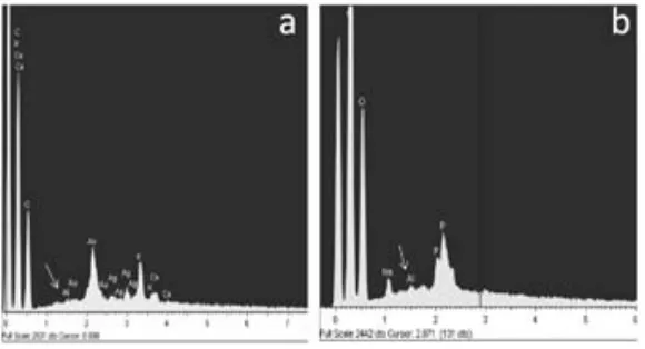
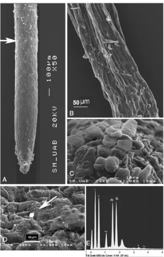
(45) 5.2). Sections were mounted on slides and sealed with 90% glicerol. The fluorescence emitted from Al-lumogallion complex was observed under a confocal laser optic microscope (TCS SP2 AOBS TCS SP2 AOBS; Leica microscopy Systems, LTD, Heidelberg) using the 488 nm excitation line from the argon laser. 2.3. Results 2.3.1. Comparison of Al-resistance between three Brachiaria species The influence of Al supply on plasma membrane integrity was followed using Evans blue staining. The technique not only allows visualizing the membrane damage, but also estimating it quantitatively (Fig. 6). Roots of B. decumbens seedlings exposed to aluminium for different time periods showed a shallow staining mainly after time 24 and 96 h. For B. brizantha there was intense staining with Evans blue. Intense colouration was mainly observed in the meristematic zone. In this species the colouration was constantly high after 48, 72 and 96 h. Root tip membrane integrity was most intensively affected by Al in B. ruziziensis (Fig. 6). Brachiaria ruziziensis showed symptoms of Al injury, stubby appearance, cracks and deformed root apices. The staining with Evans blue was most intense after 48 and 72 h of Al-treatment, while after 96 h of treatment staining intensity decreased, coincident with the appearance of new lateral roots close to the tip.. Seedlings differences in Al-tolerance could also be shown by staining with haematoxylin after 24 h exposures to nutrient solutions that contained 200 µM Al (Fig. 5). Root tips of the Al-sensitive of B. ruziziensis seedlings stained with haematoxylin exhibiting intense purple colour. This staining indicated the presence of Al in the root tip, in the meristematic zone, the transition zone and the mature root zone. In root tips of B. brizantha haematoxylin stain indicated presence of Al, in the meristematic zone and the transition zone; but staining was less intense than in B. ruziziensis (Fig. 5). In roots of B. decumbens the root remained unstained excepting numerous small spots with intense stain accumulation scattered all over the entire root from cap to the mature root zone. (Fig. 5B). 35.
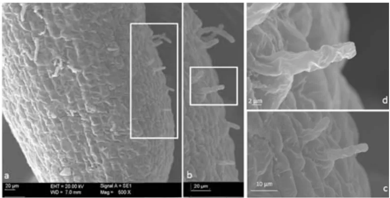
(46) 2.3.2. Localization of aluminium by lumogallion-Al, and morin-Al complex fluorescence in roots of B. decumbens and B. brizantha Morin is an Al-specific fluorochrome and is widely used to localize Al in plant tissues (Eticha et al., 2005). Roots tips of Brachiaria species stained with morin exhibiting similar distribution as observed for haematoxylin. Brachiaria brizantha showed fluorescent signal intensity much higher than that of B. decumbens (Fig. 7). In B. decumbens the green fluorescence was concentrated in structures (spots) looking like root hairs and border cells (Fig. 7). Comparisons of fluorescent and white light microscopy pictures of morin-stained root tips of B. decumbens (Fig. 8) revealed that the abundant border cells detaching from the root cap did not stain for Al (Fig. 8 A, B). As an example, the head of the red arrow in picture 8 A points exactly to the same border cell as that seen in (Fig. 8 B) and in Fig. 8 D and E at higher magnification. Morin-stainable structures were only found in close contact with the root surface (Fig. 8 A, D). Fig. 8F shows a white light microscopy picture of a root tip from B. decumbens exposed to Al for 72 h. Small root hairs emerging from the epidermal cells are clearly visible at a distance between 300 and 500 μm from the tip. Fluorescence image of morin-stained tips at this distance revealed Al accumulation in these young root hairs (Fig. 8 G). Also the older hairs at larger distance from the tips intensively stained for Al with morin (Fig. 8 C). Aluminium apparently did not restrict root hair formation in B. brizantha (not shown). When longitudinal-section were stained with lumogallion (green) and the nuclei visualized by DAPI (blue), intense fluorescence was evident in the root cell wall of B. brizantha (Fig.7 d). While in longitudinal-section of roots apex of B. decumbens lumogallion stain remained in the root cap and in the cell walls of the detaching outermost cell layer of the root tip (Fig. 7c) 2.4. Discussion 2.4.1. The integrity of the plasma membrane The plasma membrane is considered a primary target for Al toxicity. Uptake of Evans blue into the cells is as an indicator of plasma membrane of damage and of cell death (Baker and Mock 1994).. 36.
(47) Fig.5. Haematoxylin stained root tips of Brachiaria species. a) B. decumbens, b). B. brizantha and c) B. ruziziensis roots were exposed for 24 h to 200 µM AlCl3 in (32 μM 3+ Al activity) in low ionic strength nutrient solution, at pH 4.2.. a.. b.. Fig.6. Histochemical detection showed: Aluminium effect on plasma membrane integrity of Brachiaria species roots apex. After treatment of seedlings with or without 200 µM AlCl 3 for 4 times 24, 48, 72 and 96 h. a) the quantitative determination of Evans blue stain retained in a 10-mm section from root tip was performed. b) The integrity of plasma membrane of roots was examined by the degree of Evans blue uptake. All values are means ± SD (n =15). 37.
(48) Fig.7 . Morin-stained whole root tips of a) and B. decumbens. b) B. brizantha plants exposed for 24 h 3+ to 200 μM Al (32 μM Al activity) in low ionic strength nutrient solution. (Green fluorescence morinAl). Longitudinal sections of root tips stained with lumogallion-DAPI. The roots were exposed for 24 h 3+ to 200 μM Al (32 μM Al activity). From the root tip c) and B. decumbens d) B. brizantha for better visualization of cells root tip nuclei were stained with DAPI (blue) and lumogallion-Al (green). Fig.8. Root tips of B. decumbens exposed to Al for 4 h (A, B, D, E) or 72 h (C, F, G) and stained with morin. Abundant border cells either adhering to (arrow) or scattered around the root are seen in white light microscopy images (B, E), but poorly visible in fluorescent microscopy (A, D). Root hairs developing close (300–500 μm) to the tip (F, G) and in upper root zones (500 to 1500 μm from tip) (C) stained with morin.. 38.
Figure


+7


Documento similar
The observed differences in the coating layers obtained from the ethylphosphonate ionic liquid LEP102 and the methylphosphonate ionic liquids LMP101 and LMP102 could be related to
In addition, given the widespread use of prescription benzodiazepines in society, and their diversion to the illicit drug market, the increase in new benzodiazepines might also
For a short explanation of why the committee made these recommendations and how they might affect practice, see the rationale and impact section on identifying children and young
Nevertheless, a more in-depth interpretation of the results suggests that gender differences may, in general, be more acute in relation to generic issues, such as the
Although this model of DELLAs action mostly explains activated gene expression by the DELLAs, these repressors were also shown to interact with BOTRYTIS
Arsenic concentration in roots and shoots of lupin plants growing
No significant differences were seen in leaf and root total -SH contents between the control (33Mn+0Cd) and 0Mn+0Cd treatments, whereas they increased 6 and 4.9 times in the roots
communis and improved its tolerance to water stress due to the lower extreme temperatures in the root, which reduced the ratio shoot dry weight/root dry weight (S/R). 2) The use of
