¿Qué son las células troncales o “células madre”?
What are stem cells or “mother cells”?
Recibido el 25 de mayo de 2006 y aceptado el 17 de octubre de 2006.
*Sección de Histología y Biología Celular, Departamento de Morfología, Facultad de Medicina Veterinaria y Zootecnia, Universidad Nacional Autónoma de México, 04510, México D. F.
**Sección de Anatomía, Departamento de Morfología, Facultad de Medicina Veterinaria y Zootecnia, Universidad Nacional Autónoma de México, 04510, México, D. F.
***Sección de Ciencias Morfológicas Agropecuarias, Departamento de Ciencias Biológicas, Facultad de Estudios Superiores-Cuautitlán, Universidad Nacional Autónoma de México, Carretera Cuautitlán-Teoloyucan, Km 2.5, Cuautitlán Izcalli, Estado de México, México, 54714, Apartado postal 25.
†Departamento de Biomedicina Molecular, Centro de Investigación y Estudios Avanzados, Instituto Politécnico Nacional, Av. Instituto Politécnico Nacional 2508, Col. Zacatenco, 07360, México, D.F.
Correspondencia : Santiago René Anzaldúa Arce, Sección de Histología y Biología Celular, Facultad de Medicina Veterinaria y Zootecnia, Universidad Nacional Autónoma de México, 04510, México, D.F., correo electrónico: rene@fmvz.unam.mx
Abstract
This paper presents an updated review of the main general aspects of stem cells: historical background; defi nition of stem cells based on their main characteristics (degree of differentiation, pluripotent potential, self-renewing capacity and reintregration to embryonic tissue); classifi cation of stem cells by their degree of differentiation (toti-potent stem cells, pluri(toti-potent stem cells, multi(toti-potent stem cells, and tissue-specifi c stem cells); origins of pluripo-tent stem cells (embryonic carcinoma cells, embryonic stem cells, and embryonic germ cells); main methods to obtain embryonic stem cells (use of morulae, complete blastocysts and immunosurgery of inner cell mass); main methods to obtain adult stem cells (purifi cation in peripheral blood, umbilical cord blood, whole bone marrow, and nervous tissue).
Key words: STEM CELLS, EMBRYONIC STEM CELLS, ADULT STEM CELLS, MORULA, BLASTOCYST, EMBRYOBLAST, TOTIPOTENT, PLURIPOTENT, MULTIPOTENT.
Resumen
En el presente trabajo se realiza una revisión actualizada sobre los principales aspectos generales de las células troncales: antecedentes históricos, defi nición de las células troncales con base en sus principales características (grado de indiferenciación, pluripotencialidad, capacidad de autorrenovación y reintegración a tejidos embriona-rios); clasifi cación de las células troncales por su grado de diferenciación (células troncales totipotenciales, células troncales pluripotenciales, células troncales multipotenciales y células troncales de tejidos y órganos específi cos); origen de las células troncales pluripotenciales (células de carcinoma embrionario, células troncales embrionarias y células germinales embrionarias); principales métodos para obtener células troncales embrionarias (utilización de mórulas, blastocistos completos e inmunocirugía del macizo celular interno); principales métodos para obtener células troncales de tejidos adultos (purifi cación de sangre periférica, de sangre de cordón umbilical, de médula ósea completa y de tejido nervioso).
Palabras clave: CÉLULAS TRONCALES, CÉLULAS TRONCALES EMBRIONARIAS, CÉLULAS TRON-CALES ADULTAS, MÓRULA, BLASTOCISTO, EMBRIOBLASTO, TOTIPOTENCIAL, PLURIPOTENCIAL, MULTIPOTENCIAL.
Santiago René Anzaldúa Arce* María de Lourdes Juárez Mosqueda* Héctor Villaseñor Gaona* María Cristina Ríos Mas** Miguel Ángel Cornejo Cortés*** Marco Antonio Meraz Ríos†
Introducción
L
as células troncales se caracterizan por ser indi-ferenciadas, tener capacidad de autorrenovarse y, ante determinadas señales, aún poco cono-cidas, especializarse para realizar una función con-creta.1 Gran parte de los estudios se han realizado enratones e indican que en las primeras etapas del desa-rrollo embrionario surge ese tipo de células, que, al diferenciarse, forman gran número de estirpes celula-res que constituyen los tejidos del embrión.
Las células troncales, también llamadas “células madre”, se clasifi can en dos grandes grupos: tronca-les embrionarias y troncatronca-les adultas (de origen somá-tico).2,3 La diferencia principal entre ambos grupos
radica en el grado de indiferenciación o potenciali-dad que conservan para originar diferentes tipos celu-lares. Las células troncales embrionarias pueden dar origen a células de todos los tejidos del animal. De acuerdo con su grado de indiferenciación, o poten-cialidad para originar diferentes células, se clasifi can en: totipotenciales, pluripotenciales, multipotencia-les y progenitoras de tejido. Las dos primeras sólo se encuentran en el embrión, las dos últimas en el orga-nismo adulto.
La identifi cación de células troncales en diversos tejidos del cuerpo adulto y el hecho de generar célu-las troncales del embrión humano, ha despertado un enorme interés entre los científi cos, principalmente por las posibles aplicaciones terapéuticas que aqué-llas pueden tener, por ejemplo, producción de órga-nos completos para sustituir órgaórga-nos afectados, como riñón y corazón, evitando así la búsqueda de donantes o la regeneración de células en tejidos, para aplicar terapias en enfermedades neurodegenerativas, como Alzheimer y Parkinson, entre otras. Entre las aplica-ciones de las células troncales se observa la creación de animales transgénicos, como ratones, ratas, cerdos y primates, pues ellos constituyen una herramienta novedosa para producir modelos animales en donde existen las alteraciones genéticas de enfermedades del hombre, que permiten seguir la manifi estación equivalente de signos y síntomas, facilitando el hecho de probar tratamientos para desarrollar terapias más efectivas. Otro aspecto de las células troncales es su utilización en la medicina veterinaria, para imple-mentar tecnologías innovadoras en la crianza y mani-pulación genética de animales domésticos.
Sin embargo, a pesar del potencial de este tipo de células, se han generado grandes controversias en rela-ción con su investigarela-ción y uso, lo que ha ocasionado enorme confusión en cuanto a la información que se tiene sobre ellas y sus logros. Estas razones impulsa-ron la realización de este trabajo, en espera de que los
Introduction
S
tem cells are characterized by their undiffe-rentiation, self-renewing capacity and, under certain conditions, which are not well defi ned, be specialized to perform a specifi c function.1 Mostof the studies have been conducted in mice, and they indicate that these cells emerge in the early stages of embryonic development. Upon differentiation, stem cells form various cell strains that constitute the diffe-rent types of embryonic tissue.
Stem cells, also called “mother cells”, are divided into two main groups: embryonic stem cells and adult stem cells (of somatic origin).2,3 The main difference
between these groups lies in their degree of differen-tiation or potential to form different cell types. Embr-yonic stem cells can generate all types of animal tissue cells. According to their degree of differentiation or potential, they give rise to different cell types, and are classifi ed into totipotent, pluripotent, multipotent and tissue-progenitor cells. The fi rst two types are only found in embryos; the last two are found in adult organisms.
The identifi cation of stem cells in various tissues of the adult body and the human embryo’s capacity to generate stem cells have raised great interest among scientists, mainly because of the possible therapeutic applications these cells might have. Among these are the production of complete organs to replace affected organs, such as kidneys or heart, thus avoiding donor search; or cell regeneration in tissues, to be used in treatment of neurodegenerative diseases, such as Alzheimer and Parkinson. Among the applications of stem cells, there is the creation of transgenic animals, including mice, rats, pigs and primates. This constitu-tes a novel tool to produce animal models with gene-tic alterations found in human diseases, which allows to examine the equivalent manifestation of signs and symptoms, and to test treatments in order to develop more effective therapies. Stem cells can also be used in veterinary medicine, to implement innovative tech-nologies in the breeding and genetic manipulation of domestic animals.
However, despite the potential of stem cells, there has been huge controversy about their research and use. This has caused great confusion regarding their features and achievements. Those are the reasons which motivated this study. Hoping that the data con-tained in this paper will prove useful in clarifying the controversy built around this type of cells.
.
Historical background
datos presentados sirvan para esclarecer el panorama que se ha creado alrededor de este tipo de céulas.
Antecedentes históricos
El estudio de las células troncales se inició a princi-pios de la década de 1970, cuando Till y McCulloch4
y más tarde Becker et al.5 observaron cómo células
simples de la médula ósea podían generar todos los tipos de células hematopoyéticas in vivo. Este hecho propició gran cantidad de estudios, cuyas principales aportaciones se resumen en el Cuadro 1.
Defi nición
Las células troncales también se conocen como “célu-las madre” o célu“célu-las progenitoras (del inglés stem cells, tronco) y células troncales embrionarias. Pueden defi -nirse como células indiferenciadas, pluripotenciales, capaces de autorrenovarse y de incorporarse en el embrión en desarrollo, participando en la formación de todos los tejidos, cuando son inyectadas en blasto-cistos. Esta defi nición está basada en sus principales características, que se describen en el Cuadro 2.
Clasifi cación de las células troncales por su grado de diferenciación
La clasifi cación más común de las células troncales se fundamenta en la potencialidad que tienen para ori-ginar células de diferentes estirpes, lo cual está rela-cionado con el grado de diferenciación de las células troncales. Con base en lo anterior, existen células troncales totipotenciales, células troncales pluripo-tenciales, células troncales multipotenciales y células troncales de tejidos específi cos.36,50
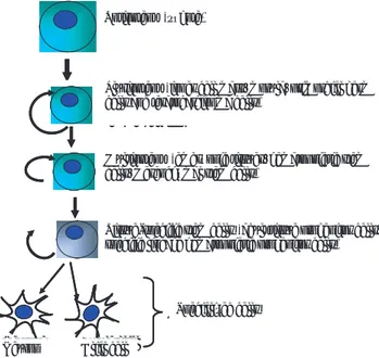
Células troncales totipotenciales
Estas células tienen la capacidad de dar origen a todas las células de un embrión completo; es decir, originan los “tejidos propios” del embrión, como el ectodermo, endodermo y mesodermo (hojas blastodérmicas), y los “tejidos extraembrionarios”: saco vitelino, alantoides, amnios y corion (membranas extrafetales). Desafortu-nadamente, no se ha demostrado que las células tron-cales totipotenciales sean autorrenovables in vivo; por el contrario, se cree que presentan autorrenovación muy limitada. Estas células tienen su origen cuando el espermatozoide fertiliza al ovocito, formando la célula diploide que se conoce como cigoto (Figura 1), la cual se caracteriza por ser totipotencial. Después de la etapa de cigoto, los embriones experimentan varias divisiones mitóticas hasta alcanzar un tamaño similar a la de las células somáticas. Las primeras células hijas that simple bone marrow cells were able to generate
all types of hematopoietic cells under in vivo condi-tions. This led to a great number of studies, whose main contributions are summarized in Table 1.
Defi nition
Stem cells are also known as “mother cells”, progenitor cells, and embryonic stem cells. They can be defi -ned as undifferentiated, pluripotent cells, capable of self-renewing and incorporating into the developing embryo, participating in the formation of all tissues when they are injected into blastocysts. This defi nition is based on their main features, as described in Table 2.
Classifi cation of stem cells by their degree of differentiation
The most common classifi cation method of stem cells is based on their potential to generate different cell strains, which is related to their degree of differentia-tion. According to this classifi cation, there are toti-potent stem cells, pluritoti-potent stem cells, multitoti-potent stem cells and tissue-specifi c stem cells.36,50
Totipotent stem cells
Species Contribution Reference
Mouse Produced pluripotent cells using ascitic conversion of teratocarcinoma.
6
Rabbit Demonstrated that embryoblast generates different tissues. 7 Mouse Produced chimeric mice by union of embryoblast and cells from a
pre-implanted blastocyst.
8
Mouse Obtained chimeric mice from blastocysts injected with pluripotent teratocarcinoma cells.
9
Mouse Obtained first marker to identify embryonic stem cells, called SSEA-1 (stage-specific embryonic antigen).
10
Mouse Developed first cultures of embryonic stem cells using blastocysts. 11, 12
Mouse
Mouse
Began using stem cells to perform transgenesis.
Postulated a similarity between primordial germ cells and embryonic stem cells.
13
14 Identified cytosine called leukemia inhibiting factor (LIF), which preserves embryonic stem cells undifferentiated phenotype.
15
Mouse
Manipulated genes specific of embryonic stem cells by homologous recombination.
Established lines of pluripotent embryonic stem cells from morulae.
16
17 Using LIF, achieved culture of embryonic stem cells without nursemaid cells.
18
Mouse Obtained chimeric animals by aggregation of embryonic stem cells with morula.
19
Human
Bovine
Isolated cells from embryoblast.
Isolated, cultured and characterized primordial germ cells.
20 21
Rabbit Informed bad chimeric formation upon injecting embryonic stem
cells into blastocyst. 22
Pig Produced chimeric animals using pluripotent cells isolated from embryos.
23
Human
Human
Obtained embryonic stem cells from embryos obtained by in vitro
fertilization.
Obtained embryonic stem cells from aborted foetus gonads.
24
25 Human Achieved differentiation of mesenchymal cells in bone,
cartilaginous, adipose, muscle tissues, and bone marrow stroma. 26
Cuadro 1
PRINCIPALES APORTACIONES EN EL ESTUDIO DE LAS CÉLULAS TRONCALES
MAIN CONTRIBUTIONS TO THE STUDY OF STEM CELLS
Year Group
1964 Kleinsmith and Pierce 1966 Cole et al. 1967 Gardner
1975 Mintz and Illmensee 1978 Solter and
Knowles 1981 Martin and
Evans; Kaufman
1986 Gossler et al.
Donova et al.
1988 Williams et al.
1989 Capecchi
Eistetter
1990 Pease et al.
1993 Wood et al.
1994 Bongso et al.
Cherny et al.
1996 Schoonjan et al.
1997 Shim et al.
1998 Thomson et al.
Shamblott et al. 1999 Pittenger et al.
2000 Iwasaki et al.
Vallilieve et al.
Bovine
Rat
Achieved birth of calves from tetraploid embryos aggregated with embryonic stem cells.
Developed culture of cells similar to embryonic stem cells.
27
28 2002 Saito et al. Equine Achieved isolation and differentiation of cells similar to
embryonic stem cells.
29
2003 Hübner et al.
Cheong et al.
Ruhnke et al.
Mouse
Human
Rat
Developed ovarian follicles, or similar structures, from embryonic stem cells.
Achieved embryonic stem cell propagation using bone marrow cells as nursemaid cells.
Achieved differentiation in embryonic stem cells obtained from blastocysts, neuronal, endothelial and hepatic lineages.
30
31
32
2004 Horiuchi et al.
Park et al.
Chicken
Human
Cloned LIF gene in bacteria and used the expressed product to culture embryonic stem cells.
Generated dopaminergic neurons from embryonic stem cells supplemented with several compounds.
33
Pluripotent stem cells
Pluripotent cells are derived from the blastocyst’s embryoblast, also called inner cell mass (Figure 2). The embryoblast’s pluripotent cells are capable of forming all the cells in the embryonic tissues, that is, any type of cells found in an individual (Figure 2), but they are not capable of generating an entire individual since they cannot differentiate into extraembryonic cells, such as the cells found in the placenta (tropho-blast) and other support tissues, which are indispen-sable for intrauterine foetus development. Pluripotent stem cells not only generate all cell types in animal tissue, but they also differentiate in vitro into endo-derm, ectoderm and mesoderm cells. Besides, when these cells grow in a liquid environment (in vitro) and undergo a process of differentiation, they form three-idimensional aggregates called embryoid bodies, which are three-dimensional structures very similar to embryos at the fi rst stages of their development.
que resultan de la división del cigoto se denominan blastómeras y son células totipotenciales con caracte-rísticas idénticas; esto último signifi ca que cualquiera de estas células puede dar origen a un individuo com-pleto. Cuando el embrión ha formado de ocho a 16 blastómeras, y en algunos casos más, se denomina mórula; después de ello, las células totipotenciales de la mórula comienzan a especializarse dando origen al blastocisto, que es una esfera hueca de células (tro-foblasto) con una masa celular (embrioblasto) locali-zada en un extremo (Figura 1).36,51
Células troncales pluripotenciales
Las células pluripotenciales se derivan del embrio-blasto, también llamado masa o macizo celular interno del blastocisto (Figura 2). Las células pluripotenciales del embrioblasto son capaces de formar todas las célu-las de los tejidos embrionarios; es decir, cualquier tipo de células que se encuentran en un individuo (Figura
Cuadro 2
DESCRIPCIÓN DE LAS CARACTERÍSTICAS QUE DEFINEN A LAS CÉLULAS TRONCALES
DESCRIPTION OF CHARACTERISTICS DEFINING STEM CELLS
Characteristic Description
Self-regeneration Cells capable of proliferating and producing identical cells, renewing themselves.35,36
Undifferentiated state Cells which have not specialized, comparable to cells found in early embryos.35-38
Pluripotent Potential Under specific conditions, they can differentiate to generate compromised cells in certain
development routes and later become specialized cells.35-37
Integration to developing
embryo
They form chimeric animals upon integrating to the embryo at morula or blastocyst stage;
animals will have cells from two different genetic backgrounds in their tissues.37,39
Formation of teratomas They produce benign tumors (teratomas) when injected subcutaneously or
intraperitoneally into immunosuppressed mice.39
In vitro undifferentiated
proliferation
Under certain requirements, they divide and grow in cell cultures, remaining stable and
undifferentiated.11,16,18,25,33,40-48
In vitro differentiation Removal of certain cytosines from culture environment leads them to constitute the so
called embryoid bodies (spherical structures similar to post-implantation embryos).36,37,49
spermatozoide Ovulo
MÓRULA
Célula totipotencial Embrioblasto
BLASTOCISTO
Trofoblasto
Blastocele
FECUNDACIÓN spermatozoide Ovulo
MÓRULA
Célula totipotencial Embrioblasto
BLASTOCISTO
Trofoblasto
Blastocele
FECUNDACIÓN
Sperm Egg Totipotent cell Embryoblast
Trophoblast
Fertilization Morula Blastocyst
Figura 1: Primeras etapas del desarrollo embrionario. Células que participan en la fecundación y estructuras que conforman la mórula y blastocisto.
Moreover, when injected into an immunodepressed animal, they are capable of forming teratomas, which are tumors containing a mixture of various types of differentiated or partially differentiated cells.36,50,52
Multipotent stem cells
They are self-renewing cells that are more differen-tiated than the previously mentioned cells (Figure 2). They are at the top of the cell hierarchy and prolife-rate in vivo to produce differentiated cells which will form a certain tissue. It is important to note that, once developed, these cells renew themselves during all the individual’s life, unlike totipotent and pluripotent stem cells, which can only be found in the embryonic stage.
In adult organisms, under in vivo conditions, mul-tipotent stem cells can divide repeatedly to repeople a tissue, as the multipotent hematopoietic stem cells found in bone marrow, which generate all the cells in the hematopoietic system (erythrocytes, leukocytes and platelets), or they can show low activity rates, as in the case of human mesenchymal stem cells, which have differentiated into connective lineages including fi broblasts, osteoblasts, condrocytes and adipocytes in vitro studies.36
Tissue-specifi c stem cells
These cells suffer a restriction in the expression pat-tern of their genome; if compared with pluripotent stem cells, they have a higher degree of differentiation and produce cell types characterisctic of the tissue in which they are found in vivo (Figure 2). These cells are often called “progenitor cells”, according to the organ
2), aunque no son capaces de originar un individuo entero porque no pueden diferenciarse en células extraembrionarias, como las de la placenta (trofo-blasto) y otros tejidos de sostén, necesarios para el desarrollo del feto dentro del útero. Las células tron-cales pluripotenciales no sólo dan origen a todos los tipos de células de los tejidos de un animal, también se diferencian in vitro en células del endodermo, ecto-dermo y mesoecto-dermo. Asimismo, cuando estas células crecen en un medio líquido (in vitro) y sufren diferen-ciación, forman agregados tridimensionales llamados cuerpos embrioides, que corresponden a estructuras tridimensionales muy semejantes a los embriones en primeros estadios de desarrollo. Además, si son inyec-tadas en un animal inmunodeprimido, son capaces de formar teratomas, que son tumores que contienen una mezcla de varios tipos de células diferenciadas o parcialmente diferenciadas.36,50,52
Células troncales multipotenciales
Son células autorrenovables más diferenciadas que las anteriores (Figura 2), se encuentran en la cima de la jerarquía celular y proliferan para producir células diferenciadas que formarán algún tejido in vivo. Es importante aclarar que estas células, una vez desa-rrolladas, se autorrenuevan durante toda la vida del individuo, a diferencia de las células troncales totipo-tenciales y pluripototipo-tenciales, que sólo se encuentran durante la etapa embrionaria.
En los organismos adultos, in vivo, las células troncales multipotenciales pueden dividirse repetida-mente para repoblar un tejido, como las células tron-cales hematopoyéticas multipotenciales de la médula ósea, que dan origen a todas las células del sistema
Multipotencial(tejidos embrionarios, células troncales hematopoyéticas, células troncales mesenquimales)
Totipotencial(cigoto).
Troncales específicas de tejidos (células progenitoras de tejidos adultos, células progenitoras hematopoyética
de linajes específicos)
Células especializadas
(macizo celular interno, mórula, células germinales primordiales y células de teratocarcinoma.
Neurona Célula glial
Totipotent (zygote)
Pluripotent (inner cell mass, morula, primordial germ cells and teratocarcinoma cells)
Multipotent (embryonic tissues, hematopoietic stem cells, mesenchymal stem cells)
Tissue-specific stem cells (adult tissue progenitor cells, specific lineage hematopoietic progenitor cells)
Specialized cells
Neuron Glial cell
Figura 2: Clasifi cación de las células troncales por su grado de diferen-ciación. Tipos de células troncales de acuerdo con el grado de indiferen-ciación que presentan. Entre paréntesis se encuentran ejemplos de célu-las que corresponden a los diferentes grados de diferenciación (imagen modifi cada de la referencia 50).
or tissue generated by them, or the organ or tissue in which they are found in vivo; for instance, specifi c lineage hematopoietic progenitor cells (erythrocytes, lymphocytes, monocytes, etc.), nervous progenitor cells, cornea progenitor cells, and undifferentiated intestinal columnar cells.36,53,54
Classifi cation of pluripotent stem cells by their origin
According to their method of obtention, stem cells are classifi ed into three types:
a) Embryonic carcinoma cells. They were the fi rst stem cells to be obtained. They are isolated by an “indi-rect route”, in which tumoral masses (teratocarcino-mas) derived from an embryo are collected (Figure 3). These masses are then disaggregated and injec-ted into an animal’s abdominal cavity to perform an “ascitic conversion”. Certain celular aggregates called “embryoid bodies” are obtained from this process; these are disaggregated with trypsin and their cells are injected into a histocompatible mouse; fi nally, plu-ripotent cells called embryonic carcinoma cells (EC) are obtained from this process.55
b) Embryonic stem cells. They were studied for the fi rst time during the 80s. In this case, there was a “second route”, in which in vitro techniques allowed to isolate directly stem cells from normal embryos, without the need to pass through a tumor state (Figure 3). These cells have been called primitive embryonic cells or embryonic stem cells (ES cells), in order to distinguish between them and embryonic carcinoma cells. Pluripotent cells can be obtained from normal embryos which have not yet been implanted into the uterus, such as disassociated morulae or intact blasto-cysts. Embryonic stem cells are the most widely used stem cells, as their capacity to form chimeric animals is enormous.56,57
hematopoyético (eritrocitos, leucocitos y plaquetas), o pueden presentar poca actividad, como en el caso de las células troncales mesenquimatosas humanas, que en ensayos in vitro, se han diferenciado en lina-jes conectivos que incluyen fi broblastos, osteoblastos, condrocitos y adipocitos.36
Células troncales específi cas de tejido
Estas células sufren una restricción en la expresión de su genoma; en comparación con las pluripotencia-les, tienen mayor grado de diferenciación y producen tipos celulares característicos del tejido en el cual se observan in vivo (Figura 2). Es común nombrar proge-nitoras a este tipo de células. según el órgano o tejido al que dieron origen, o en el que se encuentran in vivo; por ejemplo, células progenitoras hematopoyéticas de linajes específi cos (eritrocitos, linfocitos, monocitos, etc.), células progenitoras nerviosas, células progeni-toras de la córnea y células columnares indiferencia-das del intestino.36,53,54
Clasifi cación de las células troncales pluripo-tenciales por su origen
Por su fuente de obtención, las células troncales se cla-sifi can en tres tipos:
a) Células de carcinoma embrionario. Fueron las pri-meras que se obtuvieron, su aislamiento se realiza por una “ruta indirecta”, en la cual se recolectan masas tumorales (teratocarcinomas) derivadas de un embrión (Figura 3), que son disgregadas e inyectadas en la cavidad abdominal de un animal para realizar una “conversión ascítica”, a partir de ésta se obtienen agregados celulares llamados “cuerpos embrioides”; éstos son disgregados con tripsina y sus células se inyec-tan en un ratón histocompatible; fi nalmente, a partir
CELULAS DE CARCINOMA EMBRIONARIO
CELULAS GERMINALES EMBRIONARIAS CELULAS TRONCALES
EMBRIONARIAS
Teratoma
Embrioblasto Crestas Genitales
CELULAS DE CARCINOMA EMBRIONARIO
CELULAS GERMINALES EMcellsRIAS CELULAS TRONCALES
EMBRIONARIAS
Teratoma
Embrioblasto Crestas Genitales
Teratoma Embryoblast
Genital crests
Embryonic carcinoma cells
Embryonic stems cells
Embryonyc germ cells
Figura 3: Origen de las células troncales pluripotenciales. Tres clases de células troncales por su origen. Esta clasifi cación se basa en las tres rutas utilizadas para obtener las diferentes células pluri-potenciales.
EC and ES cells are similar in terms of cellular mor-phology, expression of cell surface antigens and diffe-rentiation behavior in vitro and in vivo. However, EC lines have shown important experimental limitations, since they cover a very diverse population of culture cell lines (perhaps as a result of their tumoral origin). This heterogeneity between lines is refl ected unfavora-bly in their culture differentiation capacity and in the chimeric association with an infected embryo.36,52,56,58
c) Embryonic germ cells. These cells are obtained by reprogramming primordial germ cells (PGC), to trans-form them into embryonic germ cells (EG), which are pluripotent and are precursors of mature gametes. EG cells are derived from “genital crests” in embryos with a more advanced development (Figure 3); EG cells colonize less effectively the tissues of chimeric ani-mals, as compared to embryonic stem cells.25,56
In vitro characteristics of embryonic
stem cells
During the growth of embryonic stem cells, seve-ral tests are conducted to know if they are indeed “undifferentiated”. This process is called “characteri-zation”.37 The most widely used tests in laboratories
where embryonic stem cells are cultured are:
1) Growth and subculture. This test consists in per-forming several “propagation passes” to verify if cells still retain the morphological characteristics of undi-fferentiated embryonic stem colonies (Figure 1); that is, that they continue developing colonies of spherical, compact cells over the nursemaid cells.59 At the
begin-ning, it was deemed indispensable to culture embryo-nic stem cells together with nursemaid cells, in order to avoid differentiation in stem cells; later, it was dis-covered that, in most cases, stem cells require a factor produced by nursemaid cells, which without it they differentiate. This factor is a cytosine called LIF and its expression is stimulated by stem cells. Since then, stem cells can be cultured without nursemaid cells, by adding LIF or related cytosines, such as bFGF, SF y SCF. In some species, it has been possible to obtain lines of stem cells which can remain undifferentiated without LIF.48
2) Molecular markers. There are several molecular markers to identify stem cells (Table 3). Some of the most important are:
a) Surface markers. They are specialized proteins or sugars located at the level of the cytoplasmatic mem-brane. Among these surface molecules, the one most used for detection is SSEA-1, o CD 15, FAL or trisac-charide 3-fucosyl-N-acetyl-lactosamine.58,60
b) Transcription factors. They are proteins that join the DNA and contribute to “switching on and off” specifi c genes. Among these is protein Oct-4
(trans-de éste se obtienen células pluripotenciales (trans- denomi-nadas células de carcinoma embrionario (EC).55
b) Células troncales embrionarias. Éstas se establecie-ron en la década de 1980. Aquí se marcó una segunda ruta, en la que las técnicas de cultivo in vitro permiten el aislamiento de células troncales directamente de embriones normales, sin necesidad de pasar a través de un estado de tumor (Figura 3). Estas células se han denominado células embrionarias primitivas (ES cells) o células troncales embrionarias, con el fi n de distinguirlas de las células provenientes de carcinoma embrionario. Las células pluripotenciales pueden obtenerse a partir de embriones normales que aún no se han implantado en el útero como mórulas diso-ciadas o blastocistos intactos. Las células troncales embrionarias son las más utilizadas, pues su capaci-dad para formar animales quiméricos es enorme.56,57.
Las células EC y las ES son similares en términos de morfología celular, expresión de antígenos de super-fi cie celular y comportamiento de diferenciación in vitro e in vivo, sin embargo, las líneas EC han tenido importantes limitaciones experimentales, pues com-prenden una población muy diversa de líneas celu-lares de cultivo (quizá por su origen tumoral). Esta característica heterogénea entre líneas se refl eja des-favorablemente en su capacidad de diferenciación en cultivo y en la asociación quimérica con un embrión portador.36,52,56,58
c) Células germinales embrionarias. Estas células se obtienen basándose en la reprogramación de las célu-las germinales primordiales (PGC), para programar-las como céluprogramar-las germinales embrionarias (EG), que se caracterizan por ser pluripotenciales y ser precurso-ras de los gametos maduros. Las células EG se derivan de las “crestas genitales” en embriones con un desa-rrollo más avanzado (Figura 3); las células EG coloni-zan de manera menos efectiva los tejidos de un animal quimérico, en comparación con las células troncales embrionarias.25,56
Características in vitro de las células troncales embrionarias
Durante el crecimiento de las células troncales embrio-narias se realizan diferentes pruebas para conocer si efectivamente son células troncales embrionarias “indiferenciadas”, este proceso se denomina “carac-terización”.37 Las pruebas más utilizadas en los
labo-ratorios donde se cultivan líneas de células troncales embrionarias son:
cription factor belonging to the POU famiy, which is expressed in totipotent and pluripotent cell lines, in early embryonic cells and in some germ cells lines.61
3) Cytoplasmatic enzymes. A routine test consists in detecting the presence of the alkaline fosfatase enzyme, which is expressed in great amounts in embr-yonic stem cells remaining in an undifferentiated state.62
4) Karyotype tests. These tests serve to determine if cromosomes are normal and stable. This method allows to observe if cromosomes are damaged or if the number of cromosomes has changed. However, this method cannot detect slight genetic mutations; that is, mutations at the level of DNA sequence.63
5) Differentiation. Tests are carried out to obtain cell strains from the three germ layers: ectoderm, endoderm and mesoderm. These tests show the cells’ “pluripotent potential”.64
6) Formation of embryoid bodies. Stem cells are cultu-red in a liquid environment, without nursemaid cells and without factors keeping them undifferentiated, so that they form cellular aggregates called “embryoid bodies”. These structures have multidifferentiated cells that follow routes similar to those of the embryo, but without the axial organization and the elabora-tion of a corporal development plan.49 Furthermore,
if retinoic acid is added, cell aggregates can give rise to certain type of cells, although they will invariably generate a heterogeneous cell mixture.
desarrollando colonias de células esféricas y compac-tas por encima de las células nodrizas.59 En un
prin-cipio se pensaba que era indispensable hacer crecer las células troncales embrionarias junto con células nodrizas, para evitar que las primeras se diferencia-ran; posteriormente se descubrió que, en su mayoría, las células troncales requieren de un factor producido por las células nodrizas, sin el cual se diferencian; este factor es una citocina llamada LIF y su expresión es estimulada por las células troncales. Desde entoces las células troncales pueden cultivarse en ausencia de células nodrizas, agregando LIF o citocinas relacio-nadas como bFGF, SF y SCF; en algunas especies se han obtenido líneas de células troncales que pueden mantenerse indiferenciadas sin necesidad de LIF.48
2) Marcadores moleculares. Existen diversos mar-cadores moleculares para identifi car a las células troncales (Cuadro 3), entre los más importantes se encuentran:
a) Marcadores de superfi cie. Son proteínas especiali-zadas o azúcares localizados en el nivel de la mem-brana citoplasmática. Dentro de estas moléculas de superfi cie, la más utilizada para detección es la SSEA-1, o CD 15, FAL o trisaccharide 3-fucosyl-N-acetyl-lac-tosamine.58,60
b) Factores de transcripción. Son proteínas que se unen al ADN y ayudan en el “encendido y apagado” de genes específi cos, entre éstos la proteína Oct-4 (factor de transcripción de la familia POU), que se expresa en líneas celulares totipotenciales y pluripotenciales, en células embrionarias tempranas y en algunas líneas de células germinales.61
Cuadro 3
MARCADORES PARA CÉLULAS TRONCALES68
MARKERS FOR STEM CELLS68
Marker Main Characteristics
CD133 Surface protein in nervous stem cells which generate glial cells and neurons
CD34 Surface protein in HSC
c-kit Surface receptor in bone marrow cells
CD30 Surface receptor in pluripotent stem cells
Genesis Transcription factor expressed by embryonic stem cells during undifferentiated
state
GCNF Transcription factor expressed by pluripotent stem cells
Oct-4 Transcription factor essential for establishing and maintaining totipotent and
pluripotent stem cells
SSEA-1 Glycoprotein expressed by pluripotent stem cells in early embryonic development
Sca-1 Surface protein in bone marrow cells, either HSC or MSC
In vivo characteristics of embryonic
stem cells
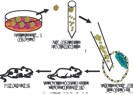
1) Formation of chimeric animals. One of the most impor-tant features of stem cells in vivo is their capacity to reintegrate to “embryogenesis”, contributing to the formation of all tissues (including gametes). Thus, they give rise to the so called “chimeric animals”, which can be produced in two ways: a) Injecting embryonic stem cells inside a blastocyst, which is then intro-duced into a female specimen acting as recipient to fi nish development (Figure 4),65-67 b) Coculturing two
embryos (in morula state) with a group of embryonic stem cells; embryos must then develop with adhered embryonic stem cells until they reach the blastocyst state; then, the blastocyst is introduced into a recipi-ent female to fi nish developmrecipi-ent.6,19
During tissue and organ formation in embryos (embryogenesis), stem cells can colonize virtually all embryonic cell types, including vitelline sac mesoderm, alantoids, amnios and gonades, but they do not contri-bute to form the trophoblast.69 These “colonization”
routes have been noted by means of transgenic mar-kers, such as galactosidase and green fl uorescent pro-tein (GFP).70 This tissue-colonization feature during
the embryonic stage has made stem cells a fundamen-tal tool in cellular physiology research, since it allows to study the functions of cell proteins, the regulation of genes in tissues, and the development of animal models for specifi c diseases.
2) Formation of teratomas. Stem cells are injected subcutaneously or intraperitoneally into an immuno-suppressed mouse, and they cause the formation of a benign tumor, called “teratoma”. Typical teratomas contain a mixture of various types of differentiated
3) Enzimas citoplasmáticas. Una prueba de rutina consiste en detectar la presencia de la enzima fosfa-tasa alcalina, que se expresa en grandes cantidades en las células troncales embrionarias que permanecen en un estado indiferenciado.62
4) Pruebas de cariotipo. Mediante esta prueba se determina si los cromosomas son normales y estables. Este método permite apreciar si los cromosomas están dañados o si el número de cromosomas ha cambiado, aunque con este método no se pueden detectar muta-ciones genéticas fi nas; es decir, a nivel de la secuencia de ADN.63
5) Diferenciación. Se realizan ensayos para obtener estirpes celulares que provengan de las tres hojas ger-minales: ectodermo, endodermo y mesodermo. Estos ensayos muestran la “pluripotencialidad”.64
6) Formación de cuerpos embrioides. Las células tronca-les se cultivan en medio de cultivo líquido, en ausencia de células nodrizas y sin factores que las mantengan indiferenciadas, con la fi nalidad de que formen agre-gados celulares denominados “cuerpos embrioides”. Estas estructuras tienen células multidiferenciadas que siguen rutas similares a las del embrión, pero sin la organización axial ni la elaboración de un plan de desarrollo corporal.49 Asimismo, si se les adiciona
ácido retinoico, los agregados celulares pueden pro-piciar cierto tipo de células, aunque invariablemente darán origen a una mezcla heterogénea de células.
Características in vivo de las células troncales embrionarias
1) Formación de animales quiméricos. Una de las carac-terísticas más importantes de las células troncales in vivo es la capacidad de reintegrarse a la “embriogé-nesis”, contribuyendo así en la formación de todos los
Cultivo de células troncales
totipotenciales Aislamiento y disgregación de células troncales
Inyección de células troncales en blastocisto Transferencia del blastocisto
a una hembra seudogestante Animal quimérico
Cultivo de células troncales
totipotenciales Aislamiento y disgregación de células troncales
Inyección de células troncales en blastocisto Transferencia del blastocisto
a una hembra seudogestante Animal quimérico
Totipotent stem cell culture
Stem cell isolation and disaggregation
Injection of stem cells into blastocyst Introduction of blastocyst into
pseudopregnant female Chimeric animal
Figura 4: Las células troncales se inyectan en un blastocisto y se tranfi eren a una hembra seudo-gestante para completar su desarrollo.
or partially differentiated cells; thus, it is possible to obtain lines of differentiated cells which have derived from stem cells. This procedure is not widely used due to the appearance of protocols with growth factors used in vitro to differentiate stem cells.71
Main methods to obtain embryonic stem cells
Nowadays, embryonic stem cells can be obtained from morulae, complete blastocysts and immunosurgery in inner cell mass.
Morula
Embryos in morula state are obtained by uterine lavage with Dulbecco’s phosphate buffered saline (DPBS), or by some method of culture with 10% or 15% of added bovine foetal serum. The pellucid zone is removed from morulae and their blastomers are separated using a pipette. When blastomers are obtai-ned, they are cultured on nursemaid cells secreting LIF. After fi ve or six days, round and compact colonies appear, which are isolated and disaggregated with trypsin; they are then sown on new nursemaid cells; thus, every three or four days colonies are checked and subcultures are performed.17
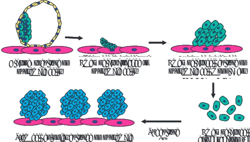
Complete blastocyst
Blastocysts are obtained by uterine lavage with Dulbecco’s phosphate buffered saline (DPBS), or by some method of culture with 10% or 15% of added bovine foetal serum. Intact blastocysts are then cul-tured on nursemaid cells secreting LIF (Figure 5). After four or fi ve days, blastocysts adhere and embryo-blast cells grow, forming round and compact colonies. These are isolated and disaggregated with trypsin; they are then sown on new nursemaid cells. After four or fi ve days, round and compact colonies are checked again and subcultures are performed (Figure 5).21, 57
Immunosurgery in inner cell mass
The technique of immunosurgery is used to obtain only embryoblast cells. This technique consists in the immediate destruction of trophoblast cells, by adding an antibody to them; the embryoblast can thus be easily separated from the remnants of trophoblastic cells. Cells are then placed on nursemaid cells. From nine to fi fteen days later, spheric and compact colo-nies are formed, which can be isolated and disassocia-ted to perform subcultures.72
tejidos (incluyendo los gametos); de esta forma dan como resultado los llamados “animales quiméricos”, que pueden producirse de dos maneras: a) Inyec-tando células troncales embrionarias dentro de un blastocisto, que se introduce en una hembra que actúa como recipiente para completar su desarrollo; (Figura 4),65-67 b) cocultivando dos embriones (en estado de
mórula) más un grupo de células troncales embriona-rias, después se dejan desarrollar los embriones con las células troncales embrionarias adheridas hasta alcanzar el estado de blastocisto, y luego se introduce en una hembra recipiente para completar su desarro-llo.6,19
Durante la formación de los tejidos y órganos en el embrión (embriogénesis), las células troncales pueden colonizar prácticamente todos los tipos celu-lares del embrión, como el mesodermo del saco vite-lino, el alantoides, el amnios y las gónadas, pero no contribuyen a formar parte del trofoblasto.69 Estas
rutas de “colonización” han sido observadas con la ayuda de marcadores transgénicos, como la galacto-sidasa o la proteína fl uorescente verde (GFP).70 Esta
propiedad de colonizar los tejidos durante la etapa embrionaria ha permitido que las células troncales se utilicen como herramienta fundamental para el estu-dio de la fi siología celular al permitir la investigación de las funciones que tienen las proteínas celulares, la regulación de los genes en los tejidos y el desarrollo de modelos animales para enfermedades específi cas.
2) Formación de teratomas. Las células troncales se inyectan vía subcutánea o en forma intraperitoneal en un ratón inmunosuprimido y provocan la formación de un tumor benigno llamado “teratoma”. Los terato-mas típicos contienen una mezcla de varios tipos de células diferenciadas o parcialmente diferenciadas, así se obtienen líneas de células diferenciadas que han derivado de células troncales. Este procedimiento se utiliza poco, debido a la aparición de protocolos con factores de crecimiento que se utilizan in vitro para diferenciar células troncales.71
Principales métodos para obtener células troncales embrionarias
Actualmente, las células troncales embrionarias se pueden obtener a partir de mórulas, blastocistos completos y por inmunocirugía del macizo celular interno.
Mórula
Adult stem cells (AST)
It is important to point out that stem cells are not exclusively found in developing embryos, but they are also present in adult tissues, where they play an impor-tant role in the organism’s homeostasis. Although it is true that cellular diversifi cation normally ends shortly after birth, organs may possess a mechanism to replace cells when they die, either by weakening, apoptosis or damage.73,74
It should also be considered that various adult tis-sues are regenerated throughout the individual’s life span by populations of stem cells, but these cells are specifi c of a particular lineage. Their only property is that they can divide symmetrically or asymmetrica-lly to produce an identical stem cell and a partiaasymmetrica-lly comrpomised progenitor cell; the latter, even if it is not completely differentiated, will have a limited deve-lopment capacity. That is why AST are considered as tissue-specifi c, capable of generating only progenitor cells corresponding to their source tissue.75
Stem cells that regenerate and repair adult tissues must be strong, capable of resisting stress of events associated with damage, such as surgical procedures, physical damage, exposition to toxic substances, or damage due to extreme cold or heat. As a matter of fact, experimental evidence suggests that stem cells residing in adult tissues are extremely adaptable to temperature and pH variations, as well as to expo-sition to toxic substances. This indicates that their requirements for survival might be minimal, and that the diffi culty encountered to isolate and successfu-lly propagate purifi ed stem cells might be related, in many culturing methods, to the presence of factors contributing to their differentiation and subsequent senescence.76
Studies where cells in mitosis are marked with bro-modeoxyuridine or other DNA precursors, not only have allowed to establish the presence of stem cells
bovino. La zona pelúcida se retira de las mórulas y sus blastómeras se separan realizando un pipeteo. Cuando se obtienen las blastómeras, se cultivan sobre células nodrizas que secreten LIF. Después de cinco o seis días aparecen colonias redondas compactas, que son aisladas y se disgregan con tripsina, para luego sembrarlas sobre células nodrizas nuevas; así, cada tres o cuatro días se buscan las colonias y se realizan subcultivos.17
Blastocisto completo
Los blastocistos se obtienen haciendo lavados del útero con DPBS o con algún medio de cutltivo adi-cionado con 10% ó 15% de suero fetal bovino. Des-pués, los blastocistos intactos se cultivan sobre células nodrizas que secreten LIF (Figura 5); a los cuatro o cinco días, los blastocistos se adhieren y las células del embrioblasto crecen formando colonias redondas y compactas, éstas son aisladas y disgregadas con trip-sina y luego se siembran sobre células nodrizas nuevas; después de tres o cuatro días, nuevamente se buscan las colonias redondas compactas y de esta manera se realizan subcultivos (Figura 5)21, 57
Inmunocirugía del macizo celular interno
Para obtener sólo las células del embrioblasto se uti-liza la técnica de “inmunocirugía”, que consiste en la destrucción mediada por el complemento de las célu-las del trofobcélu-lasto, mediante un anticuerpo que se une a dichas células; así, el embrioblasto puede ser sepa-rado fácilmente de los remanentes de las células trofo-blásticas, después se colocan las células sobre células nodrizas. De nueve a 15 días después, se forman colo-nias esféricas y compactas que pueden ser aisladas y disociadas para la realización de subcultivos.72
Cultivo de células del embrioblasto en monocapas
de células nodrizas
Disgregación de las células de embrioblastos con
Tripsina-EDTA Colonias de células troncales cultivadas
sobre monocapas de células nodrizas
Fijación del embrioblasto a células nodrizas Cultivo de blastocisto
sobre células nodrizas
Cultivo de células del embrioblasto en monocapas
de células nodrizas
Disgregación de las células de embrioblastos con
Tripsina-EDTA Colonias de células troncales cultivadas
sobre monocapas de células nodrizas
Fijación del embrioblasto a células nodrizas Cultivo de blastocisto Embryoblast attached to
nursemaid cells
Embryoblast cell culture on nursemaid cell monolayers
Embryoblast cell Subculture
es Stem cell colonies cultured on nursemaid
cell monolayers Blastocyst culture on
nursemaid cells
disaggregation with EDTA trypsin
Figura 5: Establecimiento de un cultivo de células troncales. Etapas de un cultivo de células troncales embrionarias utilizando blastocistos completos que son sembrados sobre células nodrizas (fi broblastos embrionarios).
in various body tissues, but have also provided infor-mation on their location, the phenotypes of their differentiated progeny and their cycle rates. Such stu-dies have shown a tremendous heterogeneity in the kinetics of stem cells production and in the process of differentiation among tissues.73
AST cells can be divided into two groups: a) cells that will generate only tissue- or organ-specifi c cells (tissue-progenitor cells) and b) cells that are capable of generating different cell types (multipotent stem cells).
Adult stem cells can be found in many specialized body tissues, including the brain, bone marrow, liver, skin, gastrointestinal tract, retina, cornea, and den-tine pulp. It has been thought that such tissue-specifi c stem cells vary on their degree of potentiality. Howe-ver, it is still necessary to identify, for most tissues, the possible markers retained exclusively by specifi c popu-lations of adult stem cells. For example, in some cases there is a lack of studies on genetic markers implying stem cells function in tissue maintenance.75
Hematopoietic stem cells are currently the best characterized multipotent AST. Other populations of tissue-specifi c AST cells, which have been provisiona-lly defi ned by their location or by anatomic criteria, are muscle satellite cells, epidermic stem cells, and intestinal stem cells. Recently, it was possible to iden-tify two cell populations having stem cells features in an adult mouse’s central nervous system: ependimary and astrocyte cells in the subventricular area.78
The number of stem cells and their process of differentiation should be carefully regulated to ful-fi ll the needs of regenerating tissue. Therefore, complex feedback signals have been developed to maintain an appropriate reserve of undifferentiated precursor cells and differentiated progeny. Thus, a stem cell can remain inactive and avoid entering into the cellular cycle; this may be important in order to retain a reserve of cells which may be used in stress events. Furthermore, a stem cell may suffer apopto-sis and thus not contribute to a future development; this process may be typical in such tissues as the brain, where the division of differentiated cells (neurons and glial cells) is quite limited. In general terms, a dyna-mic equilibrium between proliferation, survival and differentiation signals ensures an appropriate balance of stem cells and differentiated cells throughout the individual’s adult life.73
The macroenvironment of AST is controlled by physical properties which regulate the metabolism, including temperature and oxygen levels, and circula-tion factors such as hormones and biologically active proteins. The microenvironment of AST is quite com-plex, as there are a great number of potential signal sources which may modify cell behavior. There are
Células troncales del adulto (CTA)
Es importante enfatizar que las células troncales no son exclusivas del embrión en desarrollo, sino que están presentes también en el tejido adulto, en donde son importantes en la homeostasis del organismo. Si bien es cierto que la diversifi cación celular termina generalmente después del nacimiento, los órganos pueden poseer un mecanismo para reemplazar a las células cuando éstas mueran, ya sea por desgaste, apoptosis o daño.73,74
También debemos considerar que los diversos teji-dos en el adulto son renovateji-dos a lo largo de la vida por poblaciones de células troncales, éstas son específi cas para un linaje en particular. Su única propiedad es que pueden dividirse simétrica o asimétricamente, para producir una célula troncal idéntica y una célula progenitora parcialmente comprometida; esta última, aún cuando no esté totalmente diferenciada, tendrá una capacidad restringida de desarrollo. Por ello se toma a las CTA como tejido-específi cas, capaces de originar sólo células progenitoras correspondientes a su tejido de origen.75
Las células troncales que renuevan y reparan teji-dos del adulto deben ser fuertes, capaces de resistir el estrés de los eventos asociados con el daño, como los procedimientos quirúrgicos, daños físicos, expo-sición a tóxicos, daño debido a calor o frío extremos. De hecho, las evidencias experimentales sugieren que las células troncales residentes en los tejidos adultos son extremadamente adaptables a las variaciones de temperatura, pH y a la exposición a tóxicos. Lo ante-rior sugiere que sus requisistos para sobrevivir son quizá mínimos, y que la difi cultad para que las células troncales purifi cadas sean aisladas y propagadas exi-tosamente, podría relacionarse con la presencia, en muchos medios de cultivo, de factores que contribu-yan a su diferenciación y subsecuente senescencia.76
Estudios en que las células en mitosis son marca-das con bromodeoxi-uridina u otros precursores del ADN no sólo han permitido establecer la presencia de las células troncales en varios tejidos del cuerpo, también han proporcionado información sobre su localización, fenotipos de su progenie diferenciada y velocidad de sus ciclos. Dichos estudios han revelado una tremenda heterogeneidad en la cinética de pro-ducción de las células troncales y de diferenciación entre los tejidos.73
Las CTA pueden dividirse en dos grandes grupos: a) las que darán origen sólo a las células propias de cada tejido u órgano (células progenitoras de tejido) y b) las que son capaces de originar distintos tipos celulares (células troncales multipotenciales).
soluble molecules released into tissues through the blood vessels, and soluble factors produced by the cells constituting the tissue. There are also extrace-llular matrix molecules providing structural support to tissue cells, but also capable of releasing signals that may modify cell behavior.76 For each individual
cell, the contact it has with its neighboring cells and with extracellular matrix components create a three-dimensional microenvironment which is unique for that individual cell. Moreover, the number of potential signaling molecules in each tissue is quite high and each cell does not respond individually to all available signals; the intrinsic properties of each cell determine its capacity to respond to a certain signal or combina-tion of signals, and the degree of response of a cell might be controlled by a combination of intrinsic and extrinsic factors.76
On the other hand, in the embryonic state, instead of stem cells specifi c factors acting in a small group of genes and beginning a sequence of activation events taking place as the cell progresses through differen-tiation, many or all of the tissue-specifi c genes might be a combination target for the so called sequence-specifi c factors. In preparing genes for their expres-sion at later stages, this combination could lead to the recruitment of histone-modifying enzymes, to form closely located markers and thus generate epigenetic markers in the vicinity of genes. As ES cells differen-tiate, the combination with marked genes and other lineage-specifi c factors might lead to the recruitment of more chromatin modifi ers and other complexes capable of activating gene transcription. As a result of these factors’ activity, a vast epigenetically-modifi ed region might be established in multipotent stem cells, expanding the mark to those genes having the poten-tial to be expressed in lineages resulting from those cells, while the marks could disappear from the genes which are bound to remain inactive for a long time.79
By virtue of the prolonged life span of stem cells, they must be kept free from damage. Protection niches are composed not only of stem cells, but also of a diverse collection of different types of neighboring differentiated cells which secrete and organize a rich extracellular matrix environment, and other factors allowing stem cells to manifest their unique intrinsic properties (self-renewing and generating differen-tiated cells at terminal stage). This means that the combination of stem cells intrinsic characteristics and their microenvironment constitutes their properties and defi nes their potential.74 However, it is still
unk-nown exactly when and how certain somatic stem cells niches are formed.
In niches, regulation of an equilibrium between stem cells symmetric and asymmetric divisions is critical, both in order to maintain an appropriate
incluyendo cerebro, médula ósea, hígado, piel, tracto gastrointestinal, retina y córnea, inclusive en la pulpa de la dentina. Se ha pensado que dichas células tron-cales tejido-específi cas varían en el grado de potencia-lidad, a pesar de que aún faltan por identifi car, para la mayoría de los tejidos, los posibles marcadores rete-nidos exclusivamente por poblaciones particulares de células troncales en el adulto; como la falta, en algu-nos casos, de estudios de marcadores genéticos que impliquen el funcionamiento de las células troncales en el mantenimiento de estos tejidos.75
Las células troncales hematopoyéticas son las CTA multipotenciales mejor caracterizadas. Otras poblaciones de CTA tejido-específi cas, que han sido provisionalmente defi nidas por su localización o por un criterio anatómico, son las células satélites muscu-lares, las células troncales epidermales, y las células troncales intestinales. Hace poco se identifi caron dos poblaciones celulares con propiedades de células tron-cales en el sistema nervioso central del ratón adulto: las células ependimarias y de astrocitos de la zona sub-ventricular.78
Para satisfacer las demandas del tejido en regene-ración, el número de células troncales y el proceso de diferenciación de éstas, deben ser cuidadosamente regulados; por lo tanto, se han desarrollado señales de retroalimentación complejas para mantener una reserva apropiada de células precursoras indiferen-ciadas y de progenies diferenindiferen-ciadas. Así, una célula troncal puede permanecer inactiva y no entrar al ciclo celular, esto puede ser importante para secues-trar una reserva de células que pueden ser usadas en tiempos de estrés. También quizá una célula troncal sufra apoptosis y no contribuya a un futuro desarro-llo; este proceso puede ser la norma en tejidos como el cerebro, donde la división de las células diferencia-das (neuronas y células de la glía) es muy escasa. De manera general, un balance dinámico entre señales de proliferación, sobrevivencia y diferenciación, ase-gura que se mantenga un equilibrio apropiado de células troncales y células diferenciadas a lo largo de la vida adulta del individuo.73
number of stem cells inside the niche and to satisfy the demands to differentiate cells inside the surroun-ding tissue. Additionally, niches could orient asymme-tric divisions in order to guide the fl ux and direction of compromised progeny.74
For a daughter cell to become a stem cell, it must retain the factors allowing self-regeneration and inhi-biting differentiation. For a daughter cell to proliferate and differentiate throughout a particular lineage, it must receive at least some factors typical of stem cells, so as to remain in that state and receive or inherit differentiation factors allowing to trascend it.74
Although genetic programs related to stem cells self-regeneration and differentiation are still unk-nown to a great extent, available data indicate that the signaling paths of wingless gene, Hedgehog and Notch are involved in stem cells regulation.36
The general aspects of AST which are currently best characterized are presented in the following sec-tion.
Hematopoietic stem cells (HSC)
Biological research on this type of cells has been the most extensive and has been conducted in mice. These studies have shown that adult HSC, derived from bone marrow, express different markers, such as receptor tyrosine kinase c-Kit (CD117) and stem cells antigen Sca-1. An additional cytosine receptor expressed in HSC is c-Mp1, which binds thrombopoietin. Murine HSC express sialomucin CD34 variably, depending on development status and cell cycle stage. Studies using fl uorescent dye have also been used to defi ne HSC. For instance, rhodamine 123 dye served to demons-trate that there is a great infl ux of this fl uorocrome in the most primitive HSC, since cells not showing it presented few cell passes. Hoeschst 33342 is another fl uorescent dye used to isolate HSC fractions. Xeno-transplants have also been used to test hematopoietic cells repopulation in humans.80
Nowadays, clinical protocols including HSC enri-chment in humans often use cell expression CD34.81
Furthermore, human bone marrow also contains a second type of mesenchymal stem cells, which have been named multipotent adult progenitor cells (MAPC). MAPC apparently can differentiate not only in all cell types derived from mesoderm stem cells (MSC) (including cells similar to skeletal muscle), but also in cells having progenitor endothelial cell proper-ties.82
Brain stem cells
Until recently, it was widely accepted that neurons did not regenerate after damage due to ischemia or
modifi quen el comportamiento celular.76 Para cada
célula individual, el contacto que tiene con sus células vecinas y los componentes de la matriz extracelular crean un microambiente tridimensional único para la célula. Más aún, el número de moléculas potenciales de señalización en cada tejido es muy grande y cada célula no responde de manera individual a todas las señales disponibles; las propiedades intrínsecas de cada célula determinan su capacidad para responder a una señal dada o a una combinación de señales, y el grado en que una célula responda quzá esté con-trolado por una combinación de factores intrínsecos y extrínsecos.76
Por otra parte, en el estadio de células ES, en lugar de que los factores específi cos de células troncales actúen en un grupo pequeño de genes, e inicien la secuencia de eventos de activación que toman lugar conforme la célula progresa por la diferenciación, varios o todos los genes específi cos de tejido podrían ser blanco de unión de los llamados factores específi -cos de secuencia. En preparación de los genes para su expresión en estadios posteriores, esta unión podría llevar al reclutamiento de enzimas modifi cadoras de histonas, para formar marcadores localizados muy cercanamente, y generar así los marcadores epigenéti-cos en la vecindad de los genes. Conforme las células ES se diferencien, la unión a los genes marcados, de otros factores específi cos de linaje, podrán conducir al reclutamiento de más modifi cadores de la croma-tina y otros complejos activadores de la transcripción. Como consecuencia de la actividad de estos factores, una gran región modifi cada epigenéticamente podría ser establecida en las células troncales multipotencia-les, expandiéndose la marca a los genes que tienen el potencial para ser expresados en los linajes que se originen de esas células, mientras que las marcas podrían desaparecer de los genes que están destinados a permanecer apagados durante mucho tiempo.79
En vista del tiempo de vida tan prolongado de las células troncales, éstas deben ser mantenidas a salvo del daño. Los nichos de protección están compues-tos no sólo de células troncales, sino también de una colección diversa de diferentes tipos de células veci-nas diferenciadas que secretan y organizan un medio rico de matriz extracelular, y otros factores que permi-ten a las células troncales manifestar sus propiedades intrínsecas únicas (autorrenovarse y originar células diferenciadas en fase terminal). Esto último implica que la combinación de características intrínsecas de las células troncales y de su microambiente con-forman sus propiedades y defi nen su potencial.74 Sin
embargo, aún no se conoce exactamente cuándo y cómo se forman muchos de los nichos de las células troncales somáticas.
other traumas. Nowadays, nervous stem cells (NSC) have been identifi ed in a great number of locations in the adult brain.83 Cultured neuronal precursors in
the ventricular and subventricular area form colonies known as neurospheres. It has been demonstrated that neurosphere cultures contain peripheral nervous system cells and are capable of differentiating into neurons and glial cells.84
Muscle stem cells
It is known that, in response to stress or trauma, inac-tive skeletal muscle tissue cells can normally be acti-vated and regenerate tissue. Mononuclear myogenic progenitor cells, known as satellite cells, can be found between sarcolema and basal layer of muscle fi ber. These cells, upon being activated, generate myoblasts and begin the expression of myogenic regulating fac-tors, such as Myf5 and MyoD, generating precursor daughter cells that undergo multiple division rounds before differentiating terminally with new or preexis-ting myofi brils. Since the total number of satellite cells is constant in adult muscle, by virtue of repeated rege-neration cycles, it has been suggested that satellite cells form a population of monopotent stem cells.85 There
are studies which have identifi ed peripheral nervous sytem cells in the mouse’s skeletal muscle, involved in muscle regeneration.86
Unlike skeletal muscle, but similarly to the case of nervous tissue, it has not yet been possible to demons-trate the ability of cardiac tissue to repair itself after a heart attack, despite the detection of active cycle cells in the region surrounding the site of the attack. Nevertheless, there has been recent evidence on the existence of stem cells in the heart.87
Stem cells in other tissues
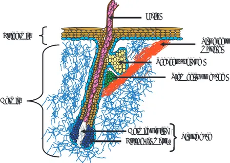
Several years have passed since the discovery of thelia like the intestinal epithelium and the skin epi-thelium, which are able to effi cently regenerate. It is also known that stem cells regenerating these tissues have a high proliferation potential. In the skin, epithe-lial cells are derived from stem cells that adhere fi rmly to the basal layer on the bulge of the pilose follicle (skin with hair) (Figure 6).88 Stem cells in the
intesti-nal epithelium are located near the base of the intes-tinal crypts (Figure 7); from that point, differentiated cells migrate through the crypts until they reach the microhair apical surface, where they repeople the intestinal epithelium and end their functional stage.89
Notably, cells derived from the intestinal epithelium have molecular features in common with blood cells, such as the signaling path c-myc.90
las divisiones simétricas y asimétricas de las células troncales es crítica, tanto para mantener un número apropiado de células troncales dentro del nicho como para cumplir las demandas para diferenciar células dentro del tejido circundante. Adicionalmente, los nichos podrían orientar las divisiones asimétricas con el fi n de guiar el fl ujo y dirección de la progenie com-prometida.74
Para que una célula hija sea una célula troncal, ésta deberá retener los factores de autorrenovación e inhi-bitorios de diferenciación. Para que una célula hija destinada a proliferar y diferenciarse a lo largo de un linaje en particular, esta célula progenitora deberá recibir cuando menos algunos factores propios de las células troncales, con el fi n de mantener este estado y recibir o heredar factores de diferenciación que lo superen.74
Aunque los programas genéticos involucrados en la autorrenovación y diferenciación de las células troncales aún se desconocen, existen datos que indi-can que las vías de señalización de wingless gene, Hed-gehog y Notch están involucradas en la regulación de las células troncales.36
A continuación se mencionan los aspectos genera-les de las CTA mejor caracterizadas.
Células troncales hematopoyéticas (HSC)
El estudio biológico de este tipo de células ha sido el más extenso y se ha llevado a cabo en el ratón. Estos estudios han revelado que las HSC adultas, derivadas de médula ósea, expresan diferentes marcadores, como el receptor tirosina-cinasa c-Kit (CD117) y el antígeno de células troncales Sca-1. Un receptor adi-cional de citocinas expresado en las HSC es c-Mp1, que une trombopoyetina. Las HSC de murino expresan de manera variable la sialomucina CD34, dependiendo del estado de desarrollo y del estadio del ciclo celu-lar. Los estudios con tinciones fl uorescentes también se han usado para defi nir a las HSC. Por ejemplo, la tinción con rodamina 123 sirivió para demostrar que se da un gran infl ujo de este fl uorocromo en las HSC más primitivas, ya que las células que no lo presenta-ron diepresenta-ron pocos pases celulares. El Hoeschst 33342 es otra tinción fl uorescente usada para el aislamiento de fracciones de HSC. También se han utilizado los xenotransplantes para ensayar la repoblación de célu-las hematopoyéticas del humano.80
Actualmente, los protocolos clínicos que incluyen el enriquecimiento de HSC del humano, general-mente utilizan la expresión celular CD34.81