RNPS 2235-145 © 2009-2016 Cardiocentro Ernesto Che Guevara, Villa Clara, Cuba. Todos los derechos reservados. 217 _________________________
Artículo Original
Doble antiagregación plaquetaria en pacientes con infarto agudo de
miocardio con elevación del segmento ST y tratamiento trombolítico
Dra. Flor de M. Castro Hernández
1,
MSc. Dra. Ana L. Alonso Mariño
2,
Dr. José I. Ramírez
Gómez
3y
MSc. Dra. Olga L. Alonso Mariño
41 Unidad de Cuidados Intensivos. Hospital Universitario Arnaldo Milián Castro. Villa Clara, Cuba. 2 Unidad de Cuidados Intensivos. Hospital Universitario Celestino Hernández Robau. Villa Clara, Cuba. 3 Servicio de Cardiología. Hospital Universitario Arnaldo Milián Castro. Villa Clara, Cuba.
4 Servicio de Hematología. Hospital Universitario Celestino Hernández Robau. Villa Clara, Cuba.
Full English text of this article is also available
INFORMACIÓN DEL ARTÍCULO
Recibido: 29 de abril de 2016 Aceptado: 10 de junio de 2016
Conflictos de intereses
Los autores declaran que no existen conflictos de intereses
Abreviaturas
IAM: infarto agudo de miocardio IAMCEST: infarto agudo de miocardio con elevación del segmento ST
SCA: síndrome coronario agudo
Versiones On-Line: Español - Inglés
AL Alonso Mariño
Hospital Universitario Celestino Hernández Robau.
Calle Cuba Nº 564, e/ Barcelona y Hospital. Santa Clara 50200. Villa Clara, Cuba. Correo electrónico:
RESUMEN
Introducción: Los síndromes coronarios agudos suelen deberse a la rotura de una placa, la activación plaquetaria y la formación de un trombo que conducen a oclu-sión coronaria y leoclu-sión miocárdica; el uso del ácido acetilsalicílico, clopidogrel y heparina de bajo peso molecular han reducido el riesgo de muerte, infarto de mio-cardio y recurrencia de la isquemia.
Objetivo: Describir la evolución clínica y los beneficios de la doble antiagregación plaquetaria asociada al tratamiento trombolítico en pacientes con infarto agudo de miocardio con elevación del segmento ST (IAMCEST).
Método: Se realizó un estudio descriptivo, transversal, contrastado, no aleatoriza-do, multicéntrico, en el período de octubre de 2012 a diciembre de 2014, en las Unidades de Cuidados Intensivos de los Hospitales Universitarios Arnaldo Milián Castro, Celestino Hernández Robau y Placetas, en Villa Clara, Cuba. La población de estudio estuvo conformada por 86 pacientes divididos en grupo estudio y con-trol, que cumplieron con los criterios de inclusión.
Resultados:El IAMCEST fue más frecuente en los pacientes del sexo masculino y entre los 70-75 años. Los factores de riesgo más frecuentes fueron el hábito de fumar y la hipertensión arterial en el 57,0% de los pacientes de ambos grupos. La localización de cara anterior presentó más complicaciones, los pacientes con localización inferior del infarto fueron los más beneficiados con la terapia antipla-quetaria dual, y en el grupo estudio se constató que a mayor precocidad del trata-miento mejor evolución y menor letalidad intrahospitalaria.
Conclusiones: La doble terapia antiplaquetaria en pacientes con IAMCEST que re-ciben tratamiento trombolítico disminuyó la frecuencia de complicaciones isqué-micas.
Palabras clave: Inhibidores de agregación plaquetaria, Fibrinolíticos, Infarto de miocardio, Evolución clínica
Dual antiplatelet therapy in patients with ST-segment elevation
acute myocardial infarction and thrombolytic treatment
ABSTRACT
let activation, and thrombus formation leading to coronary occlusion and myocar-dial injury. The use of acetylsalicylic acid, clopidogrel and low molecular weight heparin have reduced the risk of death, myocardial infarction and ischemia recur-rence
Objective: To describe the clinical course and benefits of dual antiplatelet therapy associated with thrombolytic therapy in patients with ST-segment elevation acute myocardial infarction (STEMI).
Method: A descriptive, cross-sectional, non-randomized, multicenter study was performed between October 2012 and December 2014 at the Intensive Care Units from Arnaldo Milián Castro, Celestino Hernández Robau and Placetas University Hospitals in Villa Clara, Cuba. The study population consisted of 86 patients divid-ed into study and control groups, who met the inclusion criteria.
Results: STEMI was more frequent in males and between 70-75 years. Most fre-quent risk factors were smoking and hypertension in 57.0% of patients in both groups. The anterior wall location presented more complications; patients with inferior location of the infarction were the most benefited with dual antiplatelet therapy. It was found in the study group that the earlier the treatment, the better the evolution and the lower the in-hospital mortality.
Conclusions: Dual antiplatelet therapy in patients with STEMI, receiving thrombo-lytic therapy, decreased ischemic complications frequency.
Key words: Platelet aggregation inhibitors, Fibrinolytic Agents, Myocardial Infarc-tion, Clinical evolution
INTRODUCCIÓN
Los síndromes coronarios agudos (SCA) suelen de-berse a la rotura de una placa, la activación plaque-taria y la formación de un trombo que conducen a oclusión coronaria y lesión miocárdica. El conoci-miento de su fisiopatología ha llevado al desarrollo de estrategias antitrombóticas de gran eficacia, que han reducido el riesgo de muerte, infarto y
recurren-cia de la isquemia1. La terapia antitrombótica en el
contexto del SCA incluye 3 componentes: 1) terapia antiplaquetaria que disminuye la activación y agre-gación de las plaquetas y la formación del trombo después de la ruptura de la placa, e incluye fár-macos como: la aspirina, el clopidogrel, prasugrel, ticagrelor e inhibidores de la glicoproteína (GP) IIb/ IIIa; 2) terapia anticoagulante que incluye la hepari-na no fracciohepari-nada y la de bajo peso molecular; y 3) sustancias fibrinolíticas que son usadas para la lisis del trombo e incluye la estreptoquinasa y el
activa-dor tisular del plasminógeno, entre otros2.
Es importante tener en cuenta que los beneficios obtenidos con la terapia fibrinolítica, están en mu-chas ocasiones limitados por una inadecuada reper-fusión o las reoclusiones que ocurren con posteriori-dad en estos pacientes, de allí el importante papel que juegan los antiagregantes plaquetarios, así como los anticoagulantes, en el tratamiento del infarto agudo de miocardio (IAM), los cuales influyen de
forma más protagónica en la prevención de
compli-caciones y la muerte derivada de estas3.
La aspirina actúa sobre la ciclooxigenasa 1,
in-hibe la formación de tromboxano A2 e induce una
inhibición funcional permanente de las plaquetas, por lo que no sólo resulta útil para la prevención primaria de los episodios vasculares, sino que tam-bién es eficaz en todo el espectro de los SCA y forma parte de la estrategia inicial de tratamiento en pa-cientes con sospecha de IAM con elevación del
segmento ST (IAMCEST)4. Los inhibidores de los
receptores plaquetarios del difosfato de adenosina (ADP), clopidogrel, prasugrel y ticagrelol, poseen un fuerte efecto sinérgico con la aspirina. El clopidogrel inhibe selectivamente la unión del ADP al receptor plaquetario y la subsecuente activación del com-plejo GP IIb-IIIa, mediada por el ADP, con lo cual se inhibe la agregación plaquetaria. Este fármaco modi-fica irreversiblemente el receptor plaquetario al ADP y, en consecuencia, las plaquetas expuestas son
afectadas durante todo su período de vida5.
La letalidad del IAMCEST varía entre 6-14% y está influenciada por muchos factores, entre ellos: la edad, la clase de Killip, el retraso en la aplicación del tratamiento, el tipo de tratamiento, la historia previa de IAM, la diabetes mellitus, la insuficiencia renal, así como el número de arterias coronarias
afectadas y la fracción de eyección ventricular6.
CorSalud 2016 Oct-Dic;8(4):217-226 219 la letalidad asociada al infarto miocárdico con el uso
de la doble antiagregación plaquetaria6,7, por lo que
hay razones sólidas para apoyar el uso rutinario de clopidogrel añadido a la aspirina como coadyuvante del tratamiento lítico.
En Villa Clara, la cardiopatía isquémica constitu-ye la segunda causa de muerte, sólo precedida por las neoplasias malignas, por lo que se decidió rea-lizar esta investigación con el objetivo de describir el comportamiento de la doble antiagregación pla-quetaria como terapia coadyuvante al tratamiento trombolítico en pacientes con IAMCEST ingresados en las Unidades de Cuidados Intensivos.
MÉTODO
Se realizó un estudio descriptivo y comparativo, transversal, contrastado, no aleatorizado, multicén-trico, en el período comprendido entre octubre de 2012 y diciembre de 2014, en las Unidades de Cui-dados Intensivos de los Hospitales Universitarios Arnaldo Milián Castro, Celestino Hernández Robau y Municipal de Placetas, en la provincia de Villa Clara, Cuba.
Población de estudio
Se estudiaron todos los pacientes ingresados en las UCI de dichos hospitales, con diagnóstico de IAMCEST, que recibieron tratamiento trombolítico, independientemente al lugar donde fue aplicado.
Criterios de inclusión y exclusión
Se incluyeron todos los pacientes menores de 75 años, con diagnóstico inequívoco –clínico, electro-cardiográfico y enzimático– de IAMCEST con anti-aqregación plaquetaria.
Se excluyeron a todos aquellos con riesgo de san-grado mayor, con hepatopatías crónicas, historias de diátesis hemorrágicas, embarazo y puerperio, y que habían recibido el tratamiento trombolítico con más de 12 horas de antelación.
Grupos
Los pacientes se dividieron en dos grupos. El de estudio estuvo conformado por 43 enfermos, a los cuales se les administró, posterior a la realización de la trombolisis, una dosis inicial de 300 mg de clopi-dogrel, asociada a 250 mg de aspirina; seguido de dosis diarias de 75 mg y 125 mg respectivamente, durante su estadía hospitalaria, y se les dio segui-miento electrocardiográfico, clínico, enzimático y
ecocardiográfico, hasta el día del egreso hospitala-rio.
El grupo control estuvo conformado por 43 pa-cientes, que recibieron tratamiento trombolítico, a los cuales se les administró posteriormente una dosis inicial de 250 mg de aspirina, seguido de una dosis diaria de 125 mg, como único antiagregante plaquetario.
Variables
Se estudiaron las siguientes variables: edad, sexo, factores de riesgo, localización del infarto (anterior e inferior), intervalo entre aparición de síntomas y aplicación del tratamiento trombolítico (precoz [1-3 horas], medianamente precoz [3-6 horas] y tardío [6-12 horas]), evolución, complicaciones y letalidad intrahospitalaria.
Procesamiento estadístico
La información obtenida mediante la revisión de las historias clínicas –donde se enfatizó en la evolución clínica, enzimática, electrocardiográfica y ecocardio-gráfica hasta el día del alta hospitalaria–, fue proce-sada a través de una base de datos y el uso del soft-ware de procesamiento estadístico SPSS, versión 21.0, para Windows. Esta información fue organizada en tablas de frecuencias y de contingencia, usando-se en la descripción de las mismas frecuencias abso-lutas (número de casos) y porcentajes. Los datos también fueron representados gráficamente según el tipo de información.
Para evaluar la posible asociación entre variables cualitativas se utilizó la prueba de independencia Chi Cuadrado. Con el objetivo de identificar la aso-ciación entre localización del IAM y el intervalo de aplicación del tratamiento con la presencia de complicaciones, se realizó un análisis de regresión logística binaria multivariada.
En todos los casos se fijó un intervalo de confian-za del 95%, y la significación estadística se interpretó según el siguiente criterio: si p > 0,05 no existen di-ferencias significativas, si p ≤ 0,05 la diferencia es significativa.
RESULTADOS
entre 70-75 años (41,9%), seguido por aquellos entre 60-69 años (29,1%).
Los factores de riesgo también se comportaron
de forma parecida en ambos grupos (Tabla 2), solo
existieron diferencias significativas en cuanto a la presencia de diabetes mellitus, la cual fue
predomi-nante en el grupo control (34,9 vs. 16,3%; p=0,048).
También hubo diferencias significativas ante la pre-sencia de estrés, que predominó en el grupo estudio (16,3%) con respecto al de control (2,3%) (p=0,026). Los factores de riesgo que predominaron fueron el hábito de fumar (57%) y la hipertensión arterial (57%), seguidos por los antecedentes de cardiopatía isquémica (38,3%) y diabetes mellitus (25,6%).
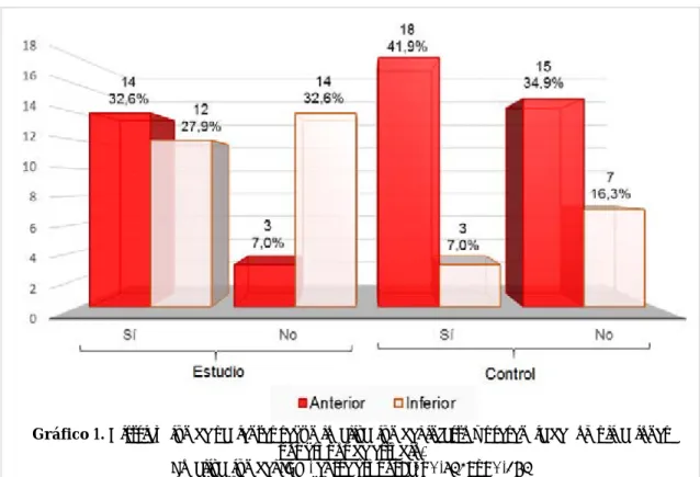
En cuanto a la localización del infarto y su
rela-ción con la aparirela-ción de complicaciones (Gráfico 1)
se observó una asociación significativa, más frecuen-tes en el IAM de cara anterior que se manifestó en un 32,6 % de los pacientes del grupo estudio, contra un 7% que no se complicó y el infarto fue de esa misma localización. Sin embargo, en este mismo gru-po, las complicaciones se presentaron en un 27,9% en pacientes con IAM de localización inferior, frente a un 32,6% que no tuvo ninguna.
En cuanto al intervalo de tratamiento y la
evolu-ción clínica (Tabla 3) de forma independiente para
ambos grupos no existieron diferencias significativas, de ahí que se hayan comportado de forma similar, al predominar la evolución no favorable a medida que aumentó el intervalo de tiempo entre la aparición de los síntomas y la aplicación del tratamiento; sin em-bargo, cuando se relacionó el mencionado intervalo Tabla 1. Distribución de pacientes según sexo y edad por grupos.
Grupos de edad
Sexo
Total
Estudio (n=43) Control (n=43)
Femenino Masculino Femenino Masculino
No % No % No % No % No %
Menor de 50 2 4,6 1 2,3 0 0,0 8 18,6 11 12,8
50 - 59 2 4,6 6 14,0 3 7,0 3 7,0 14 16,3
60 - 69 1 2,3 11 25,6 3 7,0 10 23,3 25 29,1
70 - 75 8 18,6 12 27,9 8 18,6 8 18,6 36 41,9
Total 13 30,2 30 69,8 14 32,6 29 67,4 86 100,0
χ2=3,130; p=0,372
Tabla 2. Distribución de pacientes según factores por grupos de estudio.
Factores de riesgo Estudio (n=43) Grupos de estudio Control (n=43) Total Estadígrafos
No % N° % No % χ2 p
Hábito de fumar 26 60,5 23 53,5 49 57,0 0,426 0,514
Hipertensión 24 55,8 25 58,1 49 57,0 0,047 0,827
Cardiopatía isquémica 20 46,5 13 30,2 33 15,1 2,409 0,183
Diabetes mellitus 7 16,3 15 34,9 22 25,6 3,909 0,048
Sedentarismo 8 18,6 5 11,6 13 15,1 0,816 0,366
Obesidad 5 11,6 6 14,0 11 12,8 0,104 0,747
Alcoholismo 5 11,6 5 11,6 10 11,6 0,000 1,000
Hipercolesterolemia 4 9,3 5 11,6 9 10,5 0,124 0,725
Estrés 7 16,3 1 2,3 8 9,3 4,962 0,026
CorSalud 2016 Oct-Dic;8(4):217-226 221 con la evolución de los pacientes por grupos se
observó una asociación significativa en el grupo estudio (p=0,029), donde el 37,2% presentó una evo-lución favorable, a diferencia de sólo el 25,6 % del grupo control.
Por su parte, en el grupo control no se
observa-ron diferencias significativas en su evolución con relación al tiempo de aplicación del tratamiento, pues evolucionó favorablemente solo el 7 % de los pacientes tratados precozmente; un 14% de los atendidos en un tiempo medianamente precoz, y 4,7% de los atendidos de forma tardía.
Gráfico 1. Distribución de pacientes según localización del infarto y presencia de complicaciones por grupos de estudio.
Localización del IAM, entre grupos: χ2=0,754; p=0,385 Localización del IAM, grupo estudio: χ2=5,635; p=0,0176 Localización del IAM, grupo control: χ2=1,850; p=0,1737
Tabla 3. Distribución de pacientes, según su evolución y el intervalo entre la aparición de los síntomas y la aplicación del tratamiento.
Intervalo
Evolución
Total
Grupo estudio (n=43) Grupo control (n=43)
Favorable No favorable Favorable No favorable
No % No % No % No % No %
Precoz 12 27,9 9 20,9 3 7,0 10 23,3 34 39,5
Medianamente precoz 3 7,0 12 27,9 6 14,0 14 32,6 35 40,7
Tardía 1 2,3 6 14,0 2 4,7 8 18,6 17 19,8
Total 16 37,2 27 62,8 11 25,6 32 74,4 86 100,0
Intervalo de tratamiento (Estudio): χ2=7,047; p=0,029 Intervalo de tratamiento (Control): χ2=0,412; p=0,814
Tabla 4-A. Análisis estadístico global de las complicaciones.
Complicaciones χ2Estudio p χ2Control p
Bradiarritmias 1,117 0,291 2,266 0,132 FARVR 3,888 0,049 0,717 0,392 PCR en FV 1,911 0,167 0,000 1,000 Taquicardia ventricular 3,108 0,078 1,012 0,315 Isquémicas 6,198 0,013 0,068 0,795 Fallo de bomba 3,103 0,078 0,000 1,000 Shock cardiogénico 1,117 0,291 0,212 0,645 Aneurisma ventricular - - 0,345 0,557 Otras 0,000 1,000 0,717 0,392 FA: fibrilación auricular con respuesta ventricular rápida, FV: fibrilación ventricular, PCR: parada cardiorrespiratoria
Cuando se relacionaron la localización del infarto
y las complicaciones (Tabla 4), ambos grupos se
comportaron de forma similar, sin observarse diferencias significativas entre ellos. Las complica-ciones isquémicas fueron las más frecuentes para ambos (36%), seguidas por las bradiarritmias (25,6%) y el fallo de bomba (19,8%).
Al relacionar de forma independiente cada grupo
(Tabla 4-A) se observó que en el grupo estudio la fibrilación auricular con respuesta ventricular rápida fue más frecuente en el IAM de localización anterior
que en el inferior (14,0 vs. 2,3%; p=0,049), al igual
que las complicaciones isquémicas (23,3 vs. 4,7%;
p=0,013), diferencias que fueron significativas en este grupo. El resto de las complicaciones se com-portaron de manera similar entre ellas independien-temente de la localización del IAM en ambos grupos.
La asociación de la letalidad intrahospitalaria entre ambos grupos no fue significativa. Sin embargo cuando se analizó la asociación entre la letalidad y el intervalo de tiempo para la aplicación del trata-miento, la diferencia fue significativa en el grupo es-tudio (p=0,03), donde el aumento de la letalidad fue directamente proporcional a la demora de la aplica-ción del tratamiento trombolítico. El porcentaje de fallecidos alcanzó un 7% cuando se aplicó esta tera-péutica de forma tardía, se redujo a un 2,3% cuando fue aplicada de forma medianamente precoz, y no se registraron fallecidos en los pacientes a los que se
les aplicó de forma precoz (Gráfico 2).
No hubo asociación significativa entre el interva-lo de tiempo para la aplicación del tratamiento y la letalidad en el grupo control (p=0,68). A nivel gene-ral se observó una leve disminución de la letalidad intrahospitalaria, en el grupo estudio de un 9,3% (4 pacientes), frente a un 14% (6 pacientes) en el grupo control.
Tabla 4. Distribución de pacientes, según complicaciones por grupos de estudio y localización del infarto.
Complicaciones Estudio (n=43) Localización del infarto Control (n=43) Total Estadígrafos
Anterior Inferior Anterior Inferior χ2 p
Complicaciones eléctricas
Bradiarritmias 3 (7,0) 6 (14,0) 4 (9,3) 9 (20,9) 22 (25,6) 0,977 0,323 FARVR 6 ( 14,0) 1 (2,3) 4 (9,3) 2 (4,7) 13 (15,1) 0,090 0,763 PCR en FV 4 (9,3) 1 (2,3) 1 (2,3) 1 (2,3) 7 (8,1) 1,400 0,237 Taquicardia ventricular 3 (7,0) 0 (0,0) 0 (0,0) 1 (2,3) 4 (4,7) 1,049 0,306
Isquémicas 10 (23,3) 2 (4,7) 9 (20,9) 10 (23,3) 31 (36,0) 2,472 0,116
Complicaciones mecánicas
Fallo de bomba 2 (4,7) 7 (16,3) 4 (9,3) 4 (9,3) 17 (19,8) 0,073 0,787 Shock cardiogénico 6 (14,0) 3 (7,0) 3 (7,0) 2 (4,7) 14 (16,3) 1,365 0,243 Aneurisma ventricular 0 (0,0) 0 (0,0) 2 (4,7) 1 (2,3) 3 (3,5) 3,108 0,078
Otras 1 (2,3) 1 (2,3) 4 (9,3) 2 (4,7) 8 (9,3) 2,205 0,138
CorSalud 2016 Oct-Dic;8(4):217-226 223
DISCUSIÓN
El IAM continúa siendo un problema de salud pú-blica en los países desarrollados y en vías de desarrollo, por lo que son necesarios esquemas de tratamientos efectivos y factibles8.
En esta investigación existió predominio del sexo masculino, lo que se corresponde con lo informado
por Kaul et al.2 en un estudio multicéntrico (TRACE)
realizado en la India, donde el IAMCEST fue más frecuente en pacientes mayores de 50 años, del sexo
masculino; y con lo encontrado por Correia et al.9
donde la edad media fue de 63±13 años y el 72% eran hombres. Datos similares se recogen en un
estudio realizado en Corea del Sur10, donde la edad
media de los pacientes estudiados fue de 63,9±9,6 y predominó el sexo masculino (69,7%); y en el
traba-jo de Rakowski et al.11 donde la edad media fue de
62 (56-73) años y el 69% era del sexo masculino.
Kaul et al.2 señalan a la cardiopatía isquémica
previa, la hipertensión arterial, la diabetes y la disli-pidemia como los principales factores de riesgo;
mientras que Cho et al.10 informan la hipertensión
arterial y la diabetes mellitus, lo que coincide con
los hallazgos del estudio PLATO7. Resultados
simila-res a los de esta investigación fueron encontrados
por Wang et al.12, pues el 55% de sus pacientes
fumaba y el 51% padecía de hipertensión arterial. Otros autores incluyen también dentro de los facto-res de riesgo más frecuentes el antecedente de
car-diopatía isquémica previa11.
En Estados Unidos se ha observado que la inci-dencia de pobres respondedores a la aspirina está incrementada de forma significativa en la población diabética, lo que condiciona una hiperactividad pla-quetaria y un fenotipo proaterogénico, que aumenta el riesgo en estos pacientes de sufrir una
enferme-dad cardiovascular isquémica13.
Con respecto a la localización más frecuente del
IAM, Rakowski et al.11 en Polonia informaron que el
43% de los IAMCEST eran de cara anterior y que és-tos presentaron más complicaciones, debido a una mayor afectación del músculo miocárdico;
resulta-dos similares a los registraresulta-dos por Wang et al.12
quien encontró esta localización en el 51,9% de los pacientes, y constató que cuando el IAM se asocia con diabetes mellitus y con un inicio del tratamiento de forma tardía se presenta mayor frecuencia de fa-Gráfico 2. Distribución de pacientes según intervalo entre aparición de síntomas y aplicación del
tratamiento y letalidad intrahospitalaria.
llo de bomba con Killip-Kimball mayor de I.
Además, en el estudio realizado por Rakowski et
al.11 se observa que la evolución, incluyendo las
complicaciones asociadas y la calidad de vida pos-terior al SCA, está muy ligada al tiempo de inicio de la terapia de reperfusión; a mayor rapidez en reper-fundir el vaso relacionado con el IAM, la evolución tiende a ser mejor, aunque en este estudio se obser-vó una mejor calidad de vida en aquellos pacientes a los que se les aplicó intervencionismo coronario percutáneo. En concordancia con este trabajo, en el grupo control no se observaron diferencias significa-tivas en cuanto a evolución y aplicación del tra-tamiento, y fue donde hubo mayor proporción de pacientes complicados, independientemente del tiempo de aplicación de la terapéutica.
En una investigación realizada en China12, se
ha-ce referencia a factores que influyen de forma signifi-cativa en la evolución de estos pacientes: tiempo transcurrido desde el diagnóstico hasta la aplicación del tratamiento, localización anterior del infarto, antecedentes de diabetes mellitus y ausencia de do-sis de carga de clopidogrel para la doble antiagrega-ción plaquetaria. En Estados Unidos también se han realizado estudios destacándose la importancia del tratamiento precoz en el IAM, pues en los pacientes a los que se les aplicó el tratamiento de forma rápida (porque se activó de forma prehospitalaria el siste-ma de asistencia) se observó un descenso de la
letalidad a un 6,7% vs. 9,5% de aquellos a los que no
se les brindó asistencia prehospitalaria en cuanto a
tratamiento reperfusor y antiagregante14.
En lo referido al tipo de complicaciones
presenta-das en el estudio TRACE2, se plantea que las
compli-caciones isquémicas fueron las más frecuentes,
seguidas por el shock cardiogénico, lo que
concuer-da con los resultados de esta serie; concuer-datos similares son recogidos en el trabajo realizado por Rakowski
et al.11 en Polonia, donde las complicaciones más
frecuentes fueron el fallo de bomba, seguida por las complicaciones isquémicas.
En un estudio comparativo realizado en China12
llama la atención que la mayor parte de los pacien-tes donde, además de la terapéutica reperfusora y la aspirina, no se usó una dosis de carga de clopido-grel, presentaron una mayor incidencia de fallo de bomba con una clase de Killip-Kimball mayor de I, que el grupo que usó dosis de carga seguida de 75 mg como dosis de mantenimiento en los días pos-teriores; por lo que se concluye que su uso es importante en la prevención de complicaciones secundarias al IAMCEST.
Así también, otros autores destacan que la doble antiagregación plaquetaria es muy importante para la prevención secundaria en pacientes que han sufrido un SCA15,16.
En el estudio TRACE2, el aumento de las
compli-caciones de causa isquémica apareció en los pa-cientes que se antiagregaron sólo con aspirina, con respecto a aquellos en los que se empleó clopido-grel y aspirina; y se encontró que los pacientes en los que no se administró la doble antiagregación plaquetaria presentaron un mayor riesgo de letali-dad asociada a complicaciones; lo que concuerda con los resultados obtenidos en este trabajo.
En un estudio realizado en Canadá17, se observó
una disminución del reinfarto cuando el clopidogrel se asoció con aspirina; sin embargo, se destaca la superioridad del prasugrel y el ticagrelor, aunque recomiendan hacer previamente un balance riesgo-beneficio, con respecto al riesgo de sangrado.
Saba-tine et al.3 en su serie que incluyó 3491 pacientes
con IAMCEST concluyeron que los pacientes meno-res de 75 años de edad que recibieron aspirina, clopidogrel y terapia fibrinolítica, presentaron dismi-nución de la letalidad y de la recurrencia del IAM.
Según la literatura revisada este efecto protector puede estar afectado en algunas personas por el uso de estatinas lipofílicas, bloqueadores de los canales de calcio y el hábito de fumar, la diabetes mellitus y la obesidad, ya que pueden predisponer a variacio-nes en la función plaquetaria, aunque la resistencia al efecto del clopidogrel se describe como el factor más importante que interfiere en la prevención de
complicaciones18,19. Tal es la importancia del
poli-morfismo genético y la resistencia al efecto del clopi-dogrel para la prevención de complicaciones poste-riores al evento isquémico, que se han realizado estudios en Estados Unidos donde se sugieren que la elección del antiagregante plaquetario puede ser guiado por genotipado, como una estrategia coste-efectiva en pacientes con SCA, sobre todo si a los pacientes se les realiza una intervención coronaria percutánea20.
En el estudio TRACE2 se informa mayor letalidad
en el grupo que solo utiliza aspirina como antiagre-gante plaquetario, lo que coincide con el trabajo
realizado por Husted et al.7, donde se constata una
CorSalud 2016 Oct-Dic;8(4):217-226 225 del IAM); sin embargo, el riesgo de sangrado fue
más elevado en los pacientes que utilizaron prasu-grel (2,4 vs. 1,8%). Oliver et al.21 también señalan que el ticagrelor ha demostrado eficacia superior en comparación con el clopidogrel en cuanto a la
re-ducción de la letalidad (9,0 vs. 10,7%,
respectivamen-te), debido a las características farmacodinámicas del primero, que en estos estudios ha demostrado ser más rápido y potente inhibidor de la agregación plaquetaria que el clopidogrel, porque altera la captación de la adenosina por los glóbulos rojos, lo
que pudiera influenciar en su eficacia y seguridad22.
Existen otros factores que influyen en el riesgo de letalidad, pues se incrementa en pacientes con infarto previo por la deficiente prevención secunda-ria, la no modificación del estilo de vida, o el
aban-dono del tratamiento15.
Otras complicaciones propias del uso de antiagre-gantes plaquetarios, como el sangrado o el hemato-ma renal subcapsular espontáneo, no fueron encon-tradas en esta investigación, lo que coincide con lo
informado en el estudio CONMIT6.
Limitaciones del estudio
Entre las limitaciones del estudio se encontró la im-posibilidad de seguimiento por 6-12 meses posterio-res al IAMCEST al grupo estudio, con lo que se hubiera podido tener una idea más completa de los beneficios del tratamiento a largo plazo, así como de la disminución de la letalidad tardía en estos pa-cientes.
CONCLUSIONES
La aplicación clínica de la terapia antiplaquetaria dual reflejó una disminución en la incidencia de complicaciones isquémicas posteriores al infarto agudo de miocardio con elevación del segmento ST en los pacientes que recibieron tratamiento trombo-lítico. Fueron más beneficiados aquellos que presen-taron una localización inferior del infarto. Además la letalidad intrahospitalaria asociada a complicaciones propias del infarto se redujo en el grupo estudio, influenciada principalmente por la aplicación precoz del tratamiento.
BIBLIOGRAFÍA
1. Erdem G, Flather M. Evaluación del riesgo de
he-morragia en los síndromes coronarios agudos.
Rev Esp Cardiol. 2012;65:4-6.
2. Kaul U, Sethi KK, Dalal J, Parikh K, Hiremath MS,
Mullasari AS, et al. A multicentre retrospective
study to understand anti-platelet treatment pat-terns and outcomes of acute coronary syndrome patients in India (TRACE). Indian Heart J. 2014;66: 334-9.
3. Sabatine MS, Cannon CP, Gibson CM,
López-Sen-dón JL, Montalescot G, Theroux P, et al. Addition
of clopidogrel to aspirin and fibrinolytic therapy for myocardial infarction with ST segment eleva-tion. N Engl J Med. 2005;352:1179-89.
4. Hamm CW, Bassand JP, Agewall S, Bax J,
Boers-ma E, Bueno H, et al. Guía de práctica clínica de
la ESC para el manejo del síndrome coronario agudo en pacientes sin elevación persistente del segmento ST. Rev Esp Cardiol. 2012;65:173.e1-55.
5. Fisch AS, Perry CG, Stephens SH, Horenstein RB,
Shuldiner AR. Pharmacogenomics of anti-platelet and anti-coagulation therapy. Curr Cardiol Rep. 2013;15:381.
6. Steg G, James SK, Atar D, Badano LP, Blomstrom
Lundqvist C, Borger MA, et al. Guía de práctica
clínica de la ESC para el manejo del infarto agudo de miocardio en pacientes con elevación del segmento ST. Rev Esp Cardiol. 2013;66:53.
7. Husted S, James SK, Bach RG, Becker RC, Budaj
A, Heras M, et al. The efficacy of ticagrelor is
maintained in women with acute coronary syn-dromes participating in the prospective, random-ized, PLATelet inhibition and patient Outcomes (PLATO) trial. Eur Heart J. 2014;35:1541-50.
8. Pinar E, Bardají A. Manejo del infarto agudo de
miocardio con elevación del segmento ST. Guías de actuación clínica y el mundo real. Rev Esp Cardiol Supl. 2009;09:C71-8.
9. Correia LC, García G, Kalil F, Ferreira F, Carvalhal
M, Oliveira R, et al. Prognostic value of TIMI
score versus GRACE score in ST-segment eleva-tion myocardial infarceleva-tion. Arq Bras Cardiol. 2014; 103:98-106.
10.Cho YK, Nam CW, Park HS, Yoon HJ, Kim H, Hur
SH, et al. Efficacy and safety of
antiplatelet-com-bination therapy after drug-eluting stent implanta-tion. Korean J Intern Med. 2014;29:210-6.
11.Rakowski T, Dziewierz A, Siudak Z, Kleczyński P,
Dubiel JS, Dudek D. Introduction of new oral anti-platelet drugs in myocardial infarction hospital network: initial experience. J Thromb Thrombo-lysis. 2014;37:243-5.
12.Wang X, Yu H, Li Z, Li L, Zhang Y, Gao W.
clopido-grel loading dose, and left ventricular systolic function in patients with primary percutaneous coronary intervention. Mediators Inflamm [Inter-net]. 2014 [citado 18 Abr 2016];2014:482763. Dispo-nible en:
https://www.ncbi.nlm.nih.gov/pmc/articles/PMC 4131512/pdf/MI2014-482763.pdf
13.López LR, Guyer KE, Torre IG, Pitts KR, Matsuura
E, Ames PR. Platelet thromboxane (11-dehydro-Thromboxane B2) and aspirin response in pa-tients with diabetes and coronary artery disease. World J Diabetes. 2014;5:115-27.
14.Deshpande A, Birnbaum Y. ST-segment elevation:
distinguishing ST elevation myocardial infarction from ST elevation secondary to nonischemic etiologies. World J Cardiol. 2014;610:1067-79.
15.Redfern J, Hyun K, Chew DP, Astley C, Chow C,
Aliprandi-Costa B, et al. Prescription of secondary
prevention medications, lifestyle advice, and re-ferral to rehabilitation among acute coronary syn-drome inpatients: results from a large prospective audit in Australia and New Zealand. Heart. 2014; 100:1281-8.
16.Hong KS. Dual antiplatelet therapy after
noncar-dioembolic ischemic stroke or transient ischemic attack: pros and cons. J Clin Neurol.
2014;10:189-96.
17.Chua D, Nishi C. New antiplatelet agents for
car-diovascular disease. CMAJ. 2013;185:1405-11.
18.Huber K, Bates ER, Valgimigli M, Wallentin L,
Kri-stensen SD, Anderson JL, et al. Antiplatelet and
anticoagulation agents in acute coronary syn-dromes: what is the current status and what does the future hold?. Am Heart J. 2014;168:611-21.
19.Schroeder JS, Frishman WH, Parker JD, Angiolillo
DJ, Woods C, Scirica B. Pharmacologic options for treatment of ischemic disease. In: Antman EM, Sabatine M, editors. Cardiovascular therapeutics – A Companion to Braunwald’s Heart Disease. 9th ed. Philadelphia: Saunders; 2013. p. 95-142.
20.Patel V, Lin FJ, Ojo O, Rao S, Yu S, Zhan L, et al.
Cost-utility analysis of genotype-guided antiplate-let therapy in patients with moderate-to-high risk acute coronary syndrome and planned percuta-neous coronary intervention. Pharm Pract (Gra-nada). 2014;12;438.
21.Olivier C, Diehl P, Bode C, Moser M. Thrombin
receptor antagonism in antiplatelet therapy. Cardiol Ther. 2013;2:57-68.
22.Dobesh PP, Oestreich JH. Ticagrelor:
RNPS 2235-145 © 2009-2016 Cardiocentro Ernesto Che Guevara, Villa Clara, Cuba. All rights reserved. 217 _________________________
Original Article
Dual antiplatelet therapy in patients with ST-segment elevation acute
myocardial infarction and thrombolytic treatment
Flor de M. Castro Hernández
1, MD;
Ana L. Alonso Mariño
2, MD, MSc; José I. Ramírez
Gómez
3, MD, MSc; y
Olga L. Alonso Mariño
4, MD, MSc
1 Intensive Care Unit. Hospital Universitario Arnaldo Milián Castro. Villa Clara, Cuba. 2 Intensive Care Unit. Hospital Universitario Celestino Hernández Robau. Villa Clara, Cuba. 3 Cardiology Department. Hospital Universitario Arnaldo Milián Castro. Villa Clara, Cuba. 4 Hematology Department. Hospital Universitario Celestino Hernández Robau. Villa Clara, Cuba.
Este artículo también está disponible en español
ARTICLE INFORMATION
Received: April 29, 2016 Accepted: June 10, 2016
Competing interests
The authors declare no competing interests
Acronyms
ACS: acute coronary syndrome AMI: acute myocardial infarction STEMI: ST-segment elevation acute myocardial infarction
On-Line Versions: Spanish - English
AL Alonso Mariño
Hospital Universitario Celestino Hernández Robau.
Calle Cuba Nº 564, e/ Barcelona y Hospital. Santa Clara 50200. Villa Clara, Cuba. E-mail address::
ABSTRACT
Introduction: Acute coronary syndromes are usually due to plaque rupture, plate-let activation, and thrombus formation leading to coronary occlusion and myocar-dial injury. The use of acetylsalicylic acid, clopidogrel and low molecular weight heparin have reduced the risk of death, myocardial infarction and ischemia recur-rence
Objective: To describe the clinical course and benefits of dual antiplatelet therapy associated with thrombolytic therapy in patients with ST-segment elevation acute myocardial infarction (STEMI).
Method: A descriptive, cross-sectional, non-randomized, multicenter study was performed between October 2012 and December 2014 at the Intensive Care Units from Arnaldo Milián Castro, Celestino Hernández Robau and Placetas University Hospitals in Villa Clara, Cuba. The study population consisted of 86 patients divid-ed into study and control groups, who met the inclusion criteria.
Results: STEMI was more frequent in males and between 70-75 years. Most fre-quent risk factors were smoking and hypertension in 57.0% of patients in both groups. The anterior wall location presented more complications; patients with inferior location of the infarction were the most benefited with dual antiplatelet therapy. It was found in the study group that the earlier the treatment, the better the evolution and the lower the in-hospital mortality.
Conclusions: Dual antiplatelet therapy in patients with STEMI, receiving thrombo-lytic therapy, decreased ischemic complications frequency.
Key words: Platelet aggregation inhibitors, Fibrinolytic Agents, Myocardial Infarc-tion, Clinical evolution
Doble antiagregación plaquetaria en pacientes con infarto agudo
de miocardio con elevación del segmento ST y tratamiento
trombolítico
RESUMEN
sióncoronariaylesiónmiocárdica;el usodelácidoacetilsalicílico,clopidogrely heparina de bajo peso molecular han reducido el riesgo de muerte, infarto de mio- cardio y recurrencia de la isquemia.
Objetivo: Describir la evolución clínica y los beneficios de la doble antiagregación plaquetaria asociada al tratamiento trombolítico en pacientes con infarto agudo de miocardio con elevación del segmento ST (IAMCEST).
Método: Se realizó un estudio descriptivo, transversal, contrastado, no aleatoriza-do, multicéntrico, en el período de octubre de 2012 a diciembre de 2014, en las Unidades de Cuidados Intensivos de los Hospitales Universitarios Arnaldo Milián Castro, Celestino Hernández Robau y Placetas, en Villa Clara, Cuba. La población de estudio estuvo conformada por 86 pacientes divididos en grupo estudio y con-trol, que cumplieron con los criterios de inclusión.
Resultados: El IAMCEST fue más frecuente en los pacientes del sexo masculino y entre los 70-75 años. Los factores de riesgo más frecuentes fueron el hábito de fumar y la hipertensión arterial en el 57,0% de los pacientes de ambos grupos. La localización de cara anterior presentó más complicaciones, los pacientes con localización inferior del infarto fueron los más beneficiados con la terapia antipla-quetaria dual, y en el grupo estudio se constató que a mayor precocidad del trata-miento mejor evolución y menor letalidad intrahospitalaria.
Conclusiones: La doble terapia antiplaquetaria en pacientes con IAMCEST que re-ciben tratamiento trombolítico disminuyó la frecuencia de complicaciones isqué-micas.
Palabras clave: Inhibidores de agregación plaquetaria, Fibrinolíticos, Infarto de miocardio, Evolución clínica
INTRODUCTION
Acute coronary syndromes (ACS) are usually due to plaque rupture, platelet activation, and thrombus formation leading to coronary occlusion and myo-cardial injury. Knowing its pathophysiology has led to the development of highly effective antithrom-botic strategies which have reduced the risk of
death, infarction and recurrent ischemia1.
Anti-thrombotic therapy in ACS context includes 3 components: 1) antiplatelet therapy for reducing platelets activation/aggregation and thrombus for-mation after plaque rupture, including drugs such as: aspirin, clopidogrel, prasugrel, ticagrelor and glycol-protein (GP) IIb/IIIa inhibitors; 2) anticoagulant therapy including unfractionated heparin and low molecular weight heparin, and 3) fibrinolytic sub-stances used for thrombus lysis including strep-tokinase and tissue plasminogen activator, among others.
It is important considering that the benefits ob-tained with fibrinolytic therapy are often limited by inadequate reperfusion or reocclusions that occur later in these patients, hence the important role of both antiplatelet agents and anticoagulants for acute
myocardial infarction (AMI) treatment, which have a more proactive influence in preventing
complica-tions and death derived from these3.
Aspirin acts on cyclooxygenase 1, inhibits
throm-boxane A2 formation and induces permanent
inhi-bition of platelets function, so it is not only useful for primary prevention of vascular events, but is also effective throughout ACS spectrum and is part of the initial treatment strategy in ST segment elevation acute myocardial infarction (STEMI) suspected
patients4. Platelet adenosine diphosphate (ADP)
in-hibitors, clopidogrel, prasugrel and ticagrelor, have a strong synergistic effect with aspirin. Clopidogrel selectively inhibits ADP platelet receptor binding and subsequent GP IIb-IIIa complex activation, me-diated by ADP, thereby inhibiting platelet aggrega-tion. This drug irreversibly modifies ADP platelet receptor and thus, exposed platelets are affected throughout their lifespan5.
STEMI lethality varies between 6-14% and is in-fluenced by many factors, including: age, Killip class, delayed treatment, type of treatment, previous history of AMI, diabetes mellitus, kidney disease, as well as the number of coronary arteries affected and
CorSalud 2016 Oct-Dec;8(4):217-226 219 found a lethality decrease associated with
myocar-dial infarction when using dual antiplatelet
thera-py6,7, so there are strong reasons to support the
routine use of clopidogrel added to aspirin as an adjunct to lytic treatment.
Ischemic heart disease is the second cause of death in Villa Clara, only preceded by malignant neoplasias, so it was decided to carry out this re-search to describe the behavior of dual antiplatelet therapy as adjunctive therapy to thrombolytic treatment in STEMI patients admitted to Intensive Care Units.
METHOD
A descriptive, comparative, cross-sectional, non-randomized, multicenter study was performed from October 2012 to December 2014 at the Intensive Care
Units of Arnaldo Milián Castro, Celestino Hernández
Robau and Placetas Hospitals, in the province of
Villa Clara, Cuba.
Study population
All patients admitted to the hospital’s Intensive Care Units, with a STEMI diagnosis, who received throm-bolytic treatment, regardless of the place where it was applied, were studied.
Inclusion and exclusion criteria
All patients younger than 75 years, with a (clinical, electrocardiographic and enzymatic) unequivocal diagnosis of STEMI, with antiplatelet therapy were included.
Those at risk for major bleeding, with chronic liver disease, histories of bleeding diathesis, preg-nancy and puerperium, and who had received thrombolytic treatment more than 12 hours in ad-vance were excluded.
Groups
Patients were divided into two groups. The study consisted of 43 patients, who were given, after thrombolysis, an initial dose of 300 mg clopidogrel, associated with 250 mg of aspirin; followed by daily doses of 75 mg and 125 mg, respectively, during their
hospital stay, and received electrocardiographic, clinical, enzymatic and echocardiographic follow-up until hospital discharge.
The control group consisted of 43 patients, who received thrombolytic treatment, who were given an initial dose of 250 mg aspirin, followed by a daily dose of 125 mg, as the only antiplatelet agent.
Variables
The following variables were studied: age, sex, risk factors, location of infarction (anterior and inferior), interval between onset of symptoms and application of thrombolytic treatment (early [1-3 hours], moder-ately early [3-6 hours] and late [6-12 hours]), evolu-tion, complications, and in-hospital mortality.
Statistical processing
The information obtained by reviewing clinical re-cords, emphasizing on clinical, enzymatic, electro-cardiographic and echoelectro-cardiographic evolution until hospital discharge was processed through a data-base and using SPSS statistical software, version 21.0, for Windows. This information was organized into frequency and contingency tables, using it in the description of the same absolute frequencies (number of cases) and percentages. Data was graphi-cally represented according to the type of infor-mation.
Chi Square Independence test was used to assess the possible association between qualitative varia-bles. A multivariate binary logistic regression analysis was performed to identify the association between AMI location and treatment application interval with the presence of complications.
In all cases a 95% confidence interval was set, and statistical significance was interpreted according to the following criterion: if p> 0.05 there were no significant differences, if p ≤ 0.05 difference was significant.
RESULTS
predomi-nated, about 70% of the cases, and ages between 70-75 years (41.9%), followed by those between 60-69 years (29.1%).
Risk factors behaved similarly in both groups (Table 2); there were only significant differences in the presence of diabetes mellitus, which was pre-dominant in the control group (34.9 vs. 16.3%; p= 0.048). There were also significant differences in the presence of stress, which predominated in the study group (16.3%) compared to the control group (2.3%) (p=0.026). Predominant risk factors were smoking (57%) and arterial hypertension (57%), followed by history of ischemic heart disease (38.3%) and dia-betes mellitus (25.6%).
Regarding to the infarction location and its
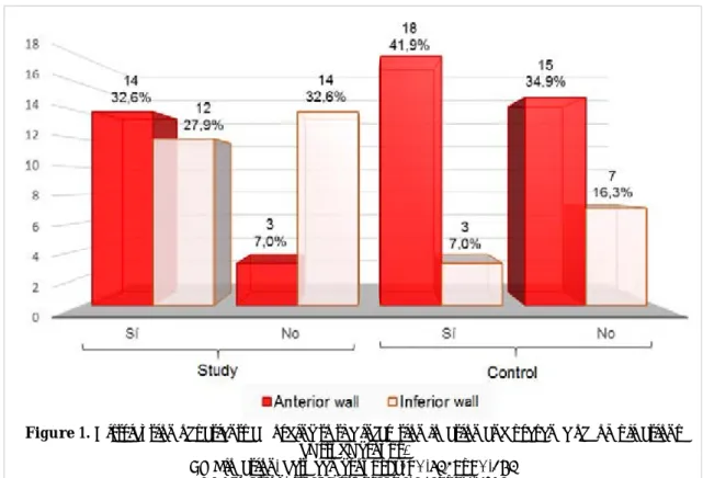
rela-tion to the appearance of complicarela-tions (Figure 1),
a significant association was observed, more fre-quent in the anterior wall AMI, which was mani-fested in 32.6% of the patients in the study group; against a 7% who did not complicate and had the same infarction location. However, in this same group, 27.9% of patients with inferior wall AMI had complications, compared to 32.6% who had none.
As for the particular treatment interval and
clini-cal evolution (Table 3) for both groups there were
no significant differences, hence they behaved si-milarly, due to the predominance of unfavorable evolution as the time interval between the onset of Table 1. Distribution of patients according to sex and age by groups.
Age groups
Sex
Total
Study (n=43) Control (n=43)
Female Male Female Male
No % No % No % No % No %
Under 50 2 4,6 1 2,3 0 0,0 8 18,6 11 12,8
50 - 59 2 4,6 6 14,0 3 7,0 3 7,0 14 16,3
60 - 69 1 2,3 11 25,6 3 7,0 10 23,3 25 29,1
70 - 75 8 18,6 12 27,9 8 18,6 8 18,6 36 41,9
Total 13 30,2 30 69,8 14 32,6 29 67,4 86 100,0
χ2=3,130; p=0,372
Table 2. Distribution of patients according to factors by study groups.
Risk Factors Study (n=43) Study Groups Control (n=43) Total Statistics
No % N° % No % χ2 p
Smoking 26 60,5 23 53,5 49 57,0 0,426 0,514
High blood pressure 24 55,8 25 58,1 49 57,0 0,047 0,827 Ischemic heart disease 20 46,5 13 30,2 33 15,1 2,409 0,183
Diabetes mellitus 7 16,3 15 34,9 22 25,6 3,909 0,048
Sedentary lifestyle 8 18,6 5 11,6 13 15,1 0,816 0,366
Obesity 5 11,6 6 14,0 11 12,8 0,104 0,747
Alcoholism 5 11,6 5 11,6 10 11,6 0,000 1,000
Hypercholesterolemia 4 9,3 5 11,6 9 10,5 0,124 0,725
Stress 7 16,3 1 2,3 8 9,3 4,962 0,026
CorSalud 2016 Oct-Dec;8(4):217-226 221 symptoms and the application of treatment
in-creased; however, when the interval was related to the patients’ evolution by groups, a significant asso-ciation was observed in the study group (p=0.029), where 37.2% presented a favorable evolution, with a
slight difference of the only 25.6% of the control group.
On the other hand, no significant differences were observed in the control group related to the time of treatment, since only 7% of early treated Figure 1. Distribution of patients according to the infarction location and presence of complications
by study groups.
AMI location, between groups: χ2=0,754; p=0,385 AMI location, study group: χ2=5,635; p=0,0176 AMI location, control group: χ2=1,850; p=0,1737
Table 3. Distribution of patients according to their evolution and the interval between onset of symptoms and treatment application.
Intervalo
Evolution
Total
Study group (n=43) Control group (n=43)
Favorable Unfavorable Favorable Unfavorable
No % No % No % No % No %
Early 12 27,9 9 20,9 3 7,0 10 23,3 34 39,5
Moderately early 3 7,0 12 27,9 6 14,0 14 32,6 35 40,7
Late 1 2,3 6 14,0 2 4,7 8 18,6 17 19,8
Total 16 37,2 27 62,8 11 25,6 32 74,4 86 100,0
Treatment interval (Study): χ2=7,047; p=0,029 Treatment interval (Control): χ2=0,412; p=0,814
Table 4-A. Global statistical analysis of complications.
Complications χ2Study p χ2Control p
Bradiarithmia 1,117 0,291 2,266 0,132 RVR 3,888 0,049 0,717 0,392 CPA in VF 1,911 0,167 0,000 1,000 Ventricular tachycardia 3,108 0,078 1,012 0,315 Ischemia 6,198 0,013 0,068 0,795 Pump failure 3,103 0,078 0,000 1,000 Cardiogenic Shock 1,117 0,291 0,212 0,645 Ventricular aneurysm - - 0,345 0,557 Other 0,000 1,000 0,717 0,392 FA: fibrilación auricular con respuesta ventricular rápida, FV: fibrilación ventricular, PCR: parada cardiorrespiratoria
patients had a favorable outcome; 14% from those treated moderately early, and 4.7% from those treated late.
When the infarction location and complications
were related (Table 4), both groups behaved
simi-larly, without observing significant differences
be-tween them. Ischemic complications were the most frequent for both (36%), followed by bradyarrhyth-mias (25.6%) and pump failure (19.8%).
When each group was independently related (Tabla 4-A), it was observed that in the study group, atrial fibrillation with rapid ventricular response was more frequent in anterior and inferior AMI (14.0 vs. 2.3%, p=0.049), as well as ischemic
complications (23.3 vs. 4.7%; p=0.013), which were
significant differences in this group. The rest of the complications behaved the same way regardless of AMI location in both groups.
The association of in-hospital lethality between both groups was not significant. However, when the association between lethality and treatment time interval application was analyzed, the difference was significant in the study group (p=0.03), where the increase in lethality was directly proportional to the thrombolytic treatment delay. Percentage of deaths reached 7% when this therapy was applied late, it was reduced to 2.3% when applied moderately early, and no deaths were recorded in patients who
re-ceived prompt treatment (Figure 2).
There was no significant association between the treatment time interval application and the control group lethality (p=0.68). Overall, a slight decrease 9.3% (4 patients) in in-hospital mortality was ob-Table 4. Distribution of patients, according to complications by study groups and infarction location.
Complications Study (n=43) Infarction location Control (n=43) Total Estadígrafos
Anterior Inferior Anterior Inferior χ2 p
Electrical Complications
Bradiarthymia 3 (7,0) 6 (14,0) 4 (9,3) 9 (20,9) 22 (25,6) 0,977 0,323 RVR 6 ( 14,0) 1 (2,3) 4 (9,3) 2 (4,7) 13 (15,1) 0,090 0,763 CPA in VF 4 (9,3) 1 (2,3) 1 (2,3) 1 (2,3) 7 (8,1) 1,400 0,237 Ventricular tachycardia 3 (7,0) 0 (0,0) 0 (0,0) 1 (2,3) 4 (4,7) 1,049 0,306
Ischemia 10 (23,3) 2 (4,7) 9 (20,9) 10 (23,3) 31 (36,0) 2,472 0,116
Mechanical Complications
Pump failure 2 (4,7) 7 (16,3) 4 (9,3) 4 (9,3) 17 (19,8) 0,073 0,787 Cardiogenic shock 6 (14,0) 3 (7,0) 3 (7,0) 2 (4,7) 14 (16,3) 1,365 0,243 Ventricular aneurysm 0 (0,0) 0 (0,0) 2 (4,7) 1 (2,3) 3 (3,5) 3,108 0,078
Other 1 (2,3) 1 (2,3) 4 (9,3) 2 (4,7) 8 (9,3) 2,205 0,138
CorSalud 2016 Oct-Dec;8(4):217-226 223 served in the study group, compared to 14% (6
pa-tients) in the control group.
DISCUSSION
AMI continues to be a public health problem in developed and developing countries, so effective
and feasible treatment schemes are needed8.
There was a male predominance in this study
which corresponds to what was reported by Kaul et
al.2 in a multicentre study (TRACE) conducted in
India, where STEMI was more frequent in 50 years old, and male patients; and with what was found by
Correia et al.9 where mean age was 63±13 years and
72% were men. Similar data are collected in a study
carried out in South Korea10, where the mean age of
the patients studied was 63.9±9.6 and male sex
predominated (69.7%); and also by Rakowski et al.11
where mean age was 62 (56-73) years and 69% were males.
Kaul et al.2 point to previous ischemic heart
dis-ease, high blood pressure, diabetes and
dyslipide-mia as the main risk factors; while Cho et al.10
reported high blood pressure and diabetes mellitus,
which coincides with PLATO7 study findings. Results
similar to our research were found by Wang et al.12,
since 55% of their patients smoked and 51% had high blood pressure. Other authors also include among the most frequent risk factors the history of previous
ischemic heart disease11.
It has been observed in the United States that the incidence of poor responders to aspirin has been significantly increased in the diabetic population, which results in a platelet hyperactivity and a pro-atherogenic phenotype, which increases the risk for
ischemic cardiovascular disease in these patients13.
Regarding the most frequent localization of AMI,
Rakowski et al.11 in Poland reported that 43% of
STEMI was on the anterior wall and that these pre-sented more complications, due to a greater affec-tion of the myocardial muscle; similar results to
those reported by Wang et al.12 who found this
location in 51.9% of the patients, and found that Figure 2. Distribution of patients according to the interval between onset of symptoms, treatment
application and in-hospital lethality.
when AMI is associated with diabetes mellitus and a late start of treatment, there is a higher frequency of pump failure with Killip-Kimball class greater than I.
In addition, the study by Rakowski et al.11 shows
that the evolution, including the associated com-plications and quality of life after ACS, is closely connected to the starting time of reperfusion thera-py; the faster the vessel reperfusion associated with AMI, the better outcome, although in this study a better quality of life was observed in those patients who underwent percutaneous coronary interven-tion. In agreement with this work, no significant differences regarding the treatment evolution and application in the control group were observed, and it had a greater proportion of patients complicated, in spite of the therapeutics time.
In a study carried out in China12, they refer to
factors that significantly influence the outcome of these patients: time elapsed from diagnosis to treat-ment application, anterior wall location of infarction, history of diabetes mellitus and absence of loading doses of clopidogrel for dual antiplatelet therapy. In the United States, studies have also been conducted highlighting the importance of early treatment in AMI, because in patients who were given a prompt treatment (because the pre-hospital care system was activated), there was a lethality decrease of 6.7% vs. 9.5% of those who were not offered pre-hospital care
for reperfusion and dual antiplatelet therapy14.
Regarding the type of complications presented in
the TRACE2 study, it is suggested that ischemic
com-plications were the most frequent, followed by cardiogenic shock, which coincides with this series
results. Similar data are collected in Rakowski et al.11
work performed in Poland, where most frequent complications were pump failure followed by is-chemic complications.
In a comparative study carried out in China12, it is
noteworthy that most patients in whom, in addition to reperfusion therapy and aspirin, loading dose of clopidogrel was not given, had a higher incidence of pump failure with a Killip-Kimball class greater than I, than the group that used loading doses followed by 75 mg as maintenance doses on subsequent days. So, it is concluded that its use is important in pre-venting secondary STEMI complications.
Also, other authors emphasize that dual anti-platelet therapy is very important for secondary
prevention in patients who have suffered ACS15,16.
In the TRACE2 study, increased ischemic-related
complications appeared in patients who received antiplatelet therapy only with aspirin, compared to those in whom clopidogrel and aspirin were used; and it was found that patients who did not receive dual antiplatelet therapy had an increased risk of lethality-related complications; which agrees with the results obtained in this work.
In a study carried out in Canada17 there was a
decrease in reinfarction when clopidogrel was asso-ciated with aspirin. However, the superiority of prasugrel and ticagrelor is emphasized, although they recommend making a risk-benefit balance with
respect to the risk of bleeding. Sabatine et al.3 in
their series that included 3491 STEMI patients concluded that patients younger than 75 years of age who received aspirin, clopidogrel and fibrinolytic therapy had a decreased lethality and recurrence of AMI.
According to the reviewed literature, this pro-tective effect may be affected in some people by the use of lipophilic statins, calcium channel blockers, and smoking, diabetes mellitus and obesity, since they may predispose to platelet function variations. Although resistance to clopidogrel effect is des-cribed as the most important factor that interferes
with the prevention of complications18,19. Such is the
importance of the genetic polymorphism and re-sistance to clopidogrel effect for the prevention of complications after the ischemic event, that studies have been carried out in the United States where it is suggested that the choice of antiplatelet agent can be guided by genotyping as a cost-effective strategy in ACS patients, especially if patients undergo
percutaneous coronary intervention20.
In the TRACE2 study, higher mortality was
re-ported in the group that only used aspirin as an antiplatelet agent, which coincides with the work
done by Husted et al.7 that shows a lethality
reduc-tion associated to AMI with the use of dual anti-platelet therapy. In the PLATO study, the use of prasugrel (9.9%) and clopidogrel (12.1%) when associated with aspirin (cardiovascular lethality due to AMI-related complications) is referred to as a lethality decrease. However, the risk of bleeding was higher in patients using prasugrel (2.4 vs. 1.8%).
Oliver et al.21 also report that ticagrelor has shown
clopi-CorSalud 2016 Oct-Dec;8(4):217-226 225 dogrel, because it alters the uptake of adenosine by
red blood cells, which may influence its efficacy and safety22.
There are other factors that influence lethality risk, as it increases in patients with previous infarction due to poor secondary prevention,
non-lifestyle modification, or treatment withdrawal15.
Other complications associated with the use of antiplatelet agents, such as spontaneous subcapsular renal bleeding or hematoma, were not found in this study, which coincides with the findings reported in
the CONMIT study6.
Study limitations
Among the study limitations was the impossibility of follow-up for 6-12 months after the STEMI in the study group, which would have given a more complete idea about long-term treatment benefits, as well as the late mortality in these patients.
CONCLUSIONS
The clinical application of dual antiplatelet therapy reflected a decrease in the incidence of ischemic complications following ST segment elevation acute myocardial infarction in patients receiving throm-bolytic therapy. Those who had an inferior infarct location were more benefited. In addition, the in-hospital mortality associated with infarction-related complications was reduced in the study group, mainly influenced by early treatment.
REFERENCES
1. Erdem G, Flather M. Evaluación del riesgo de
he-morragia en los síndromes coronarios agudos. Rev Esp Cardiol. 2012;65:4-6.
2. Kaul U, Sethi KK, Dalal J, Parikh K, Hiremath MS,
Mullasari AS, et al. A multicentre retrospective
study to understand anti-platelet treatment pat-terns and outcomes of acute coronary syndrome patients in India (TRACE). Indian Heart J. 2014;66: 334-9.
3. Sabatine MS, Cannon CP, Gibson CM,
López-Sen-dón JL, Montalescot G, Theroux P, et al. Addition
of clopidogrel to aspirin and fibrinolytic therapy for myocardial infarction with ST segment eleva-tion. N Engl J Med. 2005;352:1179-89.
4. Hamm CW, Bassand JP, Agewall S, Bax J,
Boers-ma E, Bueno H, et al. Guía de práctica clínica de
la ESC para el manejo del síndrome coronario agudo en pacientes sin elevación persistente del segmento ST. Rev Esp Cardiol. 2012;65:173.e1-55.
5. Fisch AS, Perry CG, Stephens SH, Horenstein RB,
Shuldiner AR. Pharmacogenomics of anti-platelet and anti-coagulation therapy. Curr Cardiol Rep. 2013;15:381.
6. Steg G, James SK, Atar D, Badano LP, Blomstrom
Lundqvist C, Borger MA, et al. Guía de práctica
clínica de la ESC para el manejo del infarto agudo de miocardio en pacientes con elevación del segmento ST. Rev Esp Cardiol. 2013;66:53.
7. Husted S, James SK, Bach RG, Becker RC, Budaj
A, Heras M, et al. The efficacy of ticagrelor is
maintained in women with acute coronary syn-dromes participating in the prospective, random-ized, PLATelet inhibition and patient Outcomes (PLATO) trial. Eur Heart J. 2014;35:1541-50.
8. Pinar E, Bardají A. Manejo del infarto agudo de
miocardio con elevación del segmento ST. Guías de actuación clínica y el mundo real. Rev Esp Cardiol Supl. 2009;09:C71-8.
9. Correia LC, García G, Kalil F, Ferreira F, Carvalhal
M, Oliveira R, et al. Prognostic value of TIMI
score versus GRACE score in ST-segment eleva-tion myocardial infarceleva-tion. Arq Bras Cardiol. 2014; 103:98-106.
10.Cho YK, Nam CW, Park HS, Yoon HJ, Kim H, Hur
SH, et al. Efficacy and safety of
antiplatelet-com-bination therapy after drug-eluting stent implanta-tion. Korean J Intern Med. 2014;29:210-6.
11.Rakowski T, Dziewierz A, Siudak Z, Kleczyński P,
Dubiel JS, Dudek D. Introduction of new oral anti-platelet drugs in myocardial infarction hospital network: initial experience. J Thromb Thrombo-lysis. 2014;37:243-5.
12.Wang X, Yu H, Li Z, Li L, Zhang Y, Gao W.
Asso-ciation between peak neutrophil count, clopido-grel loading dose, and left ventricular systolic function in patients with primary percutaneous coronary intervention. Mediators Inflamm [Inter-net]. 2014 [citado 18 Abr 2016];2014:482763. Dispo-nible en:
4131512/pdf/MI2014-482763.pdf
13.López LR, Guyer KE, Torre IG, Pitts KR, Matsuura
E, Ames PR. Platelet thromboxane (11-dehydro-Thromboxane B2) and aspirin response in pa-tients with diabetes and coronary artery disease. World J Diabetes. 2014;5:115-27.
14.Deshpande A, Birnbaum Y. ST-segment elevation:
distinguishing ST elevation myocardial infarction from ST elevation secondary to nonischemic etiologies. World J Cardiol. 2014;610:1067-79.
15.Redfern J, Hyun K, Chew DP, Astley C, Chow C,
Aliprandi-Costa B, et al. Prescription of secondary
prevention medications, lifestyle advice, and re-ferral to rehabilitation among acute coronary syn-drome inpatients: results from a large prospective audit in Australia and New Zealand. Heart. 2014; 100:1281-8.
16.Hong KS. Dual antiplatelet therapy after
noncar-dioembolic ischemic stroke or transient ischemic attack: pros and cons. J Clin Neurol. 2014;10:189-96.
17.Chua D, Nishi C. New antiplatelet agents for
car-diovascular disease. CMAJ. 2013;185:1405-11.
18.Huber K, Bates ER, Valgimigli M, Wallentin L,
Kri-stensen SD, Anderson JL, et al. Antiplatelet and
anticoagulation agents in acute coronary syn-dromes: what is the current status and what does the future hold?. Am Heart J. 2014;168:611-21.
19.Schroeder JS, Frishman WH, Parker JD, Angiolillo
DJ, Woods C, Scirica B. Pharmacologic options for treatment of ischemic disease. In: Antman EM, Sabatine M, editors. Cardiovascular therapeutics – A Companion to Braunwald’s Heart Disease. 9th ed. Philadelphia: Saunders; 2013. p. 95-142.
20.Patel V, Lin FJ, Ojo O, Rao S, Yu S, Zhan L, et al.
Cost-utility analysis of genotype-guided antiplate-let therapy in patients with moderate-to-high risk acute coronary syndrome and planned percuta-neous coronary intervention. Pharm Pract (Gra-nada). 2014;12;438.
21.Olivier C, Diehl P, Bode C, Moser M. Thrombin
receptor antagonism in antiplatelet therapy. Cardiol Ther. 2013;2:57-68.
22.Dobesh PP, Oestreich JH. Ticagrelor: