Other uses, including reproduction and distribution, or selling or
licensing copies, or posting to personal, institutional or third party
websites are prohibited.
In most cases authors are permitted to post their version of the
article (e.g. in Word or Tex form) to their personal website or
institutional repository. Authors requiring further information
regarding Elsevier’s archiving and manuscript policies are
encouraged to visit:
Review
Bilateral representation of language: A critical
review and analysis of some unusual cases
Byron Bernal
a, Alfredo Ardila
b,*aDepartment of Radiology/Research Institute, Miami Children’s Hospital, Miami, FL, USA
bDepartment of Communication Sciences and Disorders, Florida International University, Miami, FL, USA
a r t i c l e i n f o
Article history:
Received 19 August 2013
Received in revised form 26 September 2013 Accepted 14 October 2013
Keywords:
Brain asymmetry Language Laterality Broca’s area Wernicke’s area fMRI
a b s t r a c t
It is well known that for right-handed individuals, language is usually and mostly associated with the left hemisphere activity. The question of the potential bilateral representation of language, however, has been barely approached. The evidence regarding the bilateral representation of language taken from Wada test, PET, fMRI, tractography, and magneto-encephalography is examined. Departing from the modularity concept and dataflow computing models, two classifications –topographic and functional–of po-tential language lateralization patterns are proposed; it is pointed out that language can be bilaterally represented in different pat-terns, accordingly with the distribution of the main domains (expressive vs. receptive) and their subfunctions; and with respect to different modalities of data flow. Five illustrative cases of bilateral representation of language are presented. It is concluded that language dominance is mostly a matter of hemispheric advantage for a specific cognitive function.
Ó2013 Elsevier Ltd. All rights reserved.
1. Introduction
Over one century ago, it was well established that for right-handed individuals, language is usually and mostly associated with the left hemisphere activity (Broca, 1861, 1865; Dejerine, 1914; Wernicke, 1874). These earlyfindings were based on clinical-pathological observations but have been replicated
* Corresponding author. Department of Communication Sciences and Disorders, Florida International University, 11200 SW 8th Street, AHC3-431B, Miami, FL 33199, USA. Tel.:þ1 305 348 2750; fax:þ1 305 348 2740.
E-mail address:ardilaa@fiu.edu(A. Ardila).
Contents lists available atScienceDirect
Journal of Neurolinguistics
j o u r n a l h o m e p a g e : w w w . e l s e v i e r . c o m / l o c a t e / j ne u r ol i n g
0911-6044/$–see front matterÓ2013 Elsevier Ltd. All rights reserved.
http://dx.doi.org/10.1016/j.jneuroling.2013.10.002
in extensive studies using the intracarotid amytal test (so-called“Wada test”) (e.g.,Kurthen et al., 1994;
Loring et al., 1999;Möddel, Lineweaver, Schuele, Reinholz, & Loddenkemper, 2009; Woermann et al.,
2003). Modern modalities of non-invasive neuroimaging and electro-physiology procedures have validated those initialfindings as well: fMRI (e.g.,Binder, 2011; Holland et al., 2007; Price, 2010), near infra-red spectroscopy (Bembich, Demarini, Clarici, Massaccesi, & Grasso, 2011; Bisconti, Di Sante,
Ferrari, & Quaresima, 2012; Kennan, Kim, Maki, Koizumi, & Constable, 2002),
magneto-encephalography (Kadis et al., 2011), and diffusion tensor imaging/tractography (Matsumoto et al.,
2008; Powell et al., 2006; Rodrigo et al., 2008). However the question of bilateral representation of
language has been barely approached.
The departure question in examining the bilateral representation of language is up to what extent the right hemisphere can hold language functions.Knecht et al. (2000) measured language laterali-zation in 326 healthy individuals with functional transcranial Doppler sonography (a non invasive technique measures the velocity of bloodflow through the brain’s blood vessels using pulsed Doppler transducer - ultrasonic pulse probe that detects the reflected sound from moving blood) utilizing a word-generation task. The incidence of right hemisphere language dominance was found to increase linearly with the degree of left-handedness, from 4% in strong right-handers (right handedness score¼100) to 15% in ambidextrous individuals and 27% in strong left-handers (handedness¼ 100). However, this technique may be influenced by expertise or research bias because of the potential poor insonation conditions (Lorenz et al., 2008). Indeed, in a study with 150 healthy subjects (75 left-handers and 75 right-left-handers), left-left-handers exhibited right language dominance in 77.3% of cases while bilateral representation was observed in 14.7%, and left dominance in 8% of the subjects; 93.3% of right-handers showed left side dominance, and 6.7% showed bilateral language representation (Basic
et al., 2004).
Khedr, Hamed, Said, and Basahi (2002) assessed language lateralization in normal subjects (25
right-handed and 25 left handed) using transcranial magnetic stimulation. The authors further sub-divided the groups into strongly right-handed, moderately right-handed, strongly left-handed, moderately left-handed and ambidextrous. In the strong right handed subjects, 87% of subjects showed only language disruption with left hemisphere stimulation, while 8.2% exhibited disruption with stimulation in either hemisphere. 4.2% of subjects had disruption with stimulus in the right hemisphere. In strongly left-handed subjects, 73.7% of subjects had left hemisphere dominance, 15.8% had bilateral representation and 10.5% had right side dominance. In ambidextrous subjects bilateral representation was observed in 57% of cases. The authors concluded that speech lateralized to the left-side cerebral dominance in strongly right- and left-handed subjects, but bilateral cerebral represen-tation was frequent in mixed handedness and right-sided cerebral dominance rarely occurred.
2. Normal and anomalous right-sided lateralization of language
In 1865, at a meeting of the Société de Anthropologie de ParisPaul Broca explicitly stated: "Nous parlons avec l’hémisphère gauche"d"We speak with the left hemisphere.”(Harris, 1999). Since then, it has been accepted that the left hemisphere plays a central role in language. Furthermore,Broca (1865)
assumed that left-handers are the mirror-reverse of right-handers for cerebral control of speech, with the right hemisphere being dominant in left-handers, and the left hemisphere dominant in right-handers. This hypothesis has been referred as “conjunction hypothesis” (Harris, 1991) and was considered valid during the late XIX century. Nonetheless, it was later observed that aphasia can also be associated with right hemisphere damage in dextrals; this type of aphasia is known as “crossed aphasia”and was initially described byBramwell in 1899. Bramwell applied this term to two different conditions: (a) aphasia in a left-hander with right hemiplegia; and, (b) aphasia in a right-handed in-dividual with left hemiplegia; that is, an aphasia resulting from a cerebral lesion‘ipsilateral’to the dominant hand. Noteworthy, just a few crossed aphasics were right-handers, but most were left-handers. He considered that crossed aphasia is relatively frequent as a transient disorder, but it is extremely unusual as a permanent syndrome; in this latter case, it is only found in left-handers.Hécaen
& Albert (1978) suggested that the term “crossed aphasia”should be used only to refer to aphasia
The incidence of crossed aphasia is very low (Coppens, Hungerford, Yamaguchi, & Yamadori, 2002).
Hécaen, Mazurs, Ramier, Goldblum, and Merianne (1971) estimated an incidence of 0.38%; while
Benson and Geschwind (1973)proposed afigure of approximately 1%. In large clinical samples, it has
been found to be around 4% in the acute stage and 1% in the chronic stage (Pedersen, Jargensen,
Nakayama, Raaschou, & Olsen, 1995). It is generally accepted that crossed aphasia represents no
more than 3% of all cases of aphasia (Ha, Pyun, Hwang, & Sim, 2012). Some authors, however, have suggested that the incidence of crossed aphasia could be even lower (Ardila, 2006; Castro-Caldas &
Confraria, 1984).
Right-hemisphere lesions, however, have been found more frequently associated with aphasia in left-handed individuals (Basso & Rusconi, 1998). Some authors have reported that up to 50% of left-handers with right hemisphere lesions present aphasia, although currently the accepted percentage is notoriously lower. Indeed, left hemisphere damage in left-handers may be associated to aphasia in more than 50% of cases (Benson & Ardila, 1996). The aphasia profile in left-handers in general is similar between right and left-handers; although it has been suggested that left-handed aphasics are less impaired in comprehension and writing, but they have reading disorders more frequently than right-handed aphasics (Hécaen & Sauguet, 1971). Comparing the aphasia due to right and left hemisphere pathology in left-handed individuals, just minor differences are found. By the same token, comparing aphasia recovery in right- and left-handed individuals, only small and non-significant differences are found (Basso & Rusconi, 1998), regardless that in the past it was accepted that aphasia recovery is better in left-handers.
Left hemisphere structural pre- and peri-natal lesions (Staudt et al., 2002), developmental tumors
(Anderson et al., 2002), vascular malformations (Vikingstad et al., 2000), and focal pre- and post-natal
intractable epilepsy (Hadac, Brozová, Tintera, & Krsek, 2007;Liégeois et al., 2004) are also associated with right hemisphere activation in language tasks. Transferring of language functions from left to right hemisphere has been reported in numerous articles after stroke and tumors of the left hemisphere (e.g.,Holodny, Schulder, Ybasco, & Liu, 2002; Kosla et al., 2012; Tyler, Wright, Randall, Marslen-Wilson,
& Stamatakis, 2010; Weiller et al., 1995). All these studies show right hemisphere overtake of
homo-topic areas after injury of language eloquent areas in the left hemisphere.
Hickok and Poeppel (2004, 2007)have proposed a new framework for understanding aspects of the
functional anatomy of language. This framework assumes that early cortical stages of speech percep-tion involve auditoryfields located bilaterally–although asymmetrically –in the superior temporal gyrus. Normal language acquisition sets up a degree of bilateral representation in the superior tem-poral gyrus, contributing to the formation of more widely distributed conceptual representations. This cortical processing system then diverges into two streams, (1) a ventral stream, which is involved in mapping sound onto meaning, and (2) a dorsal stream, which is involved in mapping sound onto articulatory-based representations. The ventral stream projects toward the posterior middle temporal gyrus; while the dorsal stream projects dorso-posteriorly involving a region in the posterior Sylvian
fissure at the parietal-temporal boundary, and ultimately projecting to the frontal regions.
3. Right hemisphere involvement in language across life span
Neuroimaging studies have supported the assumption that since early in life, language is pre-dominantly processed by the left hemisphere (Dehaene-Lambertz, Dehaene, & Hertz-Pannier, 2002). However, it has also been found that the degree of lateralization increases in the first years of life.
Holland et al. (2001)studied a group of healthy children between 7 and 18 years,finding increasing left
lateralization across these ages. In a detailed study,Szaflarski, Holland, Schmithorst, and Byars (2006)
found a statistical significant positive correlation between age in children (range 5–17 years) and lateralization indexes for Broca’s area. Similarfindings of progressive lateralization of language to the left hemisphere mostly related to the expressive language system are reported byLidzba, Schwilling,
Grodd, Krägeloh-Mann, and Wilke (2011). Language progressive lateralization in childhood may be
related to the maturation of the corpus callosum (Allen, Richey, Chai, & Gorski, 1991).
Groen, Whitehouse, Badcock, and Bishop (2012)used functional transcranial Doppler ultrasound for
assessing cerebral lateralization for language production and for visuospatial memory; they selected 60 typically developing children (ages six and 16 years). The expected pattern of left-lateralized activation
for language and right-lateralized activation for visuospatial abilities was found in 58% of the children. No age-related change in direction or strength of lateralization was found for language production. The authors also tested the hypothesis whether having language and visuospatial functions in the same hemisphere was associated with poor cognitive performance; no evidence for this “functional crowding”hypothesis was found. They, however, observed that children with left-lateralized language production had higher vocabulary and nonword reading age-adjusted standard scores than other children, regardless of the laterality of visuospatial memory. They concluded that there is a link be-tween language function and left-hemisphere lateralization that cannot be explained in terms of maturational changes.
Interestingly, lateralization of language seemingly presents some changes during senescence. More activation of the right hemisphere during language comprehension and production tasks has been reported among elderly participants. This observation suggests that the degree of language laterali-zation decreases with age, and the required cognitive processes become more symmetrical over time
(Wingfield & Grossman, 2006).
Cabeza (2002)proposed the so-called HAROLD model (Hemispheric Asymmetry Reduction in Older
Adults). This model is based on functional neuroimaging and other evidence in the domains of episodic memory, semantic memory, working memory, perception, and inhibitory control. It was further pro-posed that age-related hemispheric asymmetry reduction may have a compensatory function or may reflect a dedifferentiation process, having a cognitive or neural origin.
From all the aforementioned studies it seems that the right hemisphere may also hold language functions either as a functional variant (as in some left handers), as a developmental feature (extremes of life span) or as an adaptative response to pathology.
4. Bilateral brain representation of language
Different procedures have used to analyze the brain representation of language. These procedures in general have yielded relatively similar results.
4.1. Wada test
The history of language lateralization has been partially dominated by the Intracarotid Amobarbital Test described in Japan by J.A. Wada in 1949, but most popular since the 1960s and 1970s. This technique consists of the injection of an anesthetic (sodium amobarbital) in one of the internal carotids with the aim to anesthetize a single hemisphere. The subject should experience contralateral hemi-plegia, without loss of consciousness. At this point the subject is examined to detect language and memory deficits. After a break, the other carotid artery is also injected and the language and memory tests are repeated.
Table 1summarizes a selective review offive major publications in bilateral Wada tests. It totalizes
1799 bilateral Wada studies in 1446 right-handers and 353 subjects either left-handed or ambidextrus. Although there is inhomogeneity in the procedures, classification and methods to determine brain lateralization, the review shows similar distribution of language lateralization categories. In average, bilateral representation of language is found in 10% of right-handers and in 27% of not right- handers. From these early studies, there has been attempts to formulate a classification for bilateral language dominance, that may describe the sometimes perplexing findings of the Wada test. Kurthen et al.
(1994)described five categories, based on lateralization indexes: (1) left dominant, (2) right
Table 1
Bilateral language representation by Wada studies (selective review).
Author No.
Sjcts
Hand (R) : (L) Left-H Dom %
Right H Dom %
Bilat Dom %
Suggested classification of bilateral representation
Observations
Rasmussen and Milner (1977)
262 140 : 122 96 (R) 4 (R) 0 None
70 (L) 15 (L) 15 (L)
Kurthen et al. (1994) 173 142 : 31 77 (R) 4 (R) 19 (R) Bilateral positive (12 subjects) Bilateral negative (32 subjects) General bilateral (19 subjects) -Subpatterns: (1)Interhemispheric dissociation (2)Double representation (3)Unilateral representation of subfunctions (4)Distributed representation of subfunctions
Left dominant (67.6%) Right dominant (8.7%) Incomplete left (16.2%) Incomplete right (0%) Strongly bilateral (7.5%)
23 (L) 32 (L) 45 (L)
Risse, Gates, and Fangman (1997)
368 304 : 64 87 (R) 4 (R) 9 (R) Duplicated automatic speech
Duplicated auditory comprehension Incomplete right dominance No Wada deficit in either hemisphere
11 cases were not classified. They had modality-specific limited language deficit plus automatic speech. No subjects found with interhemispheric dissocition of Broca/Wernicke type.
62 (L) 18 (L) 20 (L)
Loring et al. (1999) 551 469 : 82 86 (R) 5 (R) 9 (R) AdoptingBenbadis, Dinner, Chelune, Piedmonte, and Luders (1995)classification (based on speech arrest): Bilateral autonomous Bilateral dependent
Bilateral autonomous: little or any deficit following either left or right carotid injection. Bilateral dependent: language impairment with either carotid injection. Benbadis et al. acknowledged in an ulterior work that speech arrest is not a good sign for language lateralization (1998)
48 (L) 29 (L) 23 (L)
Möddel et al. (2009) 445 391 : 54 82 (R) 4 (R) 14 (R) Bilateral independent Bilateral dependent
69 patients (16%) with bilateral representation of language:
Bilateral dependent (6.5%) Bilateral independent (9%)
48 (L) 22 (L) 30 (L)
Total 1799 1446 : 353 86 (R) 4 (R) 10 (R)
50 (L) 23 (L) 27 (L)
Conventions: Hand, handedness. (L) stands for not right-handed, including ambidextrous. (R), right-handed; Dom, dominant.
group proposes four bilaterality sub-patterns: interhemispheric dissociation, double representation,
unilateral representation of subfunctions, and distributed representation of subfunctions between the hemispheres; this last one is observed when the patient exhibited incomplete loss of language on both Wada tests.
The study ofRisse, Gates, and Fangman (1997), based on automatic speech and auditory compre-hension only, divided the patterns of bilateral language representation in four sub-groups: (1) dupli-cation of automatic speech in the right hemisphere, (2) duplidupli-cation of auditory comprehension in the right hemisphere, (3) right dominance for all functions with some impairment with left Wada and (4) no deficit in either hemisphere.
Loring et al. (1999)adopted the classification suggested byBenbadis et al. (1995)consisting of two
types of bilateral language representation: bilateral autonomous language representation, in whose cases there is little or no language alteration in either side, andbilateral dependent language, charac-terized by language impairment with both left and right hemisphere injections.Möddel et al.’s (2009)
classification is similar although utilizing different terminology: they propose two subgroups: bilateral-dependentconsisting of those subjects presenting speech arrest after injection of any of the carotids, and speech-independentconsisting of those subjects who did not have speech arrest after either in-jection. Unfortunately, speech arrest has been proven to be a non-valid method to determine language lateralization with Wada test (Benbadis et al., 1998).
Noteworthy, all subjects of these reports are epilepsy patients, and the effects of the disease may play an important role in language reorganization. At least it can be argued that the bilateral language representation on some of these patients is a brain adaptation to a deviant trajectory of development; or a result of some mechanism of brain plasticity with re-arrangement of the language circuitry. It would explain that patients (usually intractable epilepsy patients) with bilateral language represen-tation perform worse on neuropsychological test measures obtained both pre- and postoperatively
(Pataraia et al., 2005). Nevertheless, bilateral language representation in normal subjects has been
found not to be associated to any academic achievement problem or language deficit (Knecht et al.,
2001).
4.2. Modern neuroimaging studies
Aside Wada studies, bilateral representation of language has been also found in other different techniques, including PET, fMRI, tractography and magneto-encephalography. The main advantage of these studies over Wada tests is that they may be performed on normal volunteers.
PET studies on normal volunteers have found that receptive language tasks elicit more bilateral activation than expressive language tasks (Müller et al., 1997; Papathanassiou et al., 2000), afinding confirmed by fMRI studies (e.g.,Lidzba et al., 2011). Bilateral activation of expressive areas are much less frequent but may be found in patients with brain gliomas examined with PET (Thiel et al., 1998). Tumors and epilepsy are also related to more bilateral or right language representation in fMRI studies
(Adcock, Wise, Oxbury, Oxbury, & Matthews, 2003; Springer et al., 1999) and
magneto-encephalography (Tanaka et al., 2013).
4.3. Insights from tractography
Intraoperative electrical stimulation of the left arcuate fasciculus has demonstrated its involve-ment in language transferring of phonology traits (Duffau, Gatignol, Mandonnet, Capelle, &
Taillandier, 2008; Mandonnet, Nouet, Gatignol, Capelle, & Duffau, 2007). In addition, the left
arcuate fasciculus has been reported in several articles to be associated to lateralization of language
(Bernal & Ardila, 2009; Nucifora, Verma, Melhem, Gur, & Gur, 2005; Powell et al., 2006; Rodrigo et al.,
2008). However, it has been found that the arcuate fasciculus is also seen left side dominant in subjects with proven right side language dominance (Vernooij et al., 2007), a puzzlingfinding sug-gesting a bilateral representation of language processing not evident or understood by current technology and state of the art comprehension of language processing. This finding would suggest that some trans-callosal data-flow may take place assuming that posterior (receptive) to anterior (expressive) transferring is only suitable through the dominant arcuate fasciculus. There is, however, at least two alternatives to explain a non-arcuate intrahemispheric transferring: transferring by in-termediate relay modules via U fibers, and transferring by proxy tracts utilizing other interlobar associative bundle (e.g., the inferior-occipital-frontal fasciculi). In addition to these significant fi nd-ings related to the left arcuate fasciculus, it has been described that electrical stimulation of the left ventral pathway, related to the inferior occipitofrontal fascicule, produces semantic paraphasias
(Duffau et al., 2008; Mandonet et al., 2007).
5. Toward a synthesis of bilateral representation of language
From the information collected to date and, in particular, with the insights provided by the modern neurofunctional studies, a framework emerges of facts that may allow proposing a new explanatory classification of the bilaterality of language representation.
These are the major facts:
- Expressive language is dissociated from the receptive language areas and is located in frontal areas (mostly the left inferior frontal gyrus)
- Receptive language is dissociated from the expressive language areas and is mostly located in the posterior third of the temporal lobe (mostly the left)
- There is a spectrum between left lateralization to right lateralization
- Bilateral representation of language may be found both in a limited number of patients and normal subjects.
- Someepilepsy and tumor patients show language deficit when either hemisphere is temporarily disrupted.
- Some few epilepsy and tumor patients show no significant language deficit after temporary disruption of either hemisphere.
- Some epilepsy and tumor patients show just minimal deficit in only one hemisphere when functional disruption is applied to both.
- Some degree of bilateral representation exists for the receptive function.
- Phonology has a segregated pathway (arcuate fasciculus) and it is mostly lateralized to the left hemisphere
- Semantics is also segregated in the ventral pathway (possibly through the inferior occipital-frontal fasciculus), and has a weaker lateralization.
In order to integrate these facts within the notion of “bilateral representation of language” two conceptual elements can be proposed: (a)Modularity(understood here as a cortical regional specifi -cation for a given function), and (b) Data Flow Computing Models (understood for the sake of this explanation as the transferring of a message, basically as a dichotomy between parallel vs. serial transferring). Based on modularity, seven different patterns of bilateral language representation could potentially be distinguished: (1) Bilateral expressive and receptive functions; (2) Bilateral expressive with left receptive; (3) Bilateral expressive with right side receptive; (4) Bilateral receptive with left
expressive; (5) Bilateral receptive with right side expressive, (6) Left expressive with right receptive, and (7) Right expressive with left receptive. This classification will be expanded to more levels after taking in consideration of the data-flow. These seven patterns could be expressed:
WhereE¼expressive and R¼receptive. The left side of the colon is left hemisphere and the right side refers to the right hemisphere.
The concept of“dataflow computing”stands for two different models of information processing: (a) serial, and (b) parallel. In serial processing, steps occur one after the other, in such a manner that the input for a given processor“waits”for the output of the prior processor in the chain offlow. An example of this would be that semantic processing cannot occur until the phonological and lexical analysis has taken place in another module (or processor, to keep the analogy with computers). In parallel pro-cessing, the output of any given operation is the input for two or more processing steps in the algo-rithm. An example of this would be the parallel processing of semantics of a word in conjunction with the prosody of the intonation.
A number of publications have dealt with this dichotomized model borrowed from computational sciences (Inui, Okamoto, Miki, Gunji & Kakigi, 2006; Mesulam, 1990; Townsend, 1990). Nonetheless, some disagreement about which cognitive processes are parallel and which are serial remains; neurophysiological facts and clinicalfindings support the view that auditory processing is an example of serial processing, and visual processing is a paradigmatic parallel processing, at least in its earliest stages of processing. The notion of progressive language processing from phonological decoding to syllables to words to phrases seems to empirically demonstrate the serial component of auditory language processing. This has support of event-related studies showing that semantics appear in a window of about 500 ms after word recognition (Hopf, Bader, Meng, & Bayer, 2003). However, parallel processing has been also shown in language, for example, in reading (words and sentences) utilizing event-related potentials (Dien, Frishkoff, Cerbone, & Tucker, 2003). In addition, there is parallel pro-cessing of prosody and familiar words that can be read as a whole and not in a letter-by-letter decoding fashion.
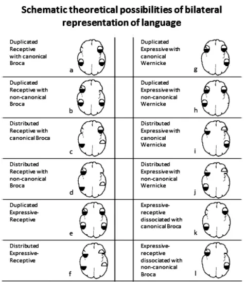
Patients with pathology may not only redistribute (reorganize) the modules in a new fashion, but also introduce changes in the dataflow. For example, some modules may become bilateral represented in a redundant manner while others may be dissociated between the hemispheres.Fig. 1illustrate the different potential subtypes: (a) Bilateral receptive language representation with canonical Broca; (b) The bilateral representation of receptive areas is accompanied by right hemisphere transferring of Broca’s area; (c) Bilateral distributed receptive language; (d) Distributed receptive with non-canonical Broca; (e) Duplicated expressive-receptive; (f) Bilateral language representation with interhemispheric dissociation of expressive and receptive sub-functions; (g) True duplication of expressive functions alone; (h) Same as prior category with receptive transferring to the right hemisphere; (i) Isolated bilateral expressive representation with interhemispheric dissociation; (j) Similar to prior case but with Wernicke’s area transferring; phonological aspects are more probable to remain in the left hemisphere; (k) Interhemispheric dissociation of language. (l) Subtype of interhemispheric dissocia-tion of language with non-canonical Broca..
5.1. Serial distribution
Serial processing distributed between the hemispheres is probably the most common of all bilateral representation of language, given the nature of language processing. The patterns of serial distribution
1. ER: ER
2. ER:E
3. E: ER
4. ER: R
5. R: RE
6. E: R
should be interpreted as an interdependence of the modules in which informationflow has a recog-nizable starting point, and from that point on each output proceeds to only one module up-stream in cognitive complexity or even reaches the final motor pathway that generates speech. A simple approach would represent that chain in the following way: phonological discrimination >>word recognition >> semantic at word level >> syntactic analysis >> working memory >> sentence processing >>semantic at sentence level >> grammatical analysis >> motor encoding >>motor response. In such a sequence no matter where the impairment is located, a final expressive (albeit Fig. 1. Possible bilateral language representation based on two by two factors: receptive vs. expressive and phonology vs. semantics. Drawings have a radiological orientation with left hemisphere represented on the right side. Ovals are divided in two halves indicating domain dissociation between phonology and semantics (see text for explanation). Left column reflects all possibilities for bilateral representation of Wernicke’s area, whereas the right column shows bilateral representations of Broca’s (otherwise not included in thefirst column) and two cases of expressive-receptive interhemispheric dissociation (k and l). Subtypes: (a) a frequent normal subtype of bilateral language representation (11 of 39 cases inRisse et al. (1997)series, group II); (b) The bilateral repre-sentation of receptive areas is accompanied by right hemisphere transferring of Broca’s area. This is an infrequent subtype; the right lateralized Broca suggests brain reorganization; (c) Bilateral distributed receptive language; this pattern includes some subtypes, accordingly with the subdomain transferred to the right hemisphere; (d) Distributed receptive with non-canonical Broca; A pattern highly suggestive of language brain reorganization. Some subtypes may emerge accordingly with the receptive dissociation; (e) This subtype represents a truly global bilateral representation of language; Wada test should fails to produce deficit in either carotid, as it was found in 2 cases of 39 in Risse’s et al. series; (f) Bilateral language representation with interhemispheric dissociation of expressive and receptive sub-functions; four subtypes may be found here: one mirroring the example, and two swapping only one domain; (g) True duplication of expressive functions alone; it is probably only theoretical as it has not yet been described; (h) Same as prior category with receptive transferring to the right hemisphere (not described); (i) Isolated bilateral expressive representation with interhemispheric dissociation; at least 2 subtypes are possible; comprehension is only affected in left Wada, but some aspects of expression are affected in each side; Wernicke’s area remain in the left side; (j) Similar to prior case but with Wernicke’s area transferring; phonological aspects are more probable to remain in the left hemisphere; (k) Interhemispheric dissociation of lan-guage. Rare condition, described in 4 of 490 epilepsy patients (Dongwook et al., 2008); (l) Subtype of interhemispheric dissociation of language; expressive functions are most likely to transfer away from the seizure focus (Dongwook et al., 2008).
disparate) deficit is warranted. Assuming that a distributed inter-hemispheric network may have modules in both sides of the brain, theflow of information would need to travel between the hemi-spheres across the corpus callosum or the anterior commissure, back and forth to link all the steps in the chain of a serialflow. Having this type of distribution, a temporary disruption of either hemisphere would produce language deficits, although they should be different and perhaps partial. Some Wada testsfindings previously reported by different authors may be explained in this way.
5.2. Parallel distribution
Patterns of parallel processing, on the other hand, should be interpreted in two different ways: (a) redundancy processing, and (b) distributed processing. Parsing instructions in a redundant manner have some analogy with algorithms of Resilient Parallel Computing, which is intended to protect processes form failures by repeating the process just in case one branch of the algorithm fails and crashes (Liu, Deters, & Zhang, 2010). Parsing instructions in a distributed processing assigns specific processors to a given function that could be executed while other processorfinishes a required process. In our analogy, the parallel redundancy would trigger two homologous brain modules, in two different hemispheres to perform the same process; whereas the parallel distributed processing modules located in different hemispheres will simultaneously process different functions,–like in the example of prosody/semantics. True parallel redundant process is probably inexistent since it would be a source of conflict messing up the cognitive dataflow; however, it is possible to imagine a redundancy between the hemispheres for a given function that theoretically would explain findings of bilateral failure on Wada tests.
The combination of the subtypes provided by modularity,and those explained bydata- flowmay theoretically explain several types of possible bilateral language representation, as illustrated inFig. 1.
6. fMRIfindings suggesting distributed bilateral processing
Clinical and fMRI studies have demonstrated the different cortical specification segmenting the expressive and receptive language functions. Further, fMRI has shown anatomical sub-specification for isolated expressive and receptive functions. The Broca’s area seems to contain two major sub-components; (a) the pars opercularis, BA 44, and the anterior insula, involved in phonological pro-cessing and direct speech production, and (b) the pars triangularis, BA 45, more involved in semantic and lexical processing (Amunts et al., 2004; Fiebach, Friederici, Müller, & von Cramon, 2002; Heim
et al., 2005; McDermott, Petersen, Watson, & Ojemann, 2003). This functional segregation has
vali-dation in the proven distinct structural connectivity that BA 44 and BA 45 exhibit in recent diffusion tensor imaging studies (Klein et al., 2007; Lemaire et al., 2012). These areas seem to have many other divergent functions beyond purely language processing (Bornkessel-Schlesewsky, Grewe, &
Schlesewsky, 2012); but of significant relevance is the dorso-ventral differentiation of the pars
oper-cularis seemingly related with a mirror neuron system (Molnar-Szakacs, Iacoboni, Koski, & Mazziotta,
2005).
Wernicke’s area sub-specialization has received less attention, in spite of encompassing a large distribution of Brodmann’s areas. Perhaps it is due to the poor anatomical landmarks delimiting the receptive language cortex. However, it is now accepted that at least transferring of language from posterior to anterior areas are carried by two different systems, (a) the dorsal system, involved in phonological processing and (b) the ventral system involved in semantic processing (Duffau et al.,
2002; Glasser & Rilling, 2008; Leclercq et al., 2010; Mandonnet et al., 2007). This subdivision
sug-gests some receptive phonological processing toward BA 40, and a more ventral and posterior semantic analysis (McDermott et al., 2003).
The subdivision of phonology/semantic domains is only an example, may be the most relevant, but not the only one. Several other subsystems are intervening in language that may have sub-specialization. The dorsal pars opercularis has been found to be involved more specifically in sequencing linguistic and non-linguistic events (Ardila & Bernal, 2007; Makuuchi, Bahlmann,
Anwander & Friederici, 2009; Willems, Ozyürek, & Hagoort, 2009), whereas the ventral part has
antonyms generation engages left lateralization regions. However, antonyms activation extends more anteriorly (Jeon, Lee, Kim, & Cho, 2009); verbal working memory (Chein, Fissell, Jacobs, & Fiez, 2002), semantic content vs. grammatical structure (Ni et al., 2000), and likely many other functions could be distinguished.
Departing from the previous information it can be inferred that serial distribution between the hemispheres can be quite complex, since a sub-specialized module may be located in one hemisphere while the rest may remain in the other. Wada tests results seem to back up this assertion.
The following images were taken from patients with intractable epilepsy, who underwent fMRI for language mapping. Some of these patterns are only evident when at least two language paradigms with different linguistic loads are given. The cases will serve to illustrate key points in bilateral lan-guage representation.
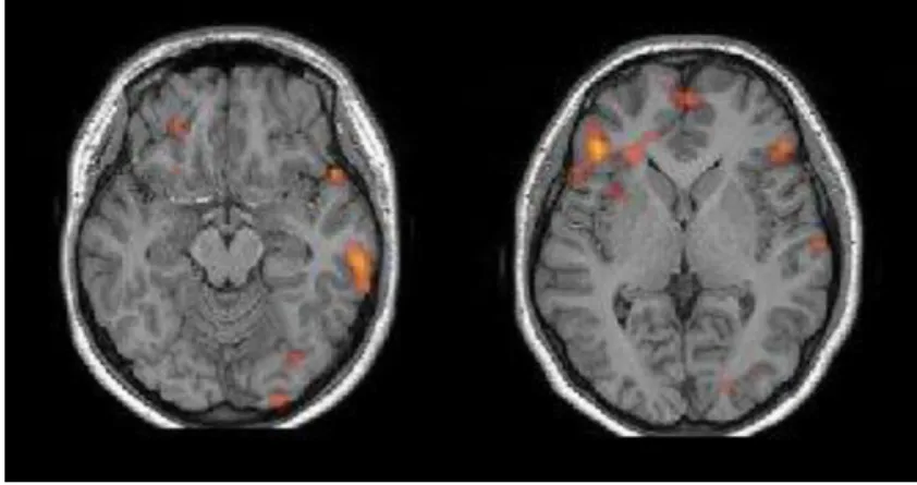
Case A (Fig. 2): Bilateral representation of language: Broca/Wernicke dissociation type. It clearly shows a bilateral representation of language with reorganization occurring only for receptive language function, most likely in keeping with the malfunction produced by the developmental lesion found in the left temporal region. A case like this will have a positive bilateral Wada test, but each side producing a different clinical picture. It would be classified byKurthen et al. (1994)as aGeneral Bilateral pattern Fig. 2. Axial T1 MRI anatomical images with functional map from an auditory description/comprehension task. Left side of the image corresponds to the right hemisphere (Radiological orientation). The left image shows activation of the posterior right tem-poral lobe corresponding to Wernicke’s area. There are only minute activations in the left hemisphere. The image on the center shows a large activation in the left inferior frontal gyrus corresponding to Broca’s area. The right images show a hypointense image located in the anterior temporal lobe (within the oval) consistent with a developmental tumor (patient 14 years old, right-handed girl). (Images courtesy of Miami Children’s Hospital, Department of Radiology).
Fig. 3. Bilateral Broca’s and left Wernicke’s area Orientation and background image as inFig. 2fMRI task: auditory description/ comprehension task. Left side of the image corresponds to the right hemisphere (Radiological orientation). Left image shows complete left lateralization for receptive language. Right image shows bilateral activation of inferior frontal gyrus slightly more prominent on the right. Patient: 11-year-old right-handed girl. (Images courtesy of Miami Children’s Hospital, Department of Radiology).
with interhemispheric dissociation. For Loring et al. (1990) and Möddel et al. (2009) this would be classified as abilateral dependent representation of language.Of note is the fact that this patient was a right-hander.
Case B (Fig. 3) shows bilateral representation of expressive language functions, with well-defined Wernicke’s left lateralization. The frontal areas are homologous, but only the right insula is involved. This subject might have abilateral positive (doublerepresentation) ofKurthen et al. (1994), where an incomplete loss of language may be expected with a left carotid injection and no impairment with a right carotid injection, provided there is redundancy of the Broca’s area. ForLoring et al. (1990)and
Möddel et al. (2009) this case will also be a bilateral dependent type. More difficult to classify
accordingly withRisse et al. (1997)subgroups of bilateral language representation, since they based it too much on impairment of automatic speech. Perhaps the case could be classified as plain“duplication of automatic speech”.
Case C (Fig. 4) is an example of global bilateral representation of language. There are some minor asymmetries, but all canonical language areas are activated in both sides. A pattern like this may explain all types ofKurthen et al. (1994), either bilateral-positive, bilateral-negative or bilateral global, or double representation, unilateral representation of subfunctions or distributed representation of subfunctions, all possible depending upon the distribution of the modules. If the bilateral condition represents redundancy of both expressive and receptive modules, a bilateral global representation of Kurthen et al.’s is obtained. If, in contrast, a modular distribution (partial or complete) between the hemispheres has taken place, either a bilateral-positive or bilateral negative is possible. Take for example, that some phonological functions remain completely lateralized in the left hemisphere while the rest of sub-modules are bilaterally represented (duplicated). In this case the right Wada will produce a partial deficit (if the reminder modules are distributed) or no deficit (if the reminder modules are duplicated), but the left Wada will produce deep language deficit as the language pro-cessing has been affected at its root. Likewise, this pattern may explain subtypes 1 (duplication of automatic speech), 2 (duplication of comprehension) and 4 (no deficit in either Wada) ofRisse et al.
(1997)but not 3 for only right sided dominant activations. Likewise, the pattern may explain Loring
et al.’s types 1 or 2 depending if the organization is just redundancy of processing (as the pattern suggests) or redistribution of sub-functions.
reflecting a different strategy of the brain working under specific semantic constrains. It seems that it is much easier for the brain to judge true that“What is on top of the house is the....roof”than to decide if
“distant–far”are synonyms, not antonyms. A case like this would be classified as“distributed repre-sentation of sub-functions”inKurthen et al. (1994), classification; bilateral autonomous byLoring et al.
(1990)andnot suitable for classificationaccording to the proposal presented byRisse et al. (1997)
Fig. 5. Redistribution of sub-functions. Task-specific-dissociated bilateral representation of language. Left side of the image corre-sponds to the right hemisphere (Radiological orientation). Upper row: fMRI with auditory description/comprehension task. Lower row: fMRI with a semantic decision task based on antonyms. Thefirst task only shows left side involvement for expressive and receptive language. The antonyms task shows bilateral activation of Broca’s area with same left sided Wernicke lateralization. Pa-tient: 13-year-old right-handed boy. (Images courtesy of Miami Children’s Hospital, Department of Radiology).
Fig. 6. Bilateral receptive–lateralized expressive. Language activation map overlaid on an FLAIR MRI axial series. Left side of the image corresponds to the right hemisphere (Radiological orientation). The subject was performing a semantic decision task based on antonyms/synonyms discrimination. Notice the bilateral symmetrical activation of secondary auditory areas in homologous func-tional areas, and the lateralization of the expressive areas to the left hemisphere. Patient was a 21-year-old man with a develop-mental tumor in the right frontal lobe. (Images courtesy of Miami Children’s Hospital, Department of Radiology).
CaseE(Fig. 6) Bilateral Wernicke’s representation has at least two different variants: with left Broca or with right Broca localization. Thefirst subtype is the most frequent of all bilateral representations, and it is probably the only found in normal right- handed patients. Bilateral representation of receptive functions is well recognized since early clinical studies described less comprehension than expressive impairment in cases of global aphasia due to hemispheric stroke (Benson & Ardila, 1996; Taylor-Sarno &
Levita, 1981). Likewise, recovery is also faster and better for receptive language than for expressive
functions. In that sense, bilateral representation of expressive functions may be seen always as a suggesting sign of language network reorganization. Still, the bilateral receptive language represen-tation may have some variants as some sub-receptive functions could be re-distributed or duplicated between the hemispheres. In fMRI studies, the homologous bilateral activation supposedly would suggest redundancy of representation, while non-homologous bilateral temporo-parietal activation would suggest a rather distributed bilateral representation. Notice at this point that Kurthen et al.’s subtypes of double representation, unilateral representation of sub-functions (distributed) and distributed representation of sub-functions are good for expressive alone, receptive alone or both, given an ample spectrum of different subtypes of functional bilateral representation for language.
7. Toward an integration
A simple classification, taking into account the different language categorical dissociations that has been found and demonstrated in Wada and fMRI studies, could be proposed from a topographic perspective (Table 2).
Any bilateral representation may be subdivided functionally in parallel (this is redundantly pro-cessed) or serial. In thefirst case, we would see those cases in which the patient does not lose the redundant function with the Wada test in neither of the carotids, while in the second (serial) he/she will exhibit partial loss of speech (for example cases mentioned byKurthen et al. (1994), Loring et al.
(1990)andRisse et al. (1997)cited before). In functional MRI, the serial processing between Broca’s
areas may be seen as a task-related dissociation of expressive language (seeFig. 4).
The same rational may apply for cases with bilateral Wernicke’s representation - a more frequent situation seen in clinical practice. Parallel processing between Wernicke’s areas would be seen as subjects not referring any comprehension impairment after injection of the Amytal in either carotid, while partial or limited comprehension may appear in both of them. Notice again that parallel pro-cessing in these examples is a sort of duplicated data flow, while the serial implies a distributed modularity.
Table 2
Proposed classification of language lateralization according to a topographic perspective. All main types and subtypes are self explanatory. The task specific dissociation subtype refers to distribution of dataflow in which some language functions reside in only one hemisphere. Functions dissociated may be explained by transferring of subfunctions (phonology or semantics) to the right hemisphere. This dissociation is only evident when the patient is presented with two or more paradigms with different linguistic loads. In this case, brain activations may appear non-congruent between tasks. We have preferred this name, to a more descriptive but less practical name like "specific-linguistic-sub-domain dissociated language representation."
Isolated Bilateral Expressive Representation Subtype I: with left Wernicke’s
Subtype II: with right Wernicke’s Isolated Bilateral Receptive Representation
Subtype I: with left Broca’s Subtype II: with right Broca’s Bilateral Global Representation Bilateral Dissociated Representation
Subtype I: Transhemispheric Broca/Wernicke dissociation Subtype I.1: Broca’s transferred
A much more complex scenario is posed by bilaterality of both Broca’s and Wernicke’s areas, since in this case the serial vs. parallel processing may be distributed in two orthogonal dimensions: anterior to posterior (receptive to expressive) and side to side between the homologous areas in which dissoci-ations of the sort phonology/semantics may occur, among many others. The multidimensionality of these intertwined factors may prompt for a quite complex and large classification. A functional clas-sification could consequently also be proposed, as presented inTable 3.
8. Conclusion
Bilateral language representation has been a very complex and intricate aspect of brain organization of cognition. It is frequent understanding that language lateralization is a matter of all or nothing. However, language dominance is mostly a matter of hemispheric advantage for a specific multi-modular cognitive function: language. As such, language in a strict sense is up to a certain point a bilateral brain function. Aside expressive/receptive and phonological/semantic dichotomies there are other many sub-functions for which we are looking for their anatomical and functional correlates with new neuroimaging techniques such as diffusion tensor imaging tractography and fMRI.
The understanding of the language network in terms of submodules and connectivity will provide ground to better understanding brain reorganization after structural and functional brain lesions.
References
Adcock, J. E., Wise, R. G., Oxbury, J. M., Oxbury, S. M., & Matthews, P. M. (2003). Quantitative fMRI assessment of the differences
in lateralization of language related brain activation in patients with temporal lobe epilepsy.Neuroimage, 8(2), 423–438.
Allen, L. S., Richey, M. F., Chai, Y. M., & Gorski, R. A. (1991). Sex differences in the corpus callosum of the living human being.
Journal of Neuroscience, 11, 933–942.
Amunts, K., Weiss, P. H., Mohlberg, H., Pieperhoff, P., Eickhoff, S., Gurd, J. M., et al. (2004). Analysis of neural mechanisms
underlying verbal fluency in cytoarchitectonically defined stereotaxic spacedthe roles of Brodmann areas 44 and 45.
Neuroimage, 22(1), 42–56.
Anderson, D. P., Harvey, A. S., Saling, M. M., Anderson, V., Kean, M., Jacobs, R., et al. (2002). Differential functional magnetic
resonance imaging language activation in twins discordant for a left frontal tumor. Journal of Child Neuralogy, 17(10),
766–769.
Ardila, A. (2006).Las afasiasAccessed March 29, 2013http://neuropsicolog.blogspot.com/2009/04/libros-de-las afasias-alfredo-ardila.html.
Ardila, A., & Bernal, B. (2007). What can be localized in the brain? towards a "factor" theory on brain organization of cognition.
International Journal of Neuroscience, 117, 935–969.
Basic, S., Hajnsek, S., Poljakovic, Z., Basic, M., Culic, V., & Zadro, I. (2004). Determination of cortical language dominance using
functional transcranial Doppler sonography in left-handers.Clinical Neurophysiology, 115(1), 154–160.
Basso, A., & Rusconi, M. L. (1998). Aphasia in left-handers. In P. Coppens, Y. Lebrun, & A. Basso (Eds.), Aphasia in atypical
populations(pp. 1–34). Mahwah, NJ: Lawrence Erlbaum Associates.
Bembich, S., Demarini, S., Clarici, A., Massaccesi, S., & Grasso, D. L. (2011). Non invasive assessment of hemispheric language
dominance by optical topography during a brief passive listening test: a pilot study.Medical Science Monitor, 17(12), CR692–
CR697.
Table 3
Proposed classification of language lateralization according to a functional perspective. Language modules may be duplicated in the other hemisphere or distributed between the hemispheres. In the first option process may occur in parallel assuming complete capability of each side, or one side should remain quiescent. Notice that, in theroy, serial flow may still happen assuming unbalanced efficiency between the duplicated modules. However, in distributed modularity (types I and II) the serial processing is obligued as data mayflow crossing the hemispheres in the cognitive up-stream trajectory.
I. Duplicated Bilateral Representation of Language (each side suffices to hold the function: parallel processing) Expressive
Receptive Both
II. Distributed Bilateral Representation of Language (each hemisphere handles a particular subset of functions: serial processing)
Expressive (phonology/semantics) Receptive (Word-level vs. sentence level) Both
III. Transhemispherical dissociated Broca/Wernicke
Benbadis, S. R., Binder, J. R., Swanson, S. J., Fischer, M., Hammeke, T. A., Morris, G. L., et al. (1998). Is speech arrest during Wada
testing a valid method for determining hemispheric representation of language?Brain and Language, 65(3), 441–446.
Benbadis, S. R., Dinner, S. D., Chelune, G. J., Piedmonte, M., & Luders, H. O. (1995). Autonomous versus dependent: a classification
of bilateral language representation by intracarotid amobarbital procedure.Journal of Epilepsy, 8, 255–263.
Benson, D. F., & Ardila, A. (1996).Aphasia: A clinical perspective. New York: Oxford University Press.
Benson, D., & Geschwind, N. (1973). Aphasia and related disturbances. In A. Baker (Ed.),Clinical Neurology(pp. 1–26). New York:
Harper and Row.
Bernal, B., & Ardila, A. (2009). The role of the arcuate fasciculus in conduction aphasia.Brain, 132(Pt 9), 2309–2316.
Binder, J. R. (2011). Functional MRI is a valid noninvasive alternative to Wada testing.Epilepsy Behavior, 20(2), 214–222.
Bisconti, S., Di Sante, G., Ferrari, M., & Quaresima, V. (2012). Functional near-infrared spectroscopy reveals heterogeneous
patterns of language lateralization over frontopolar cortex.Neuroscience Research, 73(4), 328–332.
Bornkessel-Schlesewsky, I., Grewe, T., & Schlesewsky, M. (2012). Prominence vs. aboutness in sequencing: a functional
distinction within the left inferior frontal gyrus.Brain and Language, 120(2), 96–107.
Bramwell, B. (1899). On“crossed”aphasia.Lancet, 3, 1473–1479.
Broca, P. (1861). Remarques sur le siège de la faculté du langage articulé; suivies d’une observation d’aphémie.Bulletin de la
Société d’Anthropologie, 2, 330–357.
Broca, P. (1865). Du siège de la faculté du langage articulé.Bulletin de la Société d’Anthropologie, 6, 337–393.
Cabeza, R. (2002). Hemispheric asymmetry reduction in older adults: the HAROLD model.Psychology and Aging, 17(1), 85–100.
Castro-Caldas, A., & Confraria, A. (1984). Age and type of crossed aphasia in dextral due to stroke.Brain and Language, 23, 126–133.
Chein, J. M., Fissell, K., Jacobs, S., & Fiez, J. A. (2002). Functional heterogeneity within Broca’s area during verbal working
memory.Physiology and Behavior, 77(4–5), 635–639.
Coppens, P., Hungerford, S., Yamaguchi, S., & Yamadori, A. (2002). Crossed aphasia: an analysis of the symptoms, their frequency,
and a comparison with left hemisphere aphasia symptomatology.Brain and Language, 83(3), 425–463.
Dehaene-Lambertz, N., Dehaene, S., & Hertz-Pannier, L. (2002). Functional neuroimaging of speech perception in infants.
Sci-ence, 298, 2013–2015.
Dejerine, J. (1914).Semiologie des affections du systeme nerveux. Paris: Masson.
Dien, J., Frishkoff, G. A., Cerbone, A., & Tucker, D. M. (2003). Parametric analysis of event-related potentials in semantic
comprehension: evidence for parallel brain mechanisms.Cognitive Brain Research, 15(2), 137–153.
Dongwook, L., Lee, S. J., Swanson, D. S., Sabsevitz, T. A., Hammeke, F., Winstanley, S., et al. (2008). Fmri and Wada studies in
patients with interhemispheric dissociation of language functions.Epilepsy and Behavior, 13, 350–356.
Duffau, H., Capelle, L., Sichez, N., Denvil, D., Lopes, M., Sichez, J. P., et al. (2002). Intraoperative mapping of the subcortical
language pathways using direct stimulations. An anatomo-functional study.Brain, 125(Pt 1), 199–214.
Duffau, H., Gatignol, S. T., Mandonnet, E., Capelle, L., & Taillandier, L. (2008). Intraoperative subcortical stimulation mapping of
language pathways in a consecutive series of 115 patients with Grade II glioma in the left dominant hemisphere.Journal of
Neurosurgery, 109(3), 461–471.
Fiebach, C. J., Friederici, A. D., Müller, K., & von Cramon, D. Y. (2002). fMRI evidence for dual routes to the mental lexicon in
visual word recognition.Journal of Cognitive Neurosciences, 14(1), 11–23.
Glasser, M. F., & Rilling, J. K. (2008). DTI tractography of the human brain’s language pathways.Cerebral Cortex, 18(11), 2471–
2482.
Groen, M. A., Whitehouse, A. J., Badcock, N. A., & Bishop, D. V. (2012). Does cerebral lateralization develop? A study using
functional transcranial Doppler ultrasound assessing lateralization for language production and visuospatial memory.Brain
and Behavior, 2(3), 256–269.
Hadac, J., Brozová, K., Tintera, J., & Krsek, P. (2007). Language lateralization in children with pre- and postnatal epileptogenic
lesions of the left hemisphere: an fMRI study.Epileptic Disorders, 9(Suppl. 1), S19–S27.
Ha, J. W., Pyun, S. B., Hwang, Y. M., & Sim, H. (2012). Lateralization of cognitive functions in aphasia after right brain damage.
Yonsei Medical Journal, 53(3), 486–494.
Harris, L. J. (1991). Cerebral control for speech in right-handers and left-handers: an analysis of the views of Paul Broca, his
contemporaries, and his successors.Brain and Language, 40, 1–50.
Harris, L. J. (1999). Early theory and research on hemispheric specialization.Schizophrenia Bulletin, 25(1), 11–39.
Hécaen, H., & Albert, M. L. (1978).Human neuropsychology. New York: Wiley.
Hécaen, H., Mazurs, G., Ramier, A., Goldblum, M., & Merianne, L. (1971). Aphasie croisee chez un sujet droiter bilingue.Revue
Neurologique, 1, 319–323.
Hécaen, H., & Sauguet, J. (1971). Cerebral dominance in left-handed subjects.Cortex, 7, 19–47.
Heim, S., Alter, K., Ischebeck, A. K., Amunts, K., Eickhoff, S. B., Mohlberg, H., et al. (2005). The role of the left Brodmann’s areas
and 45 in reading words and pseudowords.Cognitive Brain Research, 25(3), 982–993.
Hickok, G., & Poeppel, D. (2004). Dorsal and ventral streams: a framework for understanding aspects of the functional anatomy of language.Cognition, 92, 67–99.
Hickok, G., & Poeppel, D. (2007). The cortical organization of speech processing.Nature Reviews, 8, 393–402.
Holland, S. K., Plante, E., Weber Byars, A., Strawsburg, R. H., Schmithorst, V. J., & Ball, W. S., Jr. (2001). Normal fMRI brain
activation patterns in children performing a verb generation task.Neuroimage, 14, 837–843.
Holland, S. K., Vannest, J., Mecoli, M., Jacola, L. M., Tillema, J. M., Karunanayaka, P. R., et al. (2007). Functional MRI of language
lateralization during development in children.International Journal of Audiology, 46(9), 533–551.
Holodny, A. I., Schulder, M., Ybasco, A., & Liu, W. C. (2002). Translocation of Broca’s area to the contralateral hemisphere as the
result of the growth of a left inferior frontal glioma.Journal of Computer Assisted Tomography, 26(6), 941–943.
Hopf, J. M., Bader, M., Meng, M., & Bayer, J. (2003). Is human sentence parsing serial or parallel? Evidence from event-related
brain potentials.Cognitive Brain Research, 15(2), 165–177.
Inui, K., Okamoto, H., Miki, K., Gunji, A., & Kakigi, R. (2006). Serial and parallel processing in the human auditory cortex: a
magnetoencephalographic study.Cerebral Cortex, 16(1), 18–30.
Ishizaki, M., Ueyama, H., Nishida, Y., Imamura, S., Hirano, T., & Uchino, M. (2012). Crossed aphasia following an infarction in the
Jeon, H. A., Lee, K. M., Kim, Y. B., & Cho, Z. H. (2009). Neural substrates of semantic relationships: common and distinct
left-frontal activities for generation of synonyms vs. antonyms.Neuroimage, 48(2), 449–457.
Kadis, D. S., Pang, E. W., Mills, T., Taylor, M. J., McAndrews, M. P., & Smith, M. L. (2011). Characterizing the normal developmental
trajectory of expressive language lateralization using magnetoencephalography.Journal of the International
Neuropsycho-logical Society, 17(5), 896–904.
Kennan, R. P., Kim, D., Maki, A., Koizumi, H., & Constable, R. T. (2002). Non-invasive assessment of language lateralization by
transcranial near infrared optical topography and functional MRI.Human Brain Mapping, 16(3), 183–189.
Khedr, E. M., Hamed, E., Said, A., & Basahi, J. (2002). Handedness and language cerebral lateralization.European Journal of
Applied Physiology, 87(4–5), 469–473.
Klein, J. C., Behrens, T. E., Robson, M. D., Mackay, C. E., Higham, D. J., & Johansen-Berg, H. (2007). Connectivity-based parcellation of human cortex using diffusion MRI: establishing reproducibility, validity and observer independence in BA 44/45 and
SMA/pre-SMA.Neuroimage, 34(1), 204–211.
Knecht, S., Dräger, B., Deppe, M., Bobe, L., Lohmann, H., Flöel, A., et al. (2000). Handedness and hemispheric language
domi-nance in healthy humans.Brain, 123(12), 2512–2518.
Knecht, S., Dräger, B., Flöel, A., Lohmann, H., Breitenstein, C., Deppe, M., et al. (2001). Behavioural relevance of atypical language
lateralization in healthy subjects.Brain, 124(Pt 8), 1657–1665.
Koelsch, S., Schulze, K., Sammler, D., Fritz, T., Müller, K., & Gruber, O. (2009). Functional architecture of verbal and tonal working
memory: an FMRI study.Human Brain Mapping, 30(3), 859–873.
Kosla, K., Pfajfer, L., Bryszewski, B., Jaskólski, D., Stefanczyk, L., & Majos, A. (2012). Functional rearrangement of language areas in
patients with tumors of the central nervous system using functional magnetic resonance imaging.Polish Journal of
Radi-ology, 77(3), 39–45.
Kurthen, M., Helmstaedter, C., Linke, D. B., Hufnagel, A., Elger, C. E., & Schramm, J. (1994). Quantitative and qualitative evaluation
of patterns of cerebral language dominance. An amobarbital study.Brain and Language, 46(4), 536–564.
Leclercq, D., Duffau, H., Delmaire, C., Capelle, L., Gatignol, P., Ducros, M., et al. (2010). Comparison of diffusion tensor imaging
tractography of language tracts and intraoperative subcortical stimulations.Journal of Neurosurgery, 112(3), 503–511.
Lemaire, J. J., Golby, A., Wells, W. M., Pujol, S., Tie, Y., & Rigolo, L. (2012). Extended Broca’s area in the functional connectome of
language in adults: combined cortical and subcortical single-subject analysis using fMRI and DTI tractography. Brain
Topography, 26(3), 428–441.
Lidzba, K., Schwilling, E., Grodd, W., Krägeloh-Mann, I., & Wilke, M. (2011). Language comprehension vs. language production:
age effects on fMRI activation.Brain and Language, 119(1), 6–15.
Liégeois, F., Connelly, A., Cross, J. H., Boyd, S. G., Gadian, D. G., Vargha-Khadem, F., et al. (2004). Language reorganization in
children with early-onset lesions of the left hemisphere: an fMRI study.Brain, 127(Pt 6), 1229–1236.
Liu, D., Deters, R., & Zhang, W. J. (2010). Architectural design for resilience.Enterprise Information Systems, 4, 137–152.
Lorenz, M. W., Thoelen, N., Loesel, N., Lienerth, C., Gonzalez, M., Humpich, M., et al. (2008). Assessment of cerebral
autor-egulation with’transcranial Doppler sonography in poor bone windows using constant infusion of an ultrasound contrast
agent.Ultrasound Medical Biology, 34(3), 345–353.
Loring, D. W., Meador, K. J., Lee, G. P., Murro, A. M., Smith, J. R., Flanigin, H. F., et al. (1990). Cerebral language lateralization:
evidence from intracarotid amobarbital testing.Neuropsychologia, 28(8), 831–838.
Loring, D. W., Strauss, E., Hermann, B. P., Perrine, K., Trenerry, M. R., Barr, W. B., et al. (1999). Effects of anomalous language
representation on neuropsychological performance in temporal lobe epilepsy.Neurology, 53(2), 260–264.
Makuuchi, M., Bahlmann, J., Anwander, A., & Friederici, A. D. (2009). Segregating the core computational faculty of human
language from working memory.Proceedingof the National Academy of Sciences of U S A, 106(20), 8362–8367.
Mandonnet, E., Nouet, A., Gatignol, P., Capelle, L., & Duffau, H. (2007). Does the left inferior longitudinal fasciculus play a role in
language? A brain stimulation study.Brain., 130(Pt 3), 623–629.
Mariën, P., Paghera, B., De Deyn, P. P., & Vignolo, L. A. (2004). Adult crossed aphasia in dextrals revisited.Cortex, 40, 41–74.
Matsumoto, R., Okada, T., Mikuni, N., Mitsueda-Ono, T., Taki, J., Sawamoto, N., et al. (2008). Hemispheric asymmetry of the arcuate fasciculus: a preliminary diffusion tensor tractography study in patients with unilateral language dominance
defined by Wada test.Journal of Neurology, 255(11), 1703–1711.
McDermott, K. B., Petersen, S. E., Watson, J. M., & Ojemann, J. G. (2003). A procedure for identifying regions preferentially
activated by attention to semantic and phonological relations using functional magnetic resonance imaging.
Neuro-psychologia, 41(3), 293–303.
Mesulam, M. M. (1990). Large-scale neurocognitive networks and distributed processing for attention, language, and memory.
Annals of Neurology, 28(5), 597–613.
Möddel, G., Lineweaver, T., Schuele, S. U., Reinholz, J., & Loddenkemper, T. (2009). Atypical language lateralization in epilepsy patients.Epilepsia, 50(6), 1505–1516.
Molnar-Szakacs, I., Iacoboni, M., Koski, L., & Mazziotta, J. C. (2005). Functional segregation within pars opercularis of the inferior
frontal gyrus: evidence from fMRI studies of imitation and action observation.Cerebral Cortex, 15(7), 986–994.
Müller, R. A., Rothermel, R. D., Behen, M. E., Muzik, O., Mangner, T. J., & Chugani, H. T. (1997). Receptive and expressive language
activations for sentences: a PET study.Neuroreport, 8(17), 3767–3770.
Ni, W., Constable, R. T., Mencl, W. E., Pugh, K. R., Fulbright, R. K., Shaywitz, S. E., et al. (2000). An event-related neuroimaging
study distinguishing form and content in sentence processing.Journal of Cognitive Neurosciences, 12(1), 120–133.
Nucifora, P. G., Verma, R., Melhem, E. R., Gur, R. E., & Gur, R. C. (2005). Leftward asymmetry in relativefiber density of the arcuate
fasciculus.Neuroreport, 16(8), 791–794.
Papathanassiou, D., Etard, O., Mellet, E., Zago, L., Mazoyer, B., & Tzourio-Mazoyer, N. A. (2000). Common language network
for comprehension and production: a contribution to the definition of language epicenters with PET.Neuroimage, 11(4),
347–357.
Pataraia, E., Billingsley-Marshall, R. L., Castillo, E. M., Breier, J. I., Simos, P. G., Sarkari, S., et al. (2005). Organization of receptive
language-specific cortex before and after left temporal lobectomy.Neurology, 64(3), 481–487.
Pedersen, P. M., Jargensen, H. S., Nakayama, H., Raaschou, H. O., & Olsen, T. S. (1995). Aphasia in acute stroke: incidence,
de-terminants, and recovery.Annals of Neurology, 38, 659–666.
Powell, H. W., Parker, G. J., Alexander, D. C., Symms, M. R., Boulby, P. A., Wheeler Kingshott, C. A., et al. (2006). Hemispheric
asymmetries in language related pathways: a combined functional MRI and tractography study. Neuroimage, 32(1),
388–399.
Price, C. J. (2010). The anatomy of language: a review of 100 fMRI studies published in 2009.Annals of the New York Academy of
Sciences, 1191, 62–88.
Rasmussen, T., & Milner, B. (1977). The role of early brain injury in the lateralization of cerebral speech functions.Annals of the
New York Academy of Sciences, 299, 35–69.
Risse, G. L., Gates, J. R., & Fangman, M. C. (1997). A reconsideration of bilateral language representation based on the intracarotid
amobarbital procedure.Brain and Cognition, 33(1), 118–132.
Rodrigo, S., Oppenheim, C., Chassoux, F., Hodel, J., de Vanssay, A., Baudoin-Chial, S., et al. (2008a). Language lateralization in
temporal lobe epilepsy using functional MRI and probabilistic tractography.Epilepsia, 49(8), 1367–1376.
Springer, J. A., Binder, J. R., Hammeke, T. A., Swanson, S. J., Frost, J. A., Bellgowan, P. S., et al. (1999). Language dominance in
neurologically normal and epilepsy subjects: a functional MRI study.Brain, 122(Pt 11), 2033–2046.
Staudt, M., Lidzba, K., Grodd, W., Wildgruber, D., Erb, M., & Krägeloh, M. (2002). Right hemispheric organization of language
following early left-sided brain lesions: functional MRI topography.Neuroimage, 16(4), 954–967.
Szaflarski, J. P., Holland, S. K., Schmithorst, V. J., & Byars, A. W. (2006). fMRI study of language lateralization in children and
adults.Human Brain Mapping, 27, 202–212.
Tanaka, N., Liu, H., Reinsberger, C., Madsen, J. R., Bourgeois, B. F., Dworetzky, B. A., et al. (2013). Language lateralization
rep-resented by spatiotemporal mapping of magnetoencephalography.American Journal of Neuroradiology, 34(3), 558–563.
Taylor-Sarno, M., & Levita, E. (1981). Some observations on the nature of recovery in global aphasia after stroke.Brain and
Language, 13(1), 1–12.
Thiel, A., Herholz, K., von Stockhausen, H. M., van Leyen-Pilgram, K., Pietrzyk, U., Kessler, J., et al. (1998). Localization of
language-related cortex with 15O-labeled water PET in patients with gliomas.Neuroimage, 7(4 Pt 1), 284–295.
Townsend, J. (1990). Serial vs. Parallel processing: sometimes they Look like Tweedledum and Tweedledee but they can (and
should) Be distinguished.Psychology Science, 1, 36–53.
Tyler, L. K., Wright, P., Randall, B., Marslen-Wilson, W. D., & Stamatakis, E. A. (2010). Reorganization of syntactic processing
following left-hemisphere brain damage: does right-hemisphere activity preserve function?Brain, 133(11), 3396–3408.
Vernooij, M. W., Smits, M., Wielopolski, P. A., Houston, G. C., Krestin, G. P., & van der Lugt, A. (2007). Fiber density asymmetry of the arcuate fasciculus in relation to functional hemispheric language lateralization in both right- and left-handed healthy
subjects: a combined fMRI and DTI study.Neuroimage, 35(3), 1064–1076.
Vigneau, M., Beaucousin, V., Hervé, P. Y., Jobard, G., Petit, L., Crivello, F., et al. (2011). What is right-hemisphere contribution to
phonological, lexico-semantic, and sentence processing? Insights from a meta-analysis.Neuroimage, 54(1), 577–593.
Vikingstad, E. M., Cao, Y., Thomas, A. J., Johnson, A. F., Malik, G. M., & Welch, K. M. (2000). Language hemispheric dominance in
patients with congenital lesions of eloquent brain.Neurosurgery, 47(3), 562–570.
Weiller, C., Isensee, C., Rijntjes, M., Huber, W., Müller, S., Bier, D., et al. (1995). Recovery from Wernicke’s aphasia: a positron
emission tomographic study.Annals of Neurology, 37(6), 723–732.
Wernicke, C. (1874).Der Aphasiche Symptomencomplex. Breslau: Cohn & Weigert.
Willems, R. M., Ozyürek, A., & Hagoort, P. (2009). Differential roles for left inferior frontal and superior temporal cortex in
multimodal integration of action and language.Neuroimage, 47(4), 1992–2004.
Wingfield, A., & Grossman, M. (2006). Language and the aging brain: patterns of neural compensation revealed by functional
brain imaging.Journal of Neurophysiology, 96, 2830–2839.
Woermann, F. G., Jokeit, H., Luerding, R., Freitag, H., Schulz, R., Guertler, S., et al. (2003). Language lateralization by Wada test