Otras secciones de este sitio:
☞ ☞ ☞ ☞
☞ Índice de este número
☞ ☞ ☞ ☞
☞ Más revistas
☞ ☞ ☞ ☞
☞ Búsqueda
Others sections in this web site: ☞
☞ ☞ ☞
☞ Contents of this number ☞
☞ ☞ ☞
☞ More journals ☞
☞ ☞ ☞ ☞ Search Artículo:
Lesiones causadas por helmintos del aparato digestivo en peces estuarinos de la Laguna Tres Palos, Guerrero, México
Derechos reservados, Copyright © 2004: Facultad de Medicina Veterinaria y Zootecnia, UNAM
Veterinaria México
Número Number4
Octubre-Diciembre October-December 2 0 0 4 Volumen
Lesiones causadas por helmintos del aparato digestivo en peces
estuarinos de la Laguna Tres Palos, Guerrero, México*
Lesions caused by helminths of the digestive tract in estuarine
fi sh from the Tres Palos Lagoon, Guerrero, Mexico
Ramón Vázquez Núñez** José Ramírez Lezama*** David Osorio Sarabi*** Larisa Adriana Chávez Soriano*** Fernando Constantino Casas***
Recibido el 19 de septiembre de 2003 y aceptado el 8 de marzo de 2004. * El presente estudio fue tema de tesis de licenciatura del primer autor.
** Departamento de Patología, Facultad de Medicina Veterinaria y Zootecnia, Universidad Nacional Autónoma de México, 04510, México, D.F.
*** Laboratorio de Helmintología, Instituto de Biología, Universidad Nacional Autónoma de México, 04510, México, D. F.
Abstract
The present study was conducted to identify and compare the microscopic and gross lesions caused by helminths in the digestive tract of Ariopsis guatemalensis (“cuatete”) and Eleotris picta ( “alahuate”) from the Tres Palos Lagoon in the state of Guerrero, Mexico. Thirty individuals from each species were col-lected in the months of May, July and December 2001. Fifteen were used for the postmortem study and the other 15 were used for the parasitological exam. Three groups of helminths were identifi ed: Proteocephalus
cestodes in the “alahuate”; an acanthocephalan in the “cuatete”; and Hysterothylacium nematodes in both fi sh species. The lesions were evaluated using four categories in ascending severity of the lesions, where categories 2, 3 and 4 were the most frequent. Other lesions associated with the parasites included hem-orrhages and, less frequently, fi brosis, enteritis and necrosis. The organs that were most affected were the stomach, anterior and posterior intestines. Histologically, the areas that had the most conspicuous lesions were the submucosa and the muscular layer. Hysterothylacium sp larvae are responsible for the more evident lesions in the four categories, while the cestodes and adult nematodes without any lesions they were in the gastrointestinal lumen. Considering the size of the samples the detectable prevalence was only 10%, and can thus not necessarily be applied to all the individuals in the lagoon. Nevertheless, it is likely that Hysterothylacium sp is affecting both populations of fi sh, specially the “alahuate” in which it had a high prevalence and abundance probably associated to the fi sh’s susceptibility or the increased contact due to their alimentary habits.
Key words: ICHTHYOPATHOLOGY, COMMERCIAL FISH, GASTROINTESTINAL LESIONS, INTESTINAL HELMINTHS, HYSTEROTHYLACIUM sp.
Resumen
Se realizó el presente estudio con el fi n de identifi car y comparar las lesiones macro y microscópicas asociadas a helmintos en el tracto digestivo del Ariopsis guatemalensis (“cuatete”) y Eleotris picta (“alahuate”) de la Laguna de Tres Palos, Guerrero, México. En mayo, julio y diciembre del 2001 se colectaron 30 peces de cada especie. A 15 individuos de cada muestra se les practicó el examen histopatológico y a los 15 restantes el examen parasitológico. Se identifi caron tres grupos de helmintos: Cestodos del género Proteocephalus en el “alahuate”, acantocéfalos del género Acanthocephalus en el “cuatete” y nematodos del género Hysterothylacium en ambos peces. Se evaluaron las lesiones utilizando cuatro categorías, según su grado de severidad en orden ascendente, siendo las categorías 2, 3 y 4 las más frecuentes. Otras lesiones asociadas a los helmintos fueron hemorragias y en menor proporción fi brosis, enteritis y necrosis. Los órganos más afectados fueron: Estómago, intestino anterior e intestino posterior. A nivel histológico, los tejidos con lesiones más conspicuas fueron: Submucosa y la muscular del órgano. Las larvas de
Hysterothylacium sp son las causantes de las lesiones microscópicas evidentes dentro de las cuatro
Introducción
L
as larvas de nematodos son comunes en muchos peces teleósteos, ya que son ingeridas por el pez cuando éste se alimenta de sus presas, que a su vez fungen como huéspedes intermediarios de los gusanos. En ellos se encapsulan en vísceras o alojadas en músculo. El hospedero forma una cápsula de tejido conectivo para aislar al parásito. Los géneros de ne-matodos que se presentan en peces con importancia en salud pública son larvas de los generos: Anisakis, Pseudoterranova, Gnathostoma, Eustrongylides, Contracae-cum, Phocascaris e Hysterothylacium. Las larvas de Hys-terothylacium maduran en el intestino de los peces, permitiéndoles moverse libremente entre éste y el estómago.1 Se reconoce que el ciclo de vida de losanisáquidos marinos incluye al menos uno o dos hospederos intermediarios. El hospedero defi nitivo puede ser un pez, reptil, ave o mamífero piscívoro.2
En México se han registrado distintos endoparásitos en peces de importancia comercial, principalmente en el tubo digestivo, algunos con importancia zoonótica, como Gnathostoma sp, que se ha descrito en la musculatura esquelética de peces, incluso en los peces del presente estudio. De acuerdo con la información recopilada en Acapulco, se han presentado casos de Gnathostomiosis causada por larvas de este nematodo que comúnmente forma nódulos dentro del aparato digestivo de mamíferos.3
Los géneros de nematodos, familia Anisakidae, que se presume causan la enfermedad Anisakiasis
son: Anisakis, Phocanema, Thynnascaris Pseudoterranova
y Contracaecum.4-7 En 1955, en Holanda, se presentó el
primer caso de anisaquiosis confi rmado; sin embargo, han sido notifi cados seis casos en América del Norte.5 En 1980 se registraron lesiones causadas
por anisáquidos en un hombre de Seúl, Corea. La anisaquiosis afecto el íleon, la serosa estaba edematosa y con apariencia nodular. La infl amación era granulomatosa y con necrosis. Mudry et al.6 registraron
larvas pertenecientes a Anisakis, Thynnascaris y
Contracaecum que causaron anisaquiosis en cinco personas del norte de Francia. Los informes de los pacientes revelaron obstrucción intestinal aguda
Introduction
N
ematode larvae are common in most of the teleost fi sh since they are swallowed when fi sh eat their prey, which are the intermediary hosts of these worms that are encapsulated in viscera or muscle. The host makes a capsule of connective tissue in order to isolate the parasite. The most impor-tant nematode genera found in fi sh and also those that are important for public health are: Anisakis, Pseu-doterranova, Gnathostoma, Eustrongylides, Contracaecum, Phocascaris and Hysterothylacium. Hysterothylacium larvae grow in fi sh intestines where they move freely, even in the stomach.1 It is known that the life cycle of themarine anisakids includes at least one or two interme-diary hosts. The defi nitive host can be a fi sh, reptile, bird or a fi sh-eating mammal.2
In fi sh of commercial importance several endo-parasites, mainly in the digestive tract, have been recorded in Mexico. Some of these are zoonotic, like
Gnathostoma sp that has been found in the skeletal muscle of fi sh, even in those used in the present study. According to the information collected in Acapulco, cases of gnathostomosis have occurred caused by larvae of this nematode that usually form nodules inside the mammal digestive tract.3
The nematode genera included in the Anisakidae family that may cause anisakiasis are: Anisakis, Phocanema, Thynnascaris Pseudoterranova and Contra-caecum.4-7 In 1955 in Netherlands, the fi rst anisakiasis
case was confi rmed; nevertheless six cases have been reported in North America.5 In 1980, lesions caused
by anisakids were recorded by a man in Seul, Korea. Anisakiasis affected the ileum, the serosa was swollen with a nodular appearance; the infl ammation was granulomatous and necrotic. Mudry et al.6 studied Anisakis, Thynnascaris and Contracaecum larvae, which caused anisakiasis in fi ve people in the northern region of France; the reports of the patients revealed acute intestinal obstruction caused by stenosis and infl ammation of the ileum. In both cases, Korea and France, the lesions affected from the submucosa to the serosa.6,8 Anisakids produce severe lesions in human
stomachs and are associated to gastric neoplasia.6,9
sin producir cambios patológicos aparentes. Considerando que el tamaño de muestras sólo detecta prevalencia de más de 10% no se puede asegurar que los valores que se presenta se apliquen a todos los individuos de la Laguna. Sin embargo, es probable que Histerothylacium esté parasitando de esta manera a las poblaciones de ambas especies de peces, en especial en el “alahuate”, donde su prevalencia y abundancia son altas, asociado probablemente a que son susceptibles a los parásitos o que están en mayor contacto con los helmintos a causa de sus hábitos alimentarios.
debido a estenosis e ileítis. En ambos casos, Corea y Francia, los daños se extendían desde la submucosa hasta la serosa.6,8 Los anisáquidos producen graves
lesiones en el estómago de humanos, asociándolas con neoplasias gástricas.6,9 Si las larvas se encuentran
libres o adheridas al tracto digestivo pueden causar irritación, infl amación y ulceración.5 Además de Anisakis sp, algunas especies del género Contracaecum
causan helmintiasis en humanos.4-6,9 Histológicamente
hay una respuesta infl amatoria en el hígado con neutrófi los, macrófagos y proliferación de fi broblastos; estos últimos forman una cápsula que rodea a la larva. Los síntomas incluyen dolor gástrico o intestinal y vómito, siendo el estómago el lugar más afectado. La anisaquiosis y gnatostomosis se originan cuando el hombre consume pescado crudo o mal cocido.5,6,8,10-12
El aumento de la población humana y sus necesi-dades primarias han traído como consecuencia la sobrecarga y contaminación de los cuerpos de aguas y la fragmentación del hábitat, provocando la extinción de especies y la proliferación de otras que pudieran ser dañinas para el ambiente y para el hombre.13
En México son pocos los trabajos de investigación ictiopatológicos; considerando que existe información de los helmintos que parasitan a los peces en la Laguna de Tres Palos, Guerrero, México, resulta importante investigar cuáles son los daños que los gusanos ocasionan en el tracto digestivo de dos de las especies de peces más comerciales y así conocer más sobre el efecto de los parásitos en ellos, ya que poseen importancia por su potencial pesquero, por su valor ecológico como consumidores de organismos de interés comercial y porque potencialmente representan un factor de zoonosis en el área de estudio.
Material y métodos
En el estudio de la Laguna Tres Palos, el muestreo se realizó en tres salidas en mayo, julio y diciembre de 2001. Los peces se obtuvieron por captura comercial mediante el trasmallo.
Se colectaron 30 ejemplares de A. guatemalensis
(“cuatete”) y 30 de Eleotris picta (“alahuate”); el tamaño de los peces osciló entre 20 a 25 cm de longitud patrón, incluyendo ejemplares juveniles, así como adultos.14
Se procedió a separar los peces en 30 individuos de cada especie y se dividieron en dos grupos de 15. Al primer grupo de 15 de cada especie se le aplicó un examen helmintológico general; al siguiente grupo de 15 de cada especie se le practicó la necropsia y el examen histopatológico.
A cada uno de los individuos se les practicó la necropsia, según la técnica de Amlacker.15 Se
If larvae are loose or attached to the digestive tract, they may produce irritation, infl ammation and ulceration. Besides Anisakis sp, some species of the
Contracaecum genus cause helminthiasis in humans.4-6,9
Histologically, there is an infl ammatory response in the liver involving the proliferation of neutrophils, macrophages and fi broblasts, which create the capsule that surrounds the larva. The symptoms include gastric or intestinal pain and vomit, the stomach been the organ most affected. Anisakiasis and gnathostomosis are acquired when humans eat raw or incomplete-cocked fi sh.5,6,8,10-12
The increase in human population, along with its primary needs, have caused overload and contamination of the bodies of water and habitat fragmentation, provoking the extinction of some species and the proliferation of others that might be harmful for environment or man.13
In Mexico there are few studies on ictiopathology. Considering the existing information about the helminths that parasite fi sh in Tres Palos Lagoon, Guerrero, Mexico, it is important to investigate what lesions these worms cause in the digestive tract of two of the most commercial fi sh species and thus know more about the effect of these parasites on them, since they are important for their fi shing potential, besides the ecological value as consumers of organisms of commercial interest, and also because they represent a zoonotic risk in the studied area.
Material and methods
The study was carried out in Tres Palos Lagoon, Guer-rero, Mexico. The sampling was made three times in May, July and December 2001. Fish were obtained by commercial capture using a net.
Thirty A. guatemalensis (“cuatete”) and 30 Eleotris picta (“alahuate”) were collected. The sizes of the fi sh were between 20 and 25 cm pattern length, including juvenile ones as well as adults.14
Thirty fi sh of each species were divided into groups of 15. General helminthological exams were done to the fi rst group of each species. Necropsy and histopathological exams were performed on the second group of each species.
All necropsies were done according to the Amlacker technique.15 The fi sh were placed in a previously
colocó cada ejemplar sobre una charola de disección, debidamente desinfectada; cortando de la aleta pectoral a la pared abdominal en línea recta, haciendo un corte transversal en un punto inmediatamente anterior al ano, se obtuvo el aparato digestivo, además se colocaron inmediatamente en cajas de Petri con solución salina fi siológica al 0.65% para la revisión minuciosa de lesiones macroscópicas por helmintos bajo el microscopio estereoscópico. Se ligaron con hilo nailon los límites de las porciones intestinales anterior, media y posterior, sobre la base de una determinación cualitativa; en el caso del examen histopatológico, se colocaron en gasas y se fi jaron con formalina amortiguada al 10% con pH 7.2.
Se obtuvieron muestras del tejido para deshidra-tarlo, incluirlo en parafi na y cortarlo. Se hizo la tinción con la técnica de rutina H-E (hematoxilina-eosina) y tinciones especiales (tricrómica de Masson).16 Los
cortes histológicos fueron revisados al microscopio fotónico, identifi cando el tipo de lesión, distribución, localización, severidad y curso con base en cuatro categorías propuestas para este estudio (Cuadro 1).
En el caso del estudio parasitario, se abrió el tubo digestivo y se revisó bajo el microscopio estereoscópico, separando los gusanos con un pincel doble 0. Los helmintos encontrados fueron fi jados mediante las técnicas convencionales para su estudio e identifi cados taxonómicamente.
Los helmintos parásitos fueron recolectados, fi jados y contados con las técnicas convencionales,17 para su
posterior identifi cación taxonómica. Los parámetros poblacionales de los helmintos (prevalencia y abundancia) se basaron en las defi ciones propuestas por Margolis et al.18 Ejemplares y laminillas con los
cortes histológicos fueron depositados en la CNH (Colección Nacional de Helmintos) en los números de catálogo 4 487 a 4 493.
sections of the intestine were tied using nylon thread, under a qualitative determination base. For the histopathological exams, samples were put on gauzes and were fi xed with a buffered, 10% formalin solution with a pH 7.2.
Tissue samples were dehydrated, imbibed in paraffi n and cut. Staining was made with the conventional H&E (hematoxylin-eosine) and special methods (Masson’s thrichromic).16 The histological
sections were observed using a light microscope. The type, distribution, localization, intensity and course of the lesions were identifi ed using four levels suggested for this study (Table 1).
For the parasitologic exams, the digestive tracts were opened and checked using a stereoscopic microscope. Worms were separated using a double 0 paintbrush. The helminths were collected, fi xed and counted by convectional techniques17 so they can be
studied and taxonomically identifi ed. The population parameters of helminths (prevalence and abundance) were based on the defi nitions suggested by Margolis
et al.18 Worms and histological sections were deposited
in the NHC (National Collection of Helminths) under the catalog numbers 4 487 to 4 493.
Results
The helminthes found in the digestive tract were: adult cestodes of the Proteocephalus chamelensis species in the alahuate and acantocecophalan of the
Acanthocephalus diirus species in the cuatete. Nematodes of the Hysterothylacium sp species were the only ones found in both fi sh species. In order to establish a comparison between the alahuate and cuatete infections, prevalence and abundance were used as parameters. According to the results, prevalence values point out a greater difference among the infections
1) Parasitized section surrounded by a thin layer of fibrous connective tissue.
2) The following types of inflammatory cells are found with or without a thick layer of fibrous connective tissue:
macrophages and heterophils mainly, forming 50% or less of the total amount of cells.
3) More than 50% of the following cells, with or without a thick layer of fibrous connective tissue: heterophils, macrophages,
mononuclear cells and lymphocytes.
4) Degenerated parasitized section; epitheliod cells, fibroblasts, fibrous connective tissue proliferation or granuloma, with or
without inflammatory cells.
DESCRIPCIÓN DE LAS CUATRO CATEGORÍAS DE LESIONES MICROSCÓPICAS EN EL TRACTO DIGESTIVO DEL “CUATETE” Y “ALAHUATE”
DESCRIPTION OF THE FOUR MICROSCOPIC-LESION LEVELS IN THE DIGESTIVE TRACT OF “CUATETE” AND “ALAHUATE” FISH
of the three groups of helminths. Hysterothylacium sp reached values higher than 90%; for Proteocephalus chamelensis values were around 40% and 50% for
Acanthocephalus sp (Table 2).
Hysterothylacium sp larvae were observed in the alahuate, been more frequent in the anterior intestine, where they infected the intestine wall forming cysts or they were loose on the mucosa; a few more were seen in the posterior intestine, cloaca and less frequently in the stomach. A total of 182 nematodes were counted in the digestive tract (Figure 1).
In cuatete fi sh, the nematodes were in larval, juvenile and adult stages; most of them were in the stomach, while only a few were observed in the posterior and anterior intestine. A total of 52 nematodes were counted in the mucosa (Figure 1).
P. chamelensis (Cestoda-Proteocephalinae) was found free in the anterior intestine parasiting the alahuate fi sh. It was also observed in the posterior intestine, stomach and cloaca. A total of 35 cestodes were collected, including juvenile and adult ones.
Sixty-seven adult acantocephalan were identifi ed as Acanthocephalus dirus (Acantocephala-Echinorhyn-chidae). They were found in the cuatete parasiting the posterior intestine attached to the mucous membrane; a small quantity was also seen in the posterior intestine.
The developmental stages as well as the parasite habitats or the parasitized parts of the digestive tract are shown in Table 3.
From the histopathological observations, it was established that Hysterothylacium sp larvae caused lesions of the four levels proposed for this study. In the histological lesions included in the 1st level, the parasitized lesions were found with or without an evident proliferation of fi brous connective tissue and without infl ammatory infi ltrate in the damaged area
Resultados
Los helmintos que se encontraron en el aparato diges-tivo de los peces fueron: Cestodos adultos de la especie
Proteocephalus chamelensis en el “alahuate” y acantocé-falos del género Acanthocephalus diirus en el “cuatete”. Únicamente nematodos de Hysterothylacium sp se iden-tifi caron en ambos peces. Los parámetros utilizados para establecer una comparación entre la infección del “alahuate” y del “cuatete” fueron prevalencia y abun-dancia. De acuerdo con los resultados, los valores de prevalencia establecen mayor diferenciación entre la infección de los tres grupos de helmintos, alcanzando valores superiores a 90% para Hysterothylacium sp, del 40% para Proteocephalus chamelensis y para Acanthoceph-alus sp superior a 50%, según el hospedero corrre-spondiente (Cuadro 2).
En el “alahuate” se localizaron larvas Hysterothyla-cium sp, siendo más frecuentes en intestino anterior donde infectaron la pared del órgano formando quistes o presentándose libre en la mucosa, también se observó en intestino posterior, cloaca y siendo menos frecuente en estómago. Se contaron 182 nematodos en el aparato digestivo (Figura 1).
En el “cuatete” los nematodos que recolectaron fueron estadios larvarios, juveniles y adultos con mayor frecuencia en estómago; en el intestino posterior y en el intestino anterior fue menor. Se encontraron 52 nematodos libres en la mucosa (Figura 1).
P. chamelensis (Cestoda-Proteocephalinae) se encontró libre en el intestino anterior parasitando al “alahuate”. También se observó en intestino posterior, estómago y cloaca. Un total de 35 cestodos fueron recolectados, incluyendo juveniles y maduros.
De los 67 acantocáfalos adultos determinados como
Acanthocephalus dirus (Acantocephala-Echinorhynchidae) se encontraron parasitando al “cuatete” en la luz del
Cuadro 2
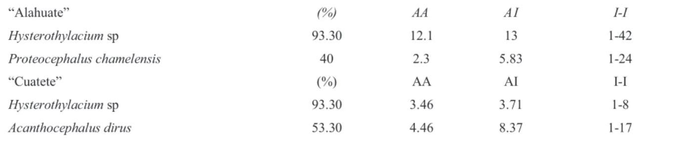
“Alahuate” (%) AA A I I-I
Hysterothylacium sp 93.30 12.1 13 1-42
Proteocephalus chamelensis 40 2.3 5.83 1-24
“Cuatete” (%) AA AI I-I
Hysterothylacium sp 93.30 3.46 3.71 1-8
Acanthocephalusdirus 53.30 4.46 8.37 1-17
Prevalence (%), Average abundance (AA), Average intensity (AI), Intensity interval (I-I).
ESPECIES Y GÉNERO DE HELMINTOS ENCONTRADOS EN EL “ALAHUATE”, E. picta
Y EL “CUATETE”, A. guatemalensis, DE LA LAGUNA TRES PALOS, GUERRERO, MÉXICO
HELMINTH SPICES AND GENERA FOUND IN “ALAHUATE” E. picta AND IN“CUATETE”,
intestino posterior adheridos a la mucosa y fue escasa su presencia en intestino posterior.
Las fases de desarrollo, así como el hábitat de los parásitos o porción del tracto digestivo parasitada, están representadas en el Cuadro 3.
De las observaciones histopatológicas realizadas en los peces, se determinó que las larvas de
Hysterothylacium sp causaron las lesiones en las cuatro categorías establecidas para este estudio. En las lesiones histológicas determinadas como categoría 1, las secciones parasitarias se encontraron en el tejido, con o sin una aparente proliferación de tejido conectivo fi broso y sin infi ltrado infl amatorio en el área dañada (Figura 2). En las lesiones determinadas en la categoría 2, las secciones parasitarias se apreciaron rodeadas por una capa gruesa o delgada de tejido conectivo fi broso y escaso infi ltrado infl amatorio (Figura 3). Las lesiones ubicadas en la categoría 3 presentaron grave reacción infl amatoria y con o sin tejido conectivo fi broso alrededor de las secciones parasitarias (Figura 4). Las lesiones en la categoría 4 presentaron proliferación de tejido conectivo fi broso, con o sin reacción granulomatosa y degeneración del parásito (Figura 5). En estas lesiones se presentaron áreas extensas de hemorragia e infl amacion con proliferación de tejido conectivo fi broso en la submucosa y en las secciones transversales de las larvas se muestra colágena rodeando al gusano en el alahuate y sin colágena en los gusanos de “cuatete”.
P. chamelensis ocasionó lesiones poco evidentes en las vellosidades de la mucosa intestinal, consistentes en atrofi a, hipertrofi a y compresión de la mucosa. (Figure 2). In the 2nd level, the parasitized sections
were surrounded by a thick or thin layer of fi brous connective tissue and scarce infl ammatory infi ltrate (Figure 3). The lesions with a serious infl ammatory reaction, with or without fi brous connective tissue were included in the 3rd level (Figure 4). Lesions in the 4th level had proliferation of fi brous connective tissue, with or without granulomatous reaction and parasite degradation (Figure 5). Extensive hemorrhagic and infl ammatory areas with proliferation of fi brous connective tissue in the submucosa were present in the lesions. A layer of collagen was observed surrounding the larvae found in the alahuate in the transversal sections. In the cuatete sections larvae lacked this collagen layer.
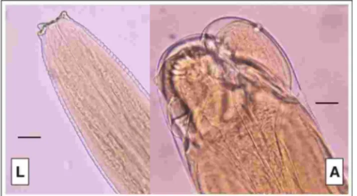
Figura 1. Región anterior de una larva L3 (L) y de un adulto
(A) de Hysterothylacium sp. Barra = 100 µm
Anterior regions of Hysterothylacium sp L3 larva (L) and adult (A). Barr = 100 µm
Host Parasite
Genus/Species Family Helminth Habitat
E. picta Anisakidae Hysterothylacium sp Larva (L3) In tissue and
lumen (intestine,
stomach)
Proteocephalinae Proteocephalus chamelensisAdults and
juveniles
Lumen (intestine)
A. guatemalensis Anisakidae Hysterothylacium sp Larvae, juve-
niles and
adults
Tissue and lumen
(stomach,
intestine)
Echinorhynchidae Acanthocephalus dirus Adults Lumen (intestine)
RESULTADOS HELMINTOLÓGICOS DE E. picta Y A. guatemalensis DE LA LAGUNA
DE TRES PALOS, GUERRERO, MÉXICO
HELMINTOLOGICAL RESULTS OF E. picta AND A. guatemalensis IN TRES PALOS
LAGOON, GUERRERO, MEXICO
Cuadro 3
P. chamelensis caused less evident lesions in the villus of the intestinal mucosa; the lesions were atrophy, hypertrophy and compression of the mucous membrane.
Discussion
There are no histological studies on the digestive tract of A. guatemalensis and E. picta; therefore a histological characterization was established in order to locate
Hysterothylacium sp larvae and, based on previous studies done on fi sh anatomy and histology;19-21 the
Discusión
Para A. guatemalensis y E. picta no existen trabajos histológicos de su aparato digestivo; por tanto, se realizó una caracterización histológica para delimitar y localizar las larvas de Hysterothylacium sp y, basándose en trabajos de anatomía e histología de peces,19-21 se
trabajó con las siguientes capas: Mucosa, submucosa, muscular del órgano y serosa. Las capas más importantes involucradas fueron la submucosa y la muscular del órgano, siendo la submucosa la capa más afectada por las larvas. No se conoce la razón, pero podrían presentarse condiciones favorables para
Figura 2. Sección de estómago del cuatete, con un corte
transversal del nematodo Hysterothylacium sp (categoría 1). Submucosa (SM), parásito (fl echa). H-E. Barra=100 µm
Stomach section of cuatete with transversal cut of
Hystero-thylacium sp (1st level) Submucosa (SM), parasite (arrow).
H&E Barr=100 µm
Figura 3. Secciones transversales de Hysterothylacium sp
rodeadas por tejido conectivo fi broso y colágena en el “ala-huate” (categoría 2). Submucosa (SM), parásito (fl echa). H-E Barra de 100 µm
Transversal sections of Hysterothylacium sp surrounded by fi brous connective tissue and collagen in alahuate (2nd level). Submucosa (SM), parasite (arrow). H&E. Barr=100 µm
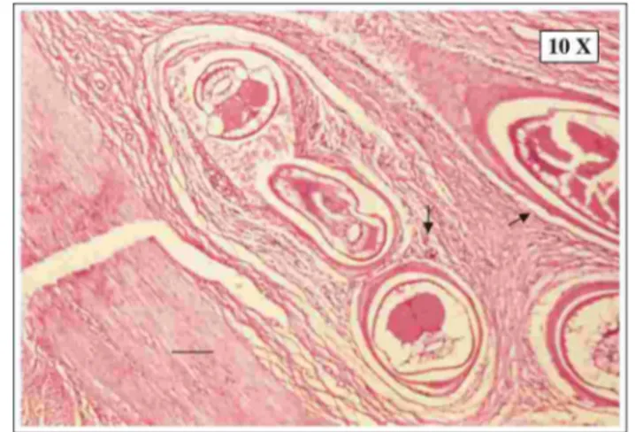
Figura 4. Sección de estómago observándose cortes
trans-versales de Hysterothylacium sp rodeados por hemorragia extensa y reacción infl amatoria grave en el “cuatete” (cate-goría 3). Submucosa (SM), hemorragia (H), parásito (fl echa). H-E. Barra 10 µm
Stomach section with transversal sections of Hysterothyla-cium sp surrounded by an extensive hemorrhage and a seri-ous infl ammatory reaction in cuatete (3rd level) Submucosa (SM), hemorrhage (H), parasite (arrow). H&E. Barr = 10 µm
Figura 5. Granuloma parasitario observando degeneración
del parásito y necrosis caseosa (categoría 4). Parásito (fl echa). H.E. Barra=10 µm
following layers were considered: mucosa, submucosa, muscle layer and serosa. The most important layers were the submucosa and the muscle layer, being the submucosa the membrane most affected by larvae. There is still no known reason for this, maybe the conditions in this layer are advantageous for larval development or, since it is the most extensive and external layer, it has the highest probabilities of being infected.
The larval stage was the most harmful stage of the parasites in fi sh. Larvae probably migrate through the digestive tract and penetrate tissue or form cysts that provoke infl ammatory reactions and hemorrhages; the most affected sections are stomach, anterior and posterior intestine.7 On the other hand, adult
nematodes in the cuatete did not produce serious pathologic lesions in the lumen of the intestine.
P. chamelensis cestodes are usually observed in the lumen of the intestine causing exclusively atrophy, hypertrophy, hyperplasia or mild compression of the goblet cells in the epithelium of the mucosa in alahuate.
Smith, Wootten10-12 and Berland, describe a host
reaction to the larval presence characterized by fi broblast proliferation, which forms a capsule of fi brous-connective and vascular tissue around the parasite. This agrees with what was observed in the present study. Hysterothylacium sp larvae are surrounded by a collagen layer and fi broblasts in the alahuate; however, in the cuatete this proliferation was not observed.
The accumulation of collagen surrounding para-sites is a host defense reaction, characteristic in this kind of infections. Because of this, it can be concluded that the defensive system of the cuatete does not respond with the same effi ciency as the alahuate does; on the other hand, it might be due to the duration of the infection. The reason why there is an absence of fi brous-connective and vascular tissue in fi sh is still unknown.
Anasakid larvae cause stomach lesions more frequently,7 as it was observed in the cuatete fi sh;
however in the alahuate these lesions were observed in the anterior intestine. In the remains of the digestive tract there was a greater quantity of lesions in the alahuate compared to the cuatete. Probably the cuatete is more resistant to Hysterothylacium sp larvae repelling a major infection since it is less susceptible or the infection involves a small number of parasites; it could also depend on the course of the disease. It is probable that nematodes have a higher biotropism to alahuates, according to their feeding habits and the contact with the parasites in the lake. For this reason, the alahuete must increase its defenses to control this infection and to create a collagen capsule in order
el desarrollo de las larvas o porque sea la capa más extensa y externa, con probabilidades altas de ser infestadas.
Las larvas fueron más dañinas para los peces, las cuales en el tracto digestivo probablemente migraban y penetraban el tejido o formaban quistes ocasionando el infi ltrado infl amatorio y hemorragias, de tal forma que producen lesiones principalmente en estómago, intestino anterior y posterior de los peces.7 En cambio,
los nematodos adultos en el “cuatete” no produjeron un cuadro patológico grave en la luz del intestino.
Por lo común, se localizaron cestodos de P. chamelensis en la luz intestinal, ocasionando exclusi-vamente atrofi a, hipertrofi a, hiperplasia o compresión leve de las células caliciformes en el epitelio de la mucosa del “alahuate”.
Smith, Wootten10-12 y Berland describen una
respuesta del hospedero ante la presencia de las larvas en el tejido, caracterizada por la proliferación de fi broblastos, los cuales forman una cápsula de tejido conectivo fi broso y vascular alrededor del parásito; esto último corresponde a lo observado en el presente estudio. Las larvas de Hysterothylacium sp están rodeadas por una capa de colágena y fi broblastos en el alahuate; sin embargo, en el “cuatete” no se presentaba esta proliferación.
La formación de colágena conteniendo al parásito es una reacción de defensa del hospedero, que es característica ante este tipo de infecciones; por lo anterior, probablemente el sistema de defensa del “cuatete” no responde con la misma efi ciencia que el del “alahuate”. Por otro lado, tal vez se deba a la cronicidad de la infestación. No se sabe a qué se deba la ausencia del tejido conectivo fi broso y vascular en peces.
to contain the parasites until they are degraded. In cuatete the lesions of the 3rd level are more common
and serious, followed by those of 4th level, which are characterized by the degradation of the parasite. In contrast, lesions of the 2nd level are more common in
the alahuate, followed by those of the 4th level. Other
lesions were evaluated qualitatively, based on the total appearance percentage for each species.
Hemorrhages are more frequent in the alahuate and less frecuent in the cuatete. The hemorrhages are probably caused by Hysterothylacium sp larvae when they cross tissue braking blood vessels. In the cuatete, fi brosis is barely found as the result of the helminth stimulation to form fi brous-connective and vascular tissue. In both species enteritis has a low percentage (6.7); it may be associated to intestinal wall perforation by hemminths and the secondary bacterial infections. In the alahuate fi sh, enteritis produces a mild heterophilic reaction. On the other hand, in the cuatete the reaction is granulomatous and non-supurative with a chronic course. Necrosis is found in a low percentage of cases (6.7) in the cuatete, but in the alahuate is slightly higher (13.3%) associated with a continuous local compression and to tissue rupture during larvae migration.
All lesions are associated to the parasitized sections; however it is possible that some of the lesions are produced by another pathogen agent, such as the bacteria found in the helminth cuticle, which were not analyzed.
Similar lesions to those caused by anisakids in the cuatete and the alahuate have been described in humans. Jackson and Cho et al. mention lesions with a granulomatous infl ammatory reaction, irritation and ulceration of the intestinal wall in humans. Mudry et al. report the formation of neoplasias. Jackson reports the development of a granuloma that surrounds and eliminates the worm. Even the histological layers that are affected by Hysterothylacium sp larvae in cuatete and alahuate are similar to those caused by anisakids in man, as is the case of the subcmucosa. According to Cho et al. and Mudry et al., the affected areas cover from the submucosa to the serosa. Considering this,
Hysterothylacium sp deserves the same attention as other anisakids since it may have zoonotic importance.
The gastrointestinal lesions caused by helminths are capable of provoking a decrease in the absorption of proteins, fats and carbohydrates, leading to a decrease in weight and size,22 affecting the commercial
value of fi sh. It is advisable to perform more studies on this kind of parasitosis, especially on those caused by Hysterothylacium sp in order to establish the pathological stages and parasitic loads through out the year and then have a better comprehension about the defensive reactions of these fi sh and also create
parásito. En cambio, en el “alahuate” la categoría más frecuente fue la 2 y le siguen lesiones de la categoría 4.
Otras lesiones que se revisaron y analizaron de manera cualitativa fueron evaluadas con base en el porcentaje de presencia para la muestra total de cada especie.
Las hemorragias son más frecuentes en el “alahuate” y menos frecuentes en el “cuatete”, probable-mente causadas por las larvas de Hysterothylacium sp al atravesar el tejido, por rupturas de vasos sanguíneos. La fi brosis sólo se presenta en este último en bajo porcentaje por la estimulación de los helmintos para formar el tejido conectivo fi broso y vascular. En la enteritis ambas especies presentan un porcentaje muy bajo (6.7); podría estar asociado con la perforación de la pared intestinal por helmintos y con infecciones secundarias bacterianas. En el “alahuate” es una enteritis heterofílica ligera; en cambio, en el “cuatete” es del tipo granulomatosa y no supurativa, por ser de curso crónico. La necrosis se presenta con un bajo porcentaje en el cuatete (6.7), pero en el “alahuate” es ligeramente mayor (13.3), asociada a la compresión local continua y a la ruptura del tejido durante la migración de las larvas.
Todas las lesiones se encuentran asociadas a las secciones parasitarias en el tejido; sin embargo, es posible que algunas de estas lesiones sean producidas por algún otro agente patógeno como bacterias en la cutícula de los helmintos, las cuales no fueron determinadas.
Se han descrito lesiones en el hombre causadas por anisáquidos similares a las encontradas en “cuatete” y “alahuate”. Jackson y Cho et al. mencionan lesiones como reacción infl amatoria granulomatosa, irritación y ulceración de las paredes intestinales en el hombre. Mudry et al. refi eren la formación de neoplasias; Jackson informa el desarrollo de un granuloma que rodea al gusano y lo elimina, e incluso las capas histológicas que son afectadas por las larvas de
Hysterothylacium en “cuatete” y “alahuate” son similares a las afectadas por otros anisáquidos en el hombre, como es el caso de la submucosa. De acuerdo con Cho
et al. y Mudry et al., las áreas del tejido afectadas se extienden de la submucosa a la serosa, de tal forma que Hysterothylacium sp no merece menos atención que otros anisáquidos, ya que puede tener importancia zoonótica.
more information about this nematode that can also affect humans.
It is important to point out that more studies on this kind of host immune response are necessary, as well as on the enzymes of the gastrointestinal tract that are affected by the presence of these parasites, since the nematodes of the Hysterothylacium genus identifi ed in this study, are potentially zoonotic.
Referencias
estados patológicos y cargas parasitarias en distintas épocas del año, para comprender mejor la reacción de defensa de estos peces y también para generar más información sobre este nematodo, que puede infectar al hombre.
Es importante destacar que se deben continuar más estudios sobre el tipo de respuesta inmunológica que tienen los hospederos antes descritos en contra de estos parásitos y qué tipo de enzimas del aparato gastrointestinal se ven afectadas al estar presente estos parásitos, ya que el nematodo del género
Hysterothylacium sp identifi cado en este estudio es potencialmente zoonótico.
Agradecimientos
Los autores agradecen a Jaime Eugenio Córdova López las tomas, revelado y sugerencias de las fotografías en este trabajo, también a Guadalupe Juárez por la realización de los cortes histológicos, así como a Emma Serrano y Germán Valero por su asesoría.
estudio de enfermedades en la conservación de fauna silvestre. Vet Méx 2000;31:223-230.
Yáñez-Arancibia A, Leyton de Yáñez V. Desarrollo del otolito embrionario, patrón de su crecimiento y comparación morfológica con otolitos juveniles y adultos del bagre marino Galeichthys caerulescens (Gunther). An Centro Cienc Mar Limnol UNAM 1977;4: 115-124. Amlacker E. Textbook of fi sh diseases. New York: Conroy and Herman, 1970.
Prophet BE, Mills B, Arrington JB, Sobin LH, M.D. Métodos histotecnológicos. Washington DC: Instituto de Patología de las Fuerzas Armadas de los Estados Unidos de América, AFIP, 1992.
Lamothe AR. Manual de técnicas para preparar y estudiar los parásitos de animales silvestres. México: AGT Editor, SA, 1997.
Margolis L, Esch GW, Holmes JC, Kurls AM, Schad GA. The use of ecological terms in parasitology (Report of an Ad Hoc committee of the american society of parasitologists). J Parasitol 1982;68: 131-133.
Schmidt GD, Roberts LS. Foundations of parasitology. Iowa State: Times Mirror/Mosby College, 1989.
Hibiya T. An atlas of fi sh histology normal and pathological features. Japan: Kodansha Ltd, 1982. Yasutake WT, Wales JH. Microscopic anatomy of salmonids: an atlas. United States of America, New York: United States Department of the Interior Fish and Wildlife Service, 1983.
Chávez SLA. Lesiones histológicas asociadas a parásitos en la mojarra “Tenguayaca” (Petenia splendida Günther,
1862) de la presa Temascal, Oaxaca, México. (tesis de licenciatura). México, México DF: Facultad de Ciencia UNAM, 1998.
Berland B. Identifi cation of larval nematodes from fi sh. In: Moller, H. editor: Nematode problems in North Atlantic fi sh. Report from a workshop in Kiel. 3-4 April 1989. Int Counc Explor Sea C. M. Norway: 1989; 6:16-22.
Cheng TC. The natural history of anisakiasis in animals. J Milk Food Technol 1976;39: 32-46.
García AP. Contribución al estudio de algunos aspectos biológicos, y determinación de los principales parásitos que atacan a Cichlasoma trimaculatum (Günter, 1868),
Galeichthys caerulences (Günter, 1864) y Mugil curema
(Valenciennes, 1830); tres especies de peces con mayor importancia comercial, que se capturan en la Laguna Tres Palos Gro (tesis de licenciatura). Acapulco (Guerrero) México: Escuela Superior de Ecología Marina. Univ Aut de Guerrero. 1999.
Myers BJ. The nematodes that cause anisakiasis. J Milk Food Technol 1975;38: 774-782.
Jackson GJ. The “new disease” status of human anisakiasis and north american cases: a review. J Milk Food Technol 1975;38: 769-773.
Mudry J, Lefebvre P, Dei-Cas E, Vernes A, Poirriez J, Débat M, et al. Anisakiase humaine: 5 cas dans le nord de la France. Gastroenterol Clin Biol 1982;10: 83-87. Beran GW, Steel JH. CRC handbook series in zoonoses. England: CRC Press, 1994.
Cho S-Y, Chi JG, Kim IS, Min Y-Y, Chun W-C, Son JH, et al. A case of human anisakiasis in Korea. Seoul J Med 1980;21: 203-208.
Miyazaki I. An Illustrated Book of Helminthic zoonoses. Japan: International Medical Foundation of Japan, Tokio. 1991.
Smith JW. Wootlen R. Pseudoterranova Larvae (Codworm) (Nematoda) in fi sh, Fiche 7 in Fiches d’ Identifi cation des Maladies et Parasites des Poissons, Crustacés et Mollusques. Denmark: NOAA National Marine Fisheries Service. 1984.
Smith JW, Wootten R. Anisakis Larvae (‘Herringworm’) (Nematoda) in fi sh. Fiche No 8 in Fiches d’ Identifi cation des Maladies et Parasites des Poissons, Crustacés et Mollusques. Denmark: NOAA National Marine Fisheries Service. 1984.
Smith JW, Wootten R. Phocascaris/Contracaecum Larvae (Nematoda) in fi sh. Fiche No 9 in Fiches d’ Identifi cation des Maladies et Parasites des Poissons, Crustacés et Mollusques. NOAA National Marine Fisheries Service. Denmark. 1984