NOTA T ´ECNICA
Vol. 33, N´um. 2, Diciembre, 2012, pp. 147-158
Development of a Virtual Simulator for Planning
Mandible Osteotomies in Orthognathic Surgeries
E.H. Govea-Valladares∗ H.I. Medell´ın-Castillo∗ T. Lim∗∗ B. Khambay ∗∗∗ M. Rodr´ıguez-Florido ∗∗∗∗ J. Ballesteros∗∗∗∗
∗Facultad de Ingenier´ıa,
Universidad Aut´onoma de San Luis Potos´ı, S.L.P. M´exico.
∗∗Innovative
Manufacturing Research Centre, Heriot-Watt University, Edinburgh, UK.
∗∗∗Dental School,
University of Glasgow, Glasgow, UK.
∗∗∗∗Instituto Tecnol´ogico
de Canarias, Las Palmas, Islas Canarias, Espa˜na.
ABSTRACT
Surgery knowledge and training is typically transmitted by the teacher-student method. In particular, the training process is carried out during real surgical interventions and under supervision of an experienced surgeon. Recent advancements in computer interaction technology and virtual environments allow a wide variety of surgical procedures to be simulated. Virtual reality (VR) applications range from art to engineering, science and medicine. In medicine, virtual simulators are being developed for pre-operative planning and training purposes. In this way, the transferring process of surgery knowledge and training can be enhanced and speed up. Medical VR simulators are characterized by their large demands on visual and physical behavior, and more recently the demand for the sense of touch, which is an essential aspect in surgery. Regarding the maxillofacial surgery, one of the most common surgical procedures is the ‘Bilateral Sagittal Split Ramus Osteotomy Mandibular” (BSSROM), which is used to relocate the jaw at the correct position, fix jaw deformities, get better functionality of the jaw and improve the patient aesthetic. In this paper the development of a 3D virtual simulator for planning mandible osteotomies in orthognathic surgeries is presented; in particular the work is focused on the BSSROM procedure. The proposed system has been developed in an open-source platform that provides a high level of realism and interaction, and where the surgeons are able to cut bone in a 3D free-form way; thus enhancing the traditional virtual osteotomy approach which is based on cutting planes. Some of the main functionalities of the system include: virtual reality environment and real-time response; 3D visualization of anatomical models and tools; free-form manipulation and interaction of cutting tool, bone, and bone fragments; simulation of single and multiple osteotomies; cutting planes osteotomies and free-form cut osteotomies. The description, development and implementation of the system are presented in this paper. The results have shown that the proposed system is practical and can be used for planning and training mandible osteotomies.
148 Revista Mexicana de Ingenier´ıa Biom´edica·volumen 33·n´umero 2·Diciembre, 2012
Correspondencia: E.H. Govea-Valladares Av. Dr. Manuel Nava #8 Edificio P Zona Universitaria, CP 78290 San Luis Potos´ı, S.L.P. M´exico, Tel/fax (444) 8-17-33-81 Correo electr´onico: [email protected]
Fecha de recepci´on: 31/Octubre/2012
Fecha de aceptaci´on: 27/Noviembre/2012
RESUMEN
El conocimiento y entrenamiento quir´urgico normalmente se transmite por el m´etodo maestro-alumno. En particular, el proceso de entrenamiento se lleva a cabo durante las intervenciones quir´urgicas reales y bajo la supervisi´on de un cirujano experimentado. Los recientes avances en la tecnolog´ıa computacional y la interacci´on con entornos virtuales han permitido que una amplia variedad de intervenciones quir´urgicas puedan ser simuladas. Las aplicaciones de la realidad virtual (VR) abarcan desde el arte hasta la ingenier´ıa, ciencia y medicina. En medicina, los simuladores virtuales est´an siendo desarrollados con prop´ositos de planeaci´on y entrenamiento pre-operatorio. De esta manera el proceso de transferencia de conocimiento y entrenamiento quir´urgico puede mejorarse y acelerarse. Los simuladores m´edicos de realidad virtual se caracterizan por sus grandes demandas visuales y comportamiento f´ısico, y m´as recientemente la demanda por el sentido del tacto, que es un aspecto esencial en la cirug´ıa. Con respecto a la cirug´ıa maxilofacial, una de las procedimientos quir´urgicos m´as comunes es la ‘Osteotom´ıa Sagital Bilateral de Rama Mandibular’ (OSBRM), la cual se utiliza para relocalizar la mand´ıbula en la posici´on correcta, corregir deformidades de la mand´ıbula, conseguir un mejor funcionamiento de la mand´ıbula y mejorar la est´etica del paciente. En este art´ıculo se presenta el desarrollo de un simulador virtual 3D para planeaci´on de cirug´ıas de osteotom´ıa mandibular ortogn´atica, en particular este trabajo se enfoca en el procedimiento OSBRM. El sistema propuesto ha sido desarrollado en una plataforma de c´odigo abierto y ofrece un sistema m´as realista e interactivo, en donde los cirujanos pueden cortar hueso de una manera libre y en 3D, mejorando as´ı el enfoque tradicional de la osteotom´ıa virtual basada en planos de corte. Algunas de las funcionalidades principales del sistema son: un entorno de realidad virtual y respuesta en tiempo real; visualizaci´on en 3D de biomodelos y herramientas; interacci´on y manipulaci´on libre de herramientas de corte, huesos y fragmentos de huesos; simulaci´on de osteotom´ıas simples y m´ultiples; osteotom´ıas con planos de corte y de forma libre. La descripci´on, el desarrollo y la aplicaci´on del sistema se presentan en este documento. Los resultados han demostrado que el sistema propuesto es pr´actico y puede ser utilizado en la planeaci´on y entrenamiento de osteotom´ıas mandibulares.
Palabras clave: simulador virtual, planificaci´on quir´urgica, OSBRM, osteotom´ıas, mand´ıbula.
INTRODUCTION
Virtual Reality (VR) is one of the main areas of knowledge that have taken advantage of the computer technological development 1. It has been used in different applications
Govea-Valladares y cols. Development of a virtual simulator for planning mandible osteotomies in orthognathic surgeries 149
The practice of medicine is performed through a teamwork and a complex decision making process 4. Practitioner abilities in medicine are gained by experience and training, which is a slow process and takes several years. To get experience and abilities, a student of medicine must be the protagonist of his/her training, taking into account as a main priority the avoidance of risks and unnecessary inconveniences for the patient 5. The use of VR in medicine may allow the student or practitioner to understand and confirm concepts and to improve skills in surgical practice, while experienced surgeons can plan or diagnostic with more accuracy 6.
In general, a VR application in medicine is composed of three main modules:
1. 3D model reconstruction module, which is responsible of generating 3D anatomical models from medical data such as CT and MRI images.
2. 3D visualization module, which is responsible of the graphics rendering of the virtual environment.
3. Manipulation module, needed to provide the interaction between the user and the virtual environment 7.
The Bilateral Sagittal Split Osteotomy Ramus Mandibular (BSSROM) procedure is a common technique used for the correction of facial deformities in an individual; it allows performing mandibular movements in different planes 8. The BSSROM represents the most commonly used procedure orthognathic surgery 9. Over the years, it has been gradually modified in terms of design, extension and instrumentation 10. These modifications make it technically friendly, predictable, biologically acceptable and versatile.
This paper presents the development of a VR system for planning and training of mandible osteotomies in orthognathic surgeries. Currently the development has been focused on the BSSROM procedure. The description, development and implementation of the proposed system are presented.
LITERATURE REVIEW
Several works related to the use of VR techniques in surgery simulation, maxillofacial surgery and virtual osteotomies, have been reported in the literature. The following paragraphs summarize some of these works.
Virtual maxillofacial surgery
Virtual surgery is an effective, definitive and objective tool for reconstruction and correction of facial deformities 11. The use of virtual surgery enhances the diagnosis and treatment planning. It can also simulate the results after performing surgery. In 12 the differences between virtual and real surgical outcomes were measured using a voxel-wise rigid registration of the cranial base with construction of pre- and post-surgery 3D models. The use of surgical templates to perform mandible reconstruction surgery according to the preoperative simulation was presented in 13. The accuracy was evaluated by means of cadaveric surgery in a 3D environment. The reconstructed mandibles showed high similarity to the surgical planning in terms of the mean translation, angular deviation, and rotation of segments of the reconstructed mandibles. A comparative analysis between the surgical outcomes achieved with Computer-Aided Surgical Simulation (CASS) and the traditional methods was presented in 14. A 3D skull model of twelve consecutive patients with Cranio Maxillofacial was generated. These models underwent 2 virtual surgeries: 1 was based on CASS (experimental group) and the other was based on traditional methods 1 year later (control group). The results showed that the surgical outcomes achieved with CASS are significantly better than those achieved with traditional planning methods.
150 Revista Mexicana de Ingenier´ıa Biom´edica·volumen 33·n´umero 2·Diciembre, 2012
mandibular yaw movements, was presented in 16. A total of 670 students were evaluated in the use of simulated patients in conjunction with anatomic and tissue task-training 17. The results suggested that the combination of simulated patients and simulation models yields to reliable scores for procedural and interpersonal skills. On the other hand, a virtual simulator to evaluate dental surgeries was presented in 18. A group of 53 dental students provided their impressions after virtual simulation. The results showed that 51 of the 53 students recommended the virtual simulation as an additional training modality in dental education. A new method to automatically compute the mid-facial plane for planning cranio-maxillofacial interventions based on 3-dimensional (3D) virtual models was presented in 18. The study included experienced and inexperienced clinicians defining the symmetry plane by a selection of landmarks; this manual definition was systematically compared with the definition resulting from the new automatic method. The results showed that the new automatic method is reliable and leads to significantly higher accuracy than the manual method when performed by inexperienced clinicians.
A virtual reality tool for CASS, named ‘The Hollowman’, was presented in 20. ‘The Hollowman’ was used in orthognatic surgery to control the translocation of the maxilla after Le Fort I osteotomy within a bimaxillary procedure. The tool was proved to be very valuable especially in complex nonlinear translocations of the maxilla because the surgeon could directly visualise the position of the mobilised bone in relation to the preoperatively planned situation. The use and combination of volumetric tomography and 3D dental software programs, as tools for oral and maxillofacial surgery, was discusses in 21. The results suggest that the combination of these technologies is useful for expanding information in dentoalveolar, preprosthetic, trauma, pathology and reconstruction, orthognathic, craniofacial, and surgical cases of esthetic implant. Moreover, the precision, accuracy, and 3D visualization capabilities of these technologies open avenues for the oral and maxillofacial
surgeon in the diagnosis, planning, and surgical management. In 22 it was presented a study to examine 3D virtual anatomical features of the sphenoid sinus and adjacent structures during virtual surgery, and to explore their relevance to actual transsphenoidal surgery. The study primarily focused on performing a virtual surgery to get measures and data needed in the real surgery. The study provided virtual anatomical information about the sphenoid sinus and important surrounding structures, which is essential for a successful real life transsphenoidal surgery.
Virtual osteotomies
The use of computer-assisted three-dimensional surgical planning in condylar reconstruction by vertical ramus osteotomy was described in 23. The results showed that the combination between surgical planning system and simulation makes the surgery more accurate and convenient, and avoids damage to vital structures. On the other hand, the amount of interference between the mandible proximal and distal segments generated by 3 different osteotomy methods using computer simulation was assessed in 24. They demonstrated no difference in severe prognathism and asymmetry cases. In 25 the surgical outcomes in free fibula mandibular reconstructions planned with virtual surgery were evaluated. From a total of 19 mandibular osteotomies, the results showed that virtual surgical planning have a positive impact on accuracy because it is possible to get more accurate measurements than manually, even by the hands of experienced surgeons.
Govea-Valladares y cols. Development of a virtual simulator for planning mandible osteotomies in orthognathic surgeries 151
biomechanical stability of bilateral sagittal split ramus osteotomies fixed by lag screws with linear and triangular configuration was presented in 27. Posterior occlusal loads were simulated to calculate the stress fields on both the segments and the fixing appliances by the MSC Marc software.
From the literature, it can be observed that virtual reality in medical applications has been widely used. Most of the surgical simulators have been designed for specific applications and they are based on a traditional manipulation of virtual models using the 2D mouse. On the other hand, virtual osteotomy simulators are based on cutting planes defined by the user, which is far from real osteotomy procedures where the bone is cut manually and the hand accuracy of the specialist is important. Thus, in this paper the development of a virtual osteotomy simulator is proposed in order to enhance the traditional virtual osteotomy approach based on cutting planes and 2D interaction. The proposed system provides a more realistic and interactive system where the surgeons are able to cut bone in a 3D free-form way. Also the system allows the free 3D manipulation of tools and anatomical models.
BSSROM PROCEDURE
The Bilateral Sagittal Split Osteotomy Ramus Mandibular (BSSROM) procedure starts with the cutting of the internal wall in the upper part of the ascending ramus of the mandible 28. The mark that leaves the tool serves as a guide to saw the rest of the jawbone 29. Figure 1 shows this procedure 29.
5
interactive system where the surgeons are able to cut bone in a 3D free-form way. Also the system allows the free 3D
manipulation of tools and anatomical models.
3. BSSROM PROCEDURE
The Bilateral Sagittal Split Osteotomy Ramus Mandibular (BSSROM) procedure starts with the cutting of the internal wall in
the upper part of the ascending ramus of the mandible 28. The mark that leaves the tool serves as a guide to saw the rest of
the jawbone 29. Figure 1 shows this procedure 29.
Figure 1: Cutting with the mill 29.
The next cut is performed following an oblique path from top to bottom and using a saw 10. The saw leaves a line mark as
showed in Figure 2. The surgeon can then change the direction of the saw to cut only about 5 mm from the bottom edge 31.
Figure 2:.Cutting with the sagittal saw 29.
The manual separation of the bone fragments is then carried out by pushing downwards and doing lateral movements along
the ramus 10. The last step is to set both parts with mini plates and screws of titanium 10, 31, Figure 3. Fig. 1. Cutting with the mill 29.
5
interactive system where the surgeons are able to cut bone in a 3D free-form way. Also the system allows the free 3D
manipulation of tools and anatomical models.
3. BSSROM PROCEDURE
The Bilateral Sagittal Split Osteotomy Ramus Mandibular (BSSROM) procedure starts with the cutting of the internal wall in
the upper part of the ascending ramus of the mandible 28. The mark that leaves the tool serves as a guide to saw the rest of
the jawbone 29. Figure 1 shows this procedure 29.
Figure 1: Cutting with the mill 29.
The next cut is performed following an oblique path from top to bottom and using a saw 10. The saw leaves a line mark as
showed in Figure 2. The surgeon can then change the direction of the saw to cut only about 5 mm from the bottom edge 31.
Figure 2:.Cutting with the sagittal saw 29.
The manual separation of the bone fragments is then carried out by pushing downwards and doing lateral movements along
the ramus 10. The last step is to set both parts with mini plates and screws of titanium 10, 31, Figure 3.
Fig. 2. Cutting with the sagittal saw 29.
The next cut is performed following an oblique path from top to bottom and using a saw 10. The saw leaves a line mark as showed in Figure 2. The surgeon can then change the direction of the saw to cut only about 5 mm from the bottom edge 31.
The manual separation of the bone fragments is then carried out by pushing downwards and doing lateral movements along the ramus 10. The last step is to set both parts with mini plates and screws of titanium 10, 31, Figure 3.
6
Figure 3: Fixation with titanium miniplates and screws 29.
4. SYSTEM DESCRIPTION
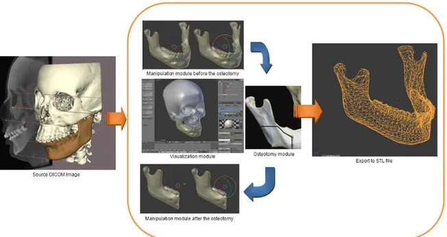
The proposed Virtual Osteotomy Simulator System (VOSS) for 3D osteotomy planning and training has the architecture shown in Figure 4. The VOSS system comprises four main modules:
1. Visualization module, responsible of carrying out the graphic rendering of virtual objects and virtual environments.
2. Osteotomy module, to perform virtual osteotomies.
3. Manipulation module, to enable 3D free-form movement and manipulation of objects and surgical tools.
4. Data exportation module, to export any information regarding the osteotomy planning (e.g. STL files of models).
Figure 4: VOSS architecture.
The GUI of VOSS is shown in Fig. 5, which has been implemented using Phyton and Blender 2.59, in a PC with a 1.73 GHz processor, 2.0GB of RAM and Windows XP. At the present, the VOSS systems allows for:
ü Virtual reality environment and real-time response.
ü 3D visualization of anatomical models and tools.
152 Revista Mexicana de Ingenier´ıa Biom´edica·volumen 33·n´umero 2·Diciembre, 2012
6
Figure 3: Fixation with titanium miniplates and screws 29.
4. SYSTEM DESCRIPTION
The proposed Virtual Osteotomy Simulator System (VOSS) for 3D osteotomy planning and training has the architecture
shown in Figure 4. The VOSS system comprises four main modules:
1. Visualization module, responsible of carrying out the graphic rendering of virtual objects and virtual environments.
2. Osteotomy module, to perform virtual osteotomies.
3. Manipulation module, to enable 3D free-form movement and manipulation of objects and surgical tools.
4. Data exportation module, to export any information regarding the osteotomy planning (e.g. STL files of models).
Figure 4: VOSS architecture.
The GUI of VOSS is shown in Fig. 5, which has been implemented using Phyton and Blender 2.59, in a PC with a 1.73 GHz
processor, 2.0GB of RAM and Windows XP. At the present, the VOSS systems allows for:
ü Virtual reality environment and real-time response.
ü 3D visualization of anatomical models and tools. Fig. 4. VOSS architecture.
SYSTEM DESCRIPTION
The proposed Virtual Osteotomy Simulator System (VOSS) for 3D osteotomy planning and training has the architecture shown in Figure 4. The VOSS system comprises four main modules:
1. Visualization module, responsible of carrying out the graphic rendering of virtual objects and virtual environments.
2. Osteotomy module, to perform virtual osteotomies.
3. Manipulation module, to enable 3D free-form movement and manipulation of objects and surgical tools.
4. Data exportation module, to export any information regarding the osteotomy planning (e.g. STL files of models).
The GUI of VOSS is shown in Fig. 5, which has been implemented using Phyton and Blender 2.59, in a PC with a 1.73 GHz processor, 2.0GB of RAM and Windows XP. At the present, the VOSS systems allows for:
• Virtual reality environment and real-time response.
• 3D visualization of anatomical models and tools.
• 3D free-form manipulation and interaction of cutting tool, bone, and bone fragments.
• Simulation of single and multiple osteotomies.
• Free-form cutting path osteotomy.
Figure 6a presents the methodology to define the VR environment in the VOSS system. This methodology comprises the following steps in Blender:
• Create a scenario: The first stage is to create a scene in Blender to generate a real environment (lights, cameras, and background image).
Govea-Valladares y cols. Development of a virtual simulator for planning mandible osteotomies in orthognathic surgeries 153
7
ü
3D free-form manipulation and interaction of cutting tool, bone, and bone fragments.
ü
Simulation of single and multiple osteotomies.
ü
Free-form cutting path osteotomy.
Figure 5: Graphic user interface (GUI).
Figure 6a presents the methodology to define the VR environment in the VOSS system. This methodology comprises the
following steps in Blender:
•
Create a scenario:
The first stage is to create a scene in Blender to generate a real environment (lights, cameras, and
background image).
•
Load skull and jaw anatomical models:
The bio-models are uploaded as STL or 3DS files, and can be obtained from
reconstruction of medical images such as CT.
•
Add texture to bone:
In order to increase the realism of the VR environment, texture to the jaw bone is added by means
of an image. Since the skull is used only as reference and visual support, the texture is set as a transparency.
•
Model cutting tools:
Cutting tools (saw and drill) are modelled using the 3D modelling commands of Blender. They can
also be imported as STL or 3DS files.
•
Add texture to tools
: The tools (drill and saw) are texturized with image to reproduce their real appearance.
•
Create sensors:
Sensors are created to establish the keyboard and/or mouse buttons to control objects in the virtual
environment.
•
Create controllers:
Controllers are used to specify the next action to be performed after a sensor is activated.
•
Create actuators:
Actuators perform the movement or manipulation of the virtual objects according to the sensors.
Movements can be linear or rotational.
The movement or manipulation of virtual models within the platform can be made using the numeric keypad, alphanumeric
keypad or the computer mouse. These movements
are defined by sensors, controllers and actuators. Figure 6b shows the
graphic interface whit all the methodology and manipulation elements.
Fig. 5. Graphic user interface (GUI).
8
(a) (b)
Figure 6: VR environment: a) methodology, b) implementation.
5. VIRTUAL OSTEOTOMY
The virtual osteotomy approach used in the VOSS corresponds to the BSSROM procedure, Figure 7. This procedure is
described in the following paragraphs.
(a) (b)
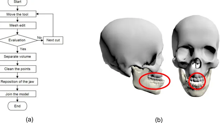
Figure 7: Virtual osteotomy: a) procedure, b) virtual model with mandibular deformation.
• Move the tool: Move the tool and place it at the point where the cut is needed. The user can select the tool to perform
the cut.
• Mesh edit: A Boolean operation is performed between the jaw (bone) and the tool models. The tool is subtracted from
the jaw. Virtual models are mesh models so they can be edited with Booleans operations.
• Evaluation: When the cut is performed with the sagittal saw, it is necessary to evaluate if the jaw was separated into two
parts. If the model is not separated yet, a new cut is performed with a new tool position.
• Separate volume: After the evaluation, the mesh model is then separated according to the volume occupied in the
154 Revista Mexicana de Ingenier´ıa Biom´edica·volumen 33·n´umero 2·Diciembre, 2012
• Add texture to bone: In order to increase the realism of the VR environment, texture to the jaw bone is added by means of an image. Since the skull is used only as reference and visual support, the texture is set as a transparency.
• Model cutting tools: Cutting tools (saw and drill) are modelled using the 3D modelling commands of Blender. They can also be imported as STL or 3DS files.
• Add texture to tools: The tools (drill and saw) are texturized with image to reproduce their real appearance.
• Create sensors: Sensors are created to establish the keyboard and/or mouse buttons to control objects in the virtual environment.
• Create controllers: Controllers are used to specify the next action to be performed after a sensor is activated.
• Create actuators: Actuators perform the movement or manipulation of the virtual objects according to the sensors. Movements can be linear or rotational.
The movement or manipulation of virtual models within the platform can be made using the numeric keypad, alphanumeric keypad or the computer mouse. These movements are defined by sensors, controllers and actuators. Figure 6b shows the graphic interface whit all the methodology and manipulation elements.
VIRTUAL OSTEOTOMY
The virtual osteotomy approach used in the VOSS corresponds to the BSSROM procedure, Figure 7. This procedure is described in the following paragraphs.
• Move the tool: Move the tool and place it at the point where the cut is needed. The user can select the tool to perform the cut.
• Mesh edit: A Boolean operation is performed between the jaw (bone) and the tool models. The tool is subtracted from the jaw. Virtual models are mesh models so they can be edited with Booleans operations.
8
(a)
(b)
Figure 6: VR environment: a) methodology, b) implementation.
5. VIRTUAL OSTEOTOMY
The virtual osteotomy approach used in the VOSS corresponds to the BSSROM procedure, Figure 7. This procedure is
described in the following paragraphs.
(a)
(b)
Figure 7: Virtual osteotomy: a) procedure, b) virtual model with mandibular deformation.
•
Move the tool:
Move the tool and place it at the point where the cut is needed. The user can select the tool to perform
the cut.
•
Mesh edit:
A Boolean operation is performed between the jaw (bone) and the tool models. The tool is subtracted from
the jaw. Virtual models are mesh models so they can be edited with Booleans operations.
•
Evaluation:
When the cut is performed with the sagittal saw, it is necessary to evaluate if the jaw was separated into two
parts. If the model is not separated yet, a new cut is performed with a new tool position.
•
Separate volume:
After the evaluation, the mesh model is then separated according to the volume occupied in the
scene. This will reduce the amount of data used in further processing.
Govea-Valladares y cols. Development of a virtual simulator for planning mandible osteotomies in orthognathic surgeries 155
• Evaluation: When the cut is performed with the sagittal saw, it is necessary to evaluate if the jaw was separated into two parts. If the model is not separated yet, a new cut is performed with a new tool position.
• Separate volume: After the evaluation, the mesh model is then separated according to the volume occupied in the scene. This will reduce the amount of data used in further processing.
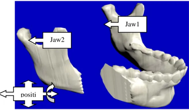
• Clean the points: Many times some points would be out of the main jaw mesh, leading to additional volumes. It is important to eliminate those volumes because the algorithm only moves the two jaw main fragments (Jaw 1 and Jaw 2).
• Reposition of the jaw: Once the jaw has been separated into two parts, the largest part (Jaw 1) is moved or manipulated to its final position at the other part (Jaw 2).
• Join the model: The final step is to align and join the two parts of the jaw.
The procedure may be also performed to the other side of the jaw bone. Normally the BSSROM procedure requires the cutting of booth jaw ramus. In this case the previous methodology is then repeated to the other jaw side.
RESULTS AND DISCUSSION
To evaluate the feasibility of the VOSS, a case study is now developed. The BSSROM procedure begins by selecting a cutting tool and placing it at the position where the first cut is mean to be made, Figure 8. Once the tool is positioned, the cut is performed and it can be repeated as many times as necessary to make a longitudinal cut along the jaw.
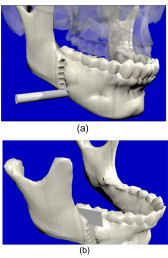
The manipulation and location of the tool can be made in a free-form way, i.e. the user can freely move the tool in any 3D path while performing the cut. Figure 9a shows the simulation of a vertical cut path using a drill,
9
•
Clean the points:
Many times some points would be out of the main jaw mesh, leading to additional volumes. It is
important to eliminate those volumes because the algorithm only moves the two jaw main fragments (Jaw 1 and Jaw 2).
•
Reposition of the jaw:
Once the jaw has been separated into two parts, the largest part (Jaw 1) is moved or manipulated
to its final position at the other part (Jaw 2).
•
Join the model:
The final step is to align and join the two parts of the jaw.
The procedure may be also performed to the other side of the jaw bone. Normally the BSSROM procedure requires the
cutting of booth jaw ramus. In this case the previous methodology is then repeated to the other jaw side.
6. RESULTS AND DISCUSSION
To evaluate the feasibility of the VOSS, a case study is now developed. The BSSROM procedure begins by selecting a
cutting tool and placing it at the position where the first cut is mean to be made, Figure 8. Once the tool is positioned, the cut
is performed and it can be repeated as many times as necessary to make a longitudinal cut along the jaw.
Figure 8: Initial positioning of the tool.
The manipulation and location of the tool can be made in a free-form way, i.e. the user can freely move the tool in any 3D
path while performing the cut. Figure 9a shows the simulation of a vertical cut path using a drill, while Figure 9b shows the cut
using a saggital saw to separate the jaw bone.
(a)
(b)
Figure 9: Available cutting process: a) drill b) sagittal saw
Fig. 8. Initial positioning of the tool.9
•
Clean the points:
Many times some points would be out of the main jaw mesh, leading to additional volumes. It is
important to eliminate those volumes because the algorithm only moves the two jaw main fragments (Jaw 1 and Jaw 2).
•
Reposition of the jaw:
Once the jaw has been separated into two parts, the largest part (Jaw 1) is moved or manipulated
to its final position at the other part (Jaw 2).
•
Join the model:
The final step is to align and join the two parts of the jaw.
The procedure may be also performed to the other side of the jaw bone. Normally the BSSROM procedure requires the
cutting of booth jaw ramus. In this case the previous methodology is then repeated to the other jaw side.
6. RESULTS AND DISCUSSION
To evaluate the feasibility of the VOSS, a case study is now developed. The BSSROM procedure begins by selecting a
cutting tool and placing it at the position where the first cut is mean to be made, Figure 8. Once the tool is positioned, the cut
is performed and it can be repeated as many times as necessary to make a longitudinal cut along the jaw.
Figure 8: Initial positioning of the tool.
The manipulation and location of the tool can be made in a free-form way, i.e. the user can freely move the tool in any 3D
path while performing the cut. Figure 9a shows the simulation of a vertical cut path using a drill, while Figure 9b shows the cut
using a saggital saw to separate the jaw bone.
(a)
(b)
Figure 9: Available cutting process: a) drill b) sagittal saw
9
• Clean the points: Many times some points would be out of the main jaw mesh, leading to additional volumes. It is
important to eliminate those volumes because the algorithm only moves the two jaw main fragments (Jaw 1 and Jaw 2).
• Reposition of the jaw: Once the jaw has been separated into two parts, the largest part (Jaw 1) is moved or manipulated
to its final position at the other part (Jaw 2).
• Join the model: The final step is to align and join the two parts of the jaw.
The procedure may be also performed to the other side of the jaw bone. Normally the BSSROM procedure requires the
cutting of booth jaw ramus. In this case the previous methodology is then repeated to the other jaw side.
6. RESULTS AND DISCUSSION
To evaluate the feasibility of the VOSS, a case study is now developed. The BSSROM procedure begins by selecting a
cutting tool and placing it at the position where the first cut is mean to be made, Figure 8. Once the tool is positioned, the cut
is performed and it can be repeated as many times as necessary to make a longitudinal cut along the jaw.
Figure 8: Initial positioning of the tool.
The manipulation and location of the tool can be made in a free-form way, i.e. the user can freely move the tool in any 3D
path while performing the cut. Figure 9a shows the simulation of a vertical cut path using a drill, while Figure 9b shows the cut
using a saggital saw to separate the jaw bone.
(a) (b)
Figure 9: Available cutting process: a) drill b) sagittal saw Fig. 9. Available cutting process: a) drill b)
156 Revista Mexicana de Ingenier´ıa Biom´edica·volumen 33·n´umero 2·Diciembre, 2012
10
As in the real procedure, the jaw can be separated into two parts which can then be moved or manipulated independently.
Figure 10 shows the bone fragments after the separation.
Figure 10: New position from jaw 1 to jaw 2.
After the manipulation and relocation of the jaw parts, a Boolean operation can be carried out to join the jaw parts. Figure 11
shows the last movement and final relocation of the mandible.
Figure 11: Final step in the virtual osteotomy.
The proposed VOSS system allowed the 3D rendering of anatomical models corresponding to an upper maxillary and a jaw.
Different textures, lighting and visual properties can be used to increase the realism level. It has been proved that 3D virtual
osteotomy procedures can be performed using the proposed system. The time performance of the cutting procedure depends
on the size of the anatomical models (in particular on the number of elements in the mesh); the average time to perform a cut
varies from 250 to 300 milliseconds, for a standard jaw and tool size. Anatomical models can be reconstructed from CT
images (DICOM) and be imported into the VOSS system as STL files.
7. CONCLUSIONS
The development of a virtual simulator for planning a Bilateral Sagittal Split Osteotomy Ramus Mandibular surgery has been
presented in this paper. The system has been developed using open-source software and without sacrificing the level of
realism. The results of the implementation and evaluation have demonstrated that it is possible to perform virtual osteotomies
using the proposed system. Future work comprises the incorporation of haptic devices to manipulate tools and virtual objects
in the VOSS system.
ACKNOWLEDGMENTS
The authors would like to acknowledge the financial support from CONACYT (National Science and Technology Council),
research grant CB-2010-01-154430. They also thank to UASLP, HWU and ITC for the support provided to this research work. Jaw1
Jaw2
positi on
Fig. 10. New position from jaw 1 to jaw 2.
10
As in the real procedure, the jaw can be separated into two parts which can then be moved or manipulated independently.
Figure 10 shows the bone fragments after the separation.
Figure 10: New position from jaw 1 to jaw 2.
After the manipulation and relocation of the jaw parts, a Boolean operation can be carried out to join the jaw parts. Figure 11
shows the last movement and final relocation of the mandible.
Figure 11: Final step in the virtual osteotomy.
The proposed VOSS system allowed the 3D rendering of anatomical models corresponding to an upper maxillary and a jaw.
Different textures, lighting and visual properties can be used to increase the realism level. It has been proved that 3D virtual
osteotomy procedures can be performed using the proposed system. The time performance of the cutting procedure depends
on the size of the anatomical models (in particular on the number of elements in the mesh); the average time to perform a cut
varies from 250 to 300 milliseconds, for a standard jaw and tool size. Anatomical models can be reconstructed from CT
images (DICOM) and be imported into the VOSS system as STL files.
7. CONCLUSIONS
The development of a virtual simulator for planning a Bilateral Sagittal Split Osteotomy Ramus Mandibular surgery has been
presented in this paper. The system has been developed using open-source software and without sacrificing the level of
realism. The results of the implementation and evaluation have demonstrated that it is possible to perform virtual osteotomies
using the proposed system. Future work comprises the incorporation of haptic devices to manipulate tools and virtual objects
in the VOSS system.
ACKNOWLEDGMENTS
The authors would like to acknowledge the financial support from CONACYT (National Science and Technology Council),
research grant CB-2010-01-154430. They also thank to UASLP, HWU and ITC for the support provided to this research work.
Jaw1 Jaw2
positi on
Fig. 11. Final step in the virtual osteotomy.
while Figure 9b shows the cut using a saggital saw to separate the jaw bone.
As in the real procedure, the jaw can be separated into two parts which can then be moved or manipulated independently. Figure 10 shows the bone fragments after the separation.
After the manipulation and relocation of the jaw parts, a Boolean operation can be carried out to join the jaw parts. Figure 11 shows the last movement and final relocation of the mandible.
The proposed VOSS system allowed the 3D rendering of anatomical models corresponding to an upper maxillary and a jaw. Different textures, lighting and visual properties can be used to increase the realism level. It has been proved that 3D virtual osteotomy procedures can be performed using the proposed system. The time performance of the cutting procedure depends on the size of the anatomical models (in particular on the number of elements in the mesh); the average time to perform a cut varies from 250 to 300 milliseconds, for a standard jaw and tool size. Anatomical models can be reconstructed from CT images (DICOM) and be imported into the VOSS system as STL files.
CONCLUSIONS
The development of a virtual simulator for planning a Bilateral Sagittal Split Osteotomy Ramus Mandibular surgery has been presented
in this paper. The system has been
developed using open-source software and without sacrificing the level of realism. The results of the implementation and evaluation have demonstrated that it is possible to perform virtual osteotomies using the proposed system. Future work comprises the incorporation of haptic devices to manipulate tools and virtual objects in the VOSS system.
ACKNOWLEDGMENTS
The authors would like to acknowledge the financial support from CONACYT (National Science and Technology Council), research grant CB-2010-01-154430. They also thank to UASLP, HWU and ITC for the support provided to this research work.
REFERENCIAS
1. Kartiko I, Kavakli M, Cheng K. Learning science in a virtual reality application: The impacts of animated-virtual actors visual complexity. Computers & Education, 2010; 55(2): 881-891.
2. Wang CL, Shen HW. Information theory in scientific visualization. Entropy, 2011; 13(1): 254-273.
3. Ruiz CA, Montagut F. Yeison J,
Heidenreich E. Algorithm for virtual modeling of organs by medical images. Vector, 2009; 4: 14-26.
4. V´azquez-Mata G. Realidad virtual y simulaci´on en el entrenamiento de los estudiantes de medicina. Educ. Med. 2008; 11(1): 29-31.
Govea-Valladares y cols. Development of a virtual simulator for planning mandible osteotomies in orthognathic surgeries 157
6. Hanna L. Simulated surgery: the virtual reality of surgical training. Surgery Medicine Publishing, 2010; 28(9): 463-468.
7. Palter VN, Grantcharov TP. Virtual reality in surgical skills training. Surgical Clinics of North America, 2010; 90(3): 605-617.
8. Ueki K, Okabe K, Miyazaki M, Mukozawa A, Marukawa K, Nakagawa K, Yamamoto E. Position of mandibular canal and ramus morphology before and after sagittal split ramus osteotomy. American Association of Oral and Maxillofacial Surgeons, 2010; 68(8): 1795-1801.
9. Yamashita Y, Otsuka T, Shigematsu M, Goto M. A long-term comparative study of two rigid internal fixation techniques in terms of masticatory function and neurosensory disturbance after mandibular correction by bilateral sagittal split ramus osteotomy. International Journal of Oral and Maxillofacial Surgery, 2011; 40(4): 360-365.
10. Quevedo R. Osteotom´ıa sagital de rama mandibular en cirug´ıa ortogn´atica. Revista Espa˜nola de Cirug´ıa Oral y Maxilofacial, 2004; 24(1):14-21.
11. Schendel SA, Jacobson R. Three-dimensional imaging and computer simulation for office-based surgery. J Oral Maxillofac Surg, 2009; 67(10): 2107-14.
12. Tucker S, Soares LH, Styner M, Kim H, Reyes M, Proffit W, Turvey T. Comparison of actual surgical outcomes and 3-dimensional surgical simulations. Journal Oral Maxillofacial Surgery, 2010; 68(10): 2412-21.
13. Guang-Sen Z, Yu-Xiong S, Gui-Ging L, Pei-Feng J,Li-Zhong L,Si-En Z, Hai-Chao L. Mandible reconstruction assisted by preoperative simulation and transferring templates: Cadaveric study of accuracy. Journal Oral Maxillofacial Surgery, 2012; 70(6): 1480-5.
14. Xia JJ, Shevchenko L, Gateno J, Teichgraeber JF, Taylor TD, Lasky RE, English JD, Kau CH., McGrory KR. Outcome study of computer-aided surgical simulation in the treatment of patients with craniomaxillofacial deformities. Journal Oral Maxillofacial Surgery, 2011; 69(7): 2014-24.
15. Marchetti C,Bianchi A, Muyldermans
L, Di Martino M, Lancellotti L,
Sarti A. Validation of new soft tissue software in orthognathic surgery planning. International Journal Oral Maxillofacial Surgery, 2011; 40(1): 26-32.
16. Alastair W, Laverick S, McIntyre GT, Epker BN. Mandibular model surgery for orthognathic surgery: The Perth technique to improve planning. Journal Oral Maxillofacial Surgery, 2011; 69(3): 950-3.
17. Isenberg GA, Berg KW, Veloski JA, Berg DD, Veloski JJ, Yeo CJ. Evaluation of the use of patient-focused simulation for student assessment in a surgery clerkship. American Journal Surgery, 2011; 201(6): 835-40.
18. Pohlenz P, Grobe A, Petersik A, Von Sternberg N, Pflesser B, Pommert A, Hohne KH, Tiede U, Springer I, Heiland M. Virtual dental surgery as a new educational tool In dental school. Journal Craniomaxillofacial Surgery, 2010; 38(8): 560-4.
19. De Momi E, Chapuis J, Pappas I, Ferrigno G, Hallermann W, Schramm A, Caversaccio M. Automatic extraction of the mid-facial plane for craniomaxillofacial surgery planning. International Journal Oral Maxillofacial Surgery. 2006, 35: 636-642.
158 Revista Mexicana de Ingenier´ıa Biom´edica·volumen 33·n´umero 2·Diciembre, 2012
21. Orentlicher G, Goldsmith D, Horowitz A. Applications of 3-dimensional virtual computerized tomography technology in oral and maxillofacial surgery: Current therapy. Journal of Oral and Maxillofacial Surgery, 2010; 68: 1933-1959.
22. Shou-Sen W, Liang X, Jun-Jie J, Ru-Mi W. Virtual reality surgical anatomy of the sphenoid sinus and adjacent structures by the transnasal approach. Journal of Cranio-Maxillofacial Surgery, 2012; 40: 494-499.
23. Yang X, Hua J, Zhua S, Liang X, Li J, Luoa E. Computer-assisted surgical planning and simulation for condylar reconstruction in patients with osteochondroma. British Journal Oral Maxillofacial Surgery, 2011; 49(3): 203-8.
24. Yang HJ, Lee WJ, Yi WJ, Hwang
SJ. Interferences between mandibular
proximal and distal segments in
orthognathic surgery for patients with asymmetric mandibular prognathism depending on different osteotomy techniques. Oral Surg Oral Med Oral Pathol Oral Radiol Endod, 2010; 110(1): 18-24.
25. Roser SM, Ramachandra S, Blair H, Grist W, Carlson GW, Christensen AM, Weimer KA, Steed MB. The Accuracy of Virtual Surgical Planning in Free Fibula Mandibular Reconstruction: Comparison of Planned and Final Results. Journal Oral Maxillofacial Surgery, 2010; 68(11): 2824-32.
26. Wittwer G, Adeyemo WL, Beinemann J, Juergens P. Evaluation of risk of injury to the inferior alveolar nerve
with classical sagittal split osteotomy technique and proposed alternative surgical techniques using computer-assisted surgery. International Journal of Oral and Maxillofacial Surgery, 2012; 41:79-86.
27. Erkmen E, Simsek Ba, Y¨ucel E, Kurt A. Three-dimensional finite element analysis used to compare methods of fixation after sagittal split ramus osteotomy: setback surgery-posterior loading. British Journal Oral Maxillofacial Surgery, 2005; 43(2): 97-104.
28. Brasileiro BF, Grempel RG, Ambrosano GM, Passeri LA. Position of mandibular canal and ramus morphology before and after sagittal split ramus osteotomy. Journal Oral Maxillofacial Surgery, 2010; 68(8): 1795-801.
29. Ueki K, Hashiba Y, Marukawa K, Okabe K, Nakagawa K, Alam S, Yamamoto E. Evaluation of Bone Formation After Sagittal Split Ramus Osteotomy With Bent Plate Fixation Using Computed Tomography. Journal Oral Maxillofacial Surgery, 2009; 67(5): 1062-8.
30. Molina JL, T´ellez M. Osteotom´ıa sagital bilateral de rama mandibular (Alternativa “momo” en el manejo f´acil de la osteotom´ıa sagital bilateral de rama mandibular. Revista Mexicana de Cirug´ıa Bucal y Maxilofacial, 2009; 5(2):52-59.