diarrhea virus (BVDV) in Nicotiana tabacum plants and
biosafety studies in mice
Melina Laguía
1, Marina Gallo
1, Cecilia Rubio
1, Yanina Langle
2, Martinez Sonia
2,
Paula Sanjuan
3, Norberto Sanjuan
3, María A Alvarez
1, Patricia Benavides
4,
Patricia L Marconi
11 Instituto de Ciencia y Tecnología Dr. César Milstein, CONICET
Saladillo 2468, Cdad. Bs As. - C1440FFX, Argentina
2 3er UA, Departamento de Biología Celular, Histología, Embriología y Genética; Facultad de Medicina,
Universidad de Buenos Aires, Ciudad de Buenos Aires, Argentina
3 Laboratorio de Patología Experimental, Instituto de Microbiología. IMPaM (UBA-CONICET),
Facultad de Medicina, Universidad de Buenos Aires, Ciudad de Buenos Aires, Argentina
4 Departamento de Química Biológica, Facultad de Farmacia y Bioquímica,
Universidad de Buenos Aires, Ciudad de Buenos Aires, Argentina marconi.patricialaura@maimonides.edu
ABSTRACT
Antigenic glycoprotein tE2 from bovine viral diarrhea virus (BVDV) and the fluorescence marker EGFP were transiently
expressed in leaves of Nicotiana tabacum using Agrobacterium tumefaciens as vector. Analyzing the Agrobacterium and EGFP RNA/protein persistence allowed us to estimate an environmental exposure to recombinant material dur
-ing 6 days after agroinfiltration. Time course analysis for the expression of EGFP shows silenc-ing after 6 days post-infiltration with recombinant Agrobacterium strain in treated leaves. The leaf extracts expressing tE2 and EGFP were clarified at 6 days after agroinfiltration. The extracts were mixed with oil or aqueous adjuvants and injected to Balb/c mice in order to establish if the plant extracts are innocuous. Four doses were applied, followed by necropsy. No significant differences between non-recombinant extracts (control treatment) or recombinant leaf extracts in clinical and biochemical analysis were observed. In all mice injected, the adjuvant caused the classical pathological lesions observed in typical inflammatory processes at the site of inoculation. Histological sections of these lesions and major organs showed no further injury.
Keywords: agroinfiltration, recombinant protein, BVDV, glycoprotein tE2, transient expression, biosafety
Biotecnología Aplicada 2016;33:2221-2226
RESUMEN
Expresión recombinante del antígeno tE2 del virus de la diarrea viral bovina (BVDV) en plantas de Nico-tiana tabacum y estudio de su bioseguridad en ratones. En el presente trabajo se expresaron el antígeno vacunal
tE2 del virus de la diarrea viral bovina (BVDV) y la proteína marcadora de fluorescencia EGFP en forma transitoria en
hojas de Nicotiana tabacum, utilizando como vector Agrobacterium tumefaciens. Analizando la supervivencia
micro-biana y la persistencia del RNA/proteína de EGFP pudimos estimar un riesgo ambiental hasta 6 días después de la agroinfiltración. La cinética de tiempo y el análisis de imágenes para EGFP muestran el silenciamiento de la expresión de la proteína heteróloga 6 días después de la infiltración con la cepa de Agrobacterium recombinante en las hojas
de tabaco tratadas. Los extractos foliares expresando tE2 fueron clarificados y mezclados con un adyuvante oleoso o acuosos. Estos candidatos a vacunas se administraron a ratones hembra Balb/c (inoculación primaria intramuscular seguida por tres dosis de refuerzo). No se observaron diferencias significativas entre los extractos no recombinantes (tratamiento control) o extractos foliares recombinantes en el análisis clínico y bioquímico. En todos los ratones inyecta
-dos, el adyuvante provocó las lesiones patológicas clásicas observadas en los procesos inflamatorios en el sitio de inoculación. Secciones histológicas de estas lesiones y de los órganos principales no mostraron lesiones adicionales.
Palabras clave: agroinfiltración, proteína recombinante, BVDV, glicoproteína tE2, expresión transitoria,
bioseguridad
I
ntroduction
The use of plants for the production of recombinant antigens is gaining interest as an alternative for the de-velopment of effective, safe and economical subunit vaccines. In particular, a transient gene expression sys-tem allows to obtain high levels of recombinant pro-teins in a short period of time without modifying the whole plant genome. One of the most common meth-ods used for transient expression is agroinfiltration, where the gene of interest is cloned into a binary vector
and transformed into an Agrobacterium host [1]. This recombinant Agrobacterium is infiltrated into the apo-plast space of the plant tissue, allowing the delivery of transgenes to cells at high copy number [2, 3]. Few days after agroinfiltration, significant amounts of re-combinant protein can be produced and harvested.
One of those proteins, the major envelope glyco-protein E2 of Bovine Viral Diarrhoea Virus (BVDV), has been expressed as truncated version of E2 (tE2) in
1. Sack M, Hofbauer A, Fischer R, Sto
-ger E. The increasing value of
plant-made proteins. Curr Opin Biotechnol. 2015;32:163-70.
2. Tremblay R, Wang D, Jevnikar AM, Ma S. Tobacco, a highly efficient green biore -actor for production of therapeutic pro-teins. Biotechnol Adv. 2010;28(2):214-21.
N. tabacum plants in our laboratory, and the immuno-genicity of recombinant plants extracts containing it evaluated in guinea pigs. Two different formulations of plant tE2 vaccines were tested, one with aqueous (Al(OH)3) and the other with oily (Montanide ISA
70) adjuvants [4]. The sera of vaccinated animals were tested by an indirect ELISA and by a Virus Neu-tralization Test. In both cases, results showed anti-bodies high titers in animals inoculated with the plant formulation, indicating a clear seroconversion of vac-cinated animals. This suggested a good immunogenic performance of the recombinant antigen [4].
Concerning the safety of using Agrobacterium strains for the purpose of heterologous protein ex-pression in plants, several studies have examined the persistence of bacterial DNA in the environment [5], but little information is available. In this work, we infiltrated N. tabacum leaves with A. tumefaciens car-rying the tE2 and EGFP sequences in its T-DNA and analyzed the time course of Agrobacterium survival and EGFP RNA/protein persistence after infiltration. Finally, in order to establish if the plant extracts are innocuous to animals, Balb/c mice were injected with clarified leaf extracts expressing tE2 and EGFP pro-teins mixed with oily or aqueous adjuvants.
M
aterials and methods
Vector construction and agroinfiltration
The pENTR-2s2-tE2-His-KDEL entry clone [4] was recombined with the pk7WG2D destination vector us-ing the Gateway clonus-ing technology (Invitrogen, USA) according to the manufacturer’s instructions. The re-sultant pK-tE2-EGFP expression clone was introduced into A. tumefaciens strain EHA101 (pTiBo542ΔT-DNA) by electroporation (Figure 1). A single colony from a fresh plate was inoculated in 10 mL YEB me-dium supplemented with rifampicin 20 mg/L, kana-mycin 50 mg/L, spectinokana-mycin 100 mg/L and 20 μM acetosyringone. Cultures were grown at 28 °C and 210 rpm until the OD600 nm reached 0.6. The bacterial
suspensions were used to agroinfiltrate the third leaf (counting from the apex) of 10 N. tabacum cv. Xanthi plants using a syringe without a needle. Leaves were infected in the apical, middle or basal (leaf petiole) sector. Neighboring leaves (upper and lower leaves) were also used for analysis. Control plants were to-bacco leaves infected with non-recombinant A. tume-faciens EHA 101 following simile pattern of infection. Microbiological assays
Samples were collected daily during 15 days after agroinfiltration from middle and basal portions of infiltrated leaves using a sterile cotton swab for each sample. Also, upper and lower neighboring leaves were sampled [6]. Each cotton swab was dipped in broth medium dispensed in agar plates (YEB medium, containing 50 μg/mL kanamycin as described [4]. The number of colonies on each plate was recorded daily during 3 days.
RT-PCR analysis
Total RNA was extracted from frozen leaf samples (100 mg) using RNeasy Plant Mini Kit (Quiagen) according to the manufacturer’s instructions. The
qual-ity of RNA was determined by agarose gel electropho-resis and quantified by spectrophotometric analysis. For cDNA synthesis, 2 μg of total RNA was reverse-transcribed using Superscript II First Strand Synthesis System (Invitrogen, USA) and random primers, fol-lowing the manufacturer’s instructions. Finally, a PCR (GenePro, BIOER) was performed using as template cDNA and EGFP specific primers (forward: 5´-catg-gtcctgctggagttcgtg-3´ and reverse 5´-cgtcgccgtc-cagctcgaccag-3´). The pK-tE2-EGFP expression clone was used as positive control and total RNA without retrotranscription was used as negative control. The PCR reaction involved a single denaturation step at 94 °C for 7 min, followed by 35 cycles at 94 °C for 60 s, 53 °C for 45 s, and 72 °C for 70 s, and a final extension at 72 °C for 5 min. The PCR products were resolved by electrophoresis in 1 % agarose gel stained with ethidium bromide in TAE 1× buffer.
Image analyzer-time course of EGFP
Leaves were peeling (squares of 5 mm2 approx.) and
mounted in water on glass slides with the abaxial side facing up and covered with glass slips. Images were taken under light or fluorescence by using an optical microscope (Nikon Eclipse E600) with an excitation filter of band pass of 450-490 nm and emission above 515 nm (B-2A filter). The fluorescence light source was provided by a 100 W high-pressure mercury bulb. Then image-processing techniques were applied us-ing ImageJ® software from the acquired images, to extract useful features that are necessary for further analysis [7]. Each image was analyzed to select the areas of interest and partitioning the digital image into multiple sets of pixels. Also, images were conven-tional single channel RGB images (composed of red, green and blue channels), and were advantageously segmented using automated thresholding procedures for the quantification of multiple sets of pixels. Fluo-rescence parameter values of all pixels within each area for RGB channel were averaged.
Samples from control agroinfiltrated leaves (to-bacco leaves infected with non-recombinant A. tu-mefaciens) were used as negative controls to prevent overestimation of pixels due to background due to chlorophyll and to normalize pigment emissions on inoculated leaves. Identical microscope power
set-3. Chen Q, Lai H, Hurtado J, Stahnke J, Leuzinger K, Dent M. Agroinfiltration as
an Effective and scalable strategy of gene delivery for production of pharmaceutical
proteins. Adv Tech Biol Med. 2013; 1:103.
4. Nelson G, Marconi P, Periolo O, La
Torre J, Alvarez MA. Immunocompetent
truncated E2 glycoprotein of bovine viral diarrhea virus (BVDV) expressed in Nico-tiana tabacum plants: a candidate antigen
for new generation of veterinary vaccines.
Vaccine. 2012;30(30):4499-504.
5. Nielsen KM, Johnsen PJ, Bensasson D,
Daffonchio D. Release and persistence of extracellular DNA min the environment. Env Biosafety Res. 2007;6(1-2):37-53.
6. Brenner DJ, Krieg NR, Staley JR.
Bergey’s Manual® of Systematic
Bacte-riology: Volume 2: The Proteobacteria, Part B: The Gammaproteobacteria. New York: Springer Science & Business Media;
2007, p. 1106.
7. Schindelin J, Arganda-Carreras I, Frise E, Kaynig V, Longair M, Pietzsch T,
et al. Fiji: an open-source platform for
biological-image analysis. Nat Methods. 2012;9(7):676-82.
egfp
RB LB
nptII
35S terminator
KDEL
His tag
2S2 CaMV35S
tE2
1092 bp
Figure 1. Schematic representation of T-DNA region of the pk7-tE2-EGFP expression clone. LB, RB: T-DNA left and right border; CaMV35S: cauliflower mosaic virus 35S promoter; 35S Terminator, cauliflower mosaic virus 35S transcription terminator; tE2: truncated E2
version lacking its transmembrane domain fused to the signal peptide of the Arabidopsis thaliana 2S2 seed storage protein for driving the protein to the apoplast, and to the KDEL retention signal for protein accumulation in the endoplasmic reticulum; egfp: enhanced
green fluorescent protein gene. In addition, a C-terminal hexahistidine tag was introduced (His-tag). Vector used (pK7WG2D, Gateway Technology) include neomycin phosphotrans
tings were applied for all individual images of a fig-ure, to allow comparison of fluorescence intensities between samples.
Protein extraction and western blot analysis
Agroinfiltrated leaves (0.5 g) were disrupted with 0.5 mL PBS extraction buffer (0.24 g/L KH2PO4, 1.44 g/L
Na2HPO4, 0.2 g/L KCl, 8 g/L NaCl, leupeptin 10 μg/
mL, pH 7.0-7.2) using a tissue homogenizer (Polytron PT 10-35, Kinematica). The extract was centrifuged at 14 000 g and 4 °C for 20 min and the supernatant used for expression analysis. Total protein concen-tration was determined according to Bradford [8]. Proteins were separated in 10 % SDS-PAGE under non-reducing conditions and transferred to a poly-vinylidene fluoride membrane (Immobilon-P, Mil-lipore). Western Blot was performed using a mouse monoclonal antibody anti-E2 (kindly provided by ICT Milstein, Argentina) diluted at 1:100 (titer higher than 1:4000), and a goat anti-gamma mouse chain-conju-gated peroxidase antibody (Sigma) at a 1:500 dilution according to manufacturer instructions. The immune complexes were detected after incubation with Super-signal West Pico Chemiluminescent Substrate (Pierce Biotechnology, USA).
A calibration curve was generated to estimate the tE2 accumulation in agroinfiltrated leaves, with a se-ries of known concentration of baculovirus-derived tE2 (bE2) [9]. The amounts of tE2 protein were de-termined in leaf extracts by comparison with the bE2 calibration curve using the Gel-Pro analyzer software (Media Cybernetics, Rockville, MD). tE2 accumula-tion was estimated as tE2 μg per percentage of total soluble protein (TSP).
Balb/c Mice immunization schedule
Mice (28 day-old female, average weight 24 ± 2 g, n = 10 per treatment) were kept in cages (1144B, Tecni-plast, Touch SLIM PLUS, Italy) at 22 ± 2 ºC 12/12 h, 55 ± 10 % relative humidity, light/dark photoperiod (12/12 h), and fed on pellets ad libitum [10]. Animals were clinically evaluated on a daily basis, including weight control. Each mouse was immunized intra-muscular with 0.2 mL of a solution containing the mineral oil base adjuvant Montanide ISA 70 SEPPIC and 20 µg tE2 in the plant extract (adjuvant:antigen ratio 60:40) followed by 3 boosters at days 14, 28 and 35. When an aqueous adjuvant (Al(OH)3-Hydragel;
Sigma) was used, either 20 µg of tE2 plant extract (adjuvant:antigen ratio 10:90) were injected intramus-cular (0.2 mL of vaccine). Additional boosters were performed at days 14, 28 and 35. Control animals were primed and boosted with 0.2 mL of wild-type tobacco leaf extract (tobacco leaves infected with non-recom-binant A. tumefaciens EHA 101) plus each of the two adjuvants mentioned above.
Necropsies
Animals were sacrificed by excess of ether anaesthe-sia, and complete necropsies were performed 42 days after the first injection. Samples were collected from heart, lungs, liver, spleen, genital tract and from skin covering the injection site with adjacent subcutane-ous tissue and muscle. Tissues were fixed in Bouin’s fluid, routinely embedded in paraffin, and sections
were stained with haematoxylin-eosin. This study was carried out in compliance with The Animal Welfare Regulations of the National Research Council of Ar-gentina (CONICET).
Blood studies
Whole blood samples were collected at the time of immunization (day 0) and after 42 days post-vaccina-tion. Sera from all animals were used to perform virus neutralization testing.
Blood hematology
Blood samples were collected from retro-orbital bleeding at the time of necropsy (day 42) in ethyl-enediaminetetraacetic acid (EDTA) vacuum blood collection tubes and immediately shaken to mix the blood with the EDTA-anticoagulant inside the tubes for automated haematology analyses. Blood samples were analyzed using an automated haematology ana-lyzer (Beckman Coulter LH 750, USA) to measure total white blood cells (WBC), packed red blood cells (haematocrit), and number of platelets.
Blood biochemistry
Blood samples were drawn from mice by retro-orbital bleeding and recovered in serum vacuum blood col-lection tubes. Blood samples were centrifuged at 5000 rpm for 5 min and then sera were collected in Eppen-dorf tubes and stored at –20 ºC. Liver and pancreas functionalities were tested by measuring AST (aspar-tate aminotransferease), ALT (alanine amintransfer-ase), LDH (lactic dehydrogenamintransfer-ase), amylase enzyme activities as well as glucose concentration. Serum samples were analyzed using an automated biochem-istry analyzer (Hitachi Cobas c311, Hitachi, Japan) following the manufacturer’s instructions. Standard controls were run before each determination.
Statistical analyses
All assays were repeated thrice. Results of weight and serological assays were compared using analysis of variance (ANOVA). A P-value of less than 0.05 was considered significant.
R
esults and discussion
tE2 expressionThe tE2 expression was confirmed in agroinfiltrated leaves by Western blot using a monoclonal antibody against E2 antigen (Figure 2). A band of approxi-mately 80 kDa corresponding to the tE2 dimer was observed in protein extracts from leaves harvested 4 days post-infiltration (dpi). In addition, another band of 35 kDa was observed which corresponded to the expected molecular weight of the tE2 monomer. No immunoreactive bands were detected in samples from control plants (Figure 2A and B). The amount of tE2 accumulated in agroinfiltrated leaves was esti-mated by comparing the intensity of the bands corre-sponding to tE2 with a standard curve of baculovirus-derived tE2 (bE2)(Figure 2C). The tE2 expression level reach up 20 μg/g, representing 5.6 % respect to total soluble protein (322 μg/g TSP) (Figure 2A). A similar pattern and concentration for the recombinant protein was reported by Nelson et al. [4].
8. Bradford MM. A rapid and sensitive
method for the quantitation of microgram quantities of protein utilizing the principle
of protein-dye binding. Anal Biochem. 1976;72:248-54.
9. Marzocca MP, Seki C, Giambiagi SM, Robiolo B, Schauer R, Dus Santos MJ, et al. Truncated E2 of bovine viral diarrhea
virus (BVDV) expressed in Drosophila
me-lanogaster cells: a candidate antigen for a
BVDV ELISA. J Virol Methods.
2007;144(1-2):49-56.
10. Suckow MA, Danneman P, Brayton C. The laboratory mouse. Boca Raton: CRC
Microbiological assays of Agrobacterium persistence
The transient expression is a secure methodology considering that agroinfiltration does not induce any alteration in the plant genome. However, Agrobacte-rium is a transgenic organism, covered by the cur-rently existing legislation and risk assessment as a ge-netically modified organism (GMO) [5, 11]. In order to evaluate the bacterial persistence, samples were collected from leaves surface and colony-forming units were recorder during 15 dpi. Samples collected from agroinfiltrated zones during the first and second dpi produced growing colonies after 24 h in culture. After 3 dpi, no colony development was observed in samples obtained using cotton swabs from agroinfil-trated zones. These negative results were obtained from samples taken in the upper and lower neighbor-ing leaf surfaces or control plants (tobacco leaves in-fected with non-recombinant A. tumefaciens) during 15 dpi (data not shown).
EGFP RNA persistence by RT-PCR analysis
Bacterial DNA can be released either through active secretion by living cells or passive release from dead cells or it could be present in agroinfiltrated leaves [5]. We also evaluated the persistence of EGFP RNA in agroinfiltrated leaves. RT-PCR analysis was per-formed in infiltrated leaves and in neighboring leaves. PCR products showed a band of approximately 700 bp in agroinfiltrated leaves (apical, basal and middle zones) at 2, 3 and 4 dpi (Figure 3). The intensity of the band decrease from day 5 and at day 7 becomes undetectable. The molecular size of these bands is coincident with the EGFP used as template (positive control). No amplification was observed in neighbor-ing leaves (data not shown).
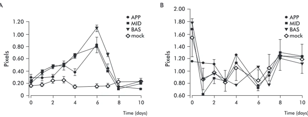
Time course image analysis of EGFP expression
The time course of EGFP production was determined by image analysis using a fluorescence microscope and appropriate software for 10 dpi (Figure 4). Each value corresponded to the mean of the given area for samples obtained from 5 different agroinfiltrated leaves. The conventional image could be quantified as the sum of pixels from each RGB channel. The high-est EGFP expression (green channel) was obtained at
6 dpi at injection site (basal zone) and spread to ad-jacent areas of injection (middle and apical) (Figure 4A). EGFP content was higher in vascular tissue than in the lamina as previously reported [12]. After 6 dpi, the expression was reduced, which would be related to RNA silencing mechanisms [13, 14] (Figure 4A). Likewise, the fluorescence level of infiltrated leaves decayed to that of control leaves on day eight (Figure 4A). Chlorophyll fluorescence (red channel) demon-strated to be a good control as it was not sensitive to environmental light variations or EGFP expression, and provided single-channel images [15]. As shown in Figure 4B, the red channel had no significant dif-ferences among agroinfiltrated leaves as compared to controls.
Balb/c mice immunization with recombinant
foliar extracts
Mice were maintained in good health conditions during all the experimental treatments. Clinical ob-servations revealed no significant differences in the average of body weight of mice throughout the experiments. Similar weight curves with a steady weight gain were obtained in animals injected with recombinant proteins and in control animals injected with control foliar extracts without any expressions of recombinant proteins (data not shown).
11. Sparrow P, Broer I, Hood EE, Eversole K, Hartung F, Schiemann J. Risk assess -ment and regulation of molecular farming
- a comparison between Europe and US.
Curr Pharm Des. 2013;19(31):5513-30.
12. Imlau A, Truernit E, Sauer N. Cell-to-cell and long-distance trafficking of the green fluorescent protein in the
phloem and symplastic unloading of
the protein into sink tissues. Plant Cell.
1999;11(3):309-22.
13. Mishra NS, Mukherjee SK. A Peep into the plant miRNA World. Open Plant Sci J.
2007;1:1-9.
14. Chau BL, Lee KA. Function and anato -my of plant siRNA pools derived from hair-pin transgenes. Plant Methods. 2007;3:13.
15. Walter A, Liebisch F, Hund A. Plant phenotyping: from bean weighing to im -age analysis. Plant Methods. 2015;11:14.
bE2 tE2 C MW kDa bE2 tE2 C MW kDa 5 20 30 30 80 30 80 MW
70 55 40
80 60 50 40
70 55 40
kDa
bE2 Sample 1 Sample 2tE2
A B C
Figure 2. Expression of tE2 in N. tabacum leaves 4 days after agroinfiltration. A) Western blot analysis. 5 μg of total proteins (tE2) was loaded in a 10 % SDS-PAGE gel, further transferred to a PVDF membrane and immu
-noblotted with an anti-tE2 monoclonal antibody. The arrowhead indicates the expected position for the dimer form. B) A Coomassie blue-stained SDS-PAGE gel was used as a loading control. C) Semi-quantification of tE2 by Western blot. A dilution series of baculovirus-derived tE2 (5, 20 and 30 ng) was used as a standard curve for tE2 quantification from two samples (30 and 80 μg of TSP). bE2: 10 μg baculovirus-derived tE2; C:control plants; MW: PageRuler Prestained Protein Ladder for western blot and Novex Sharp Unstained Protein Standard for Coomassie Blue (ThermoFisher).
Figure 3. RT-PCR of tE2 gene visualized on 1 % agarose gel. Total RNA was isolated from agroinfiltrated Nicotiana tabacum leaves using RNeasy Plant Mini Kit (Qiagen, Germany).
The first DNA strand was synthesized from 2 μg RNA using random primer and Super
-script II (Invitrogen) retrotran-scriptase. RNAs from the tE2 gene were amplified using spe
-cific primers. dpi, days post-infiltration; A: apical; B: basal; M: middle; P and N: positive (pk7-tE2-EGFP) and negative (total RNA) controls, respectively; MW: 100 bp DNA ladder (Invitrogen, USA). Data was analyzed for up to 10 dpi, with no variation from 7 dpi on.
1000 1500 bp
500
MW A
2 dpi 3 dpi 4 dpi 7 dpi
A A A
Macroscopic analysis of injection sites: in the case of the control or extracts expressing tE2 with oil ad-juvant, rounded, cystic, 0.2 -0.8 cm width structures containing a milky fluid were evident (Figure 5A). After histological examination of injection sites, dis-crete inflammatory foci were found which coexisted with an infiltrate mainly composed of plasma cells in all the animals treated. Absence of bacterial colo-nies and the kind of inflammatory cells, especially surrounding injection sites, suggested a reactive cystic structure rather than a collection of infectious abscess. Macrophages with foamy cytoplasm, poly-morphonuclear neutrophil leukocytes, edema, and diffuse deposits of fibrin and necrotic, and apoptotic cells were also observed as a component of inflam-matory lesions (Figure 5B). These infiltrates were self-limited, with only minimal compromise of deep soft tissues.
Animals injected with tE2, containing Al(OH)3
Hi-dragel®Adjuvant, showed cutaneous injuries. In all samples, the histological examination revealed a well delimited site with an inflammatory pustular injury composed of leukocytes, necrotic areas with fibrin deposition, and mild calcifications (Figure 5C).
Hematological and biochemical parameters were tested, to investigate the potential damage caused in mice by the recombinant proteins delivered in the foliar extracts. Blood hematology comprised hae-matocrit levels, WBC and platelet counts to evaluate anaemia, and possible pathological and inflamma-tory changes in connection with normal value ranges. Clinical and hematological blood values obtained from mice, either from controls or from those treated with plant foliar extracts, did not show significant dif-ferences compared to normal reference clinical and haematological values, regardless of the adjuvant used [16]. Total platelet counts showed to be lower than the reference normal range only in mice injected with Al(OH)3 as adjuvant (Table).
The injuries observed at injection sites have been described [17-21], thus elucidating that the toxicity of the adjuvant administered is the cause of these in-flammatory reactions. Moreover, DeGregorio et al.
[22] demonstrated in a knockout mice model that inflammation components were present at injection sites and required adjuvanticity. Furthermore, mice immunized with these extracts showed no apparent adverse effects, beyond the injection site, in contrast to the well-known and severe inflammatory lesions observed following immunization with Freund’s ad-juvant in rats [23].
Pixels
1.20 1.00
0.40 0.20 0.60 0.80
0
0 A
Time (days)
2 4 6 8 10
Pixels
1.80 2.00
1.60
1.00 0.80 1.20 1.40
0.60
0 B
Time (days)
2 4 6 8 10
APP APP
MID MID
BAS BAS
mock mock
Figure 4. Time course of pixels in samples of Nicotiana tabacum leaves (apical, middle and basal zones; APP, MID and BAS, respectively) agroinfiltrated with Agrobacterium tumefaciens harboring the pK-2S2-tE2-His-KDEL vector carrying the EGFP under an independent promoter and the control leaf (mock) sample. Images were obtained by using a microscope, an image processing software and appropriate filters (see Materials and methods). A) Pixels taken in the green channel. B) Pixels taken in the red channel.
A B
C
Figure 5. Macroscopic and histology sections of injected areas in female Balb/c mice im
-munized intramuscularly with foliar extracts of agroinfiltrated Nicotiana tabacum plants expressing the glycoprotein tE2. A) Macroscopic observation representative of mice
in-oculated with oily adjuvants; a diffuse, homogeneous infiltrate of plasma cells is shown (arrow). B) Histology section of a mouse inoculated with oily adjuvant. Inflammatory lesions at subcutaneous levels was found at low magnification. C) Histology section of a mouse inoculated with an aqueous adjuvant. Foamy macrophages surrounded by neutrophil polymorphonuclear leucocytes were detected (arrow). Magnification: 400×.
16. Wirth-Dzięciołowska E, Karasze
-wska J, Sadowski T, Pyęniak K, Gaje-wska
M. Selected blood serum biochemical
indicators in twelve inbred strains of
In the last years, transient gene expression technol-ogy in plants with binary or viral vectors have gained interest due to rapid and large amounts of protein production obtained even achieving industrial scale levels. Nevertheless, a commercial process requires high productivity and maximum product yield with minimum cost [24] and must follow regulations like GMP, GLP and risk assessment. Agroinfiltration, is an economic biotechnology method for transiently express a gene during a short period of time without introducing a permanent genetic modification in the plant genome [25]. Also, a variety of strategies have been develop during the last decade with the aim to increase the protein yield and to obtain an efficient transient platform production. There are several re-ports regarding the use of antigens produced in plants as plant extracts to immunize animals that are able to induced protection against diverse diseases [26-28]. Besides, the potential toxicity of these plant extracts
in the immunized animals was not evaluated for biosafety purposes. Previous experiments were con-ducted following basic concepts in biological safety and national codes of practice for the safe handling of pathogenic microorganisms in laboratories.
It is important to highlight that the testing methods used in this study could not avoid the use of animals. Hence, following the 3R principles, we designed the minimum experiments to test our hypothesis. By using the minimum animals as possible, it is considered that the results obtained herein provide enough evidence to affirm that recombinant foliar extracts containing the BVDV tE2 glycoprotein are safe to be applied in ani-mals after 4 dpi, thus providing a new tool to deliver antigens for vaccination without protein purification.
A
cknowledgements
The authors would like to thank Dr. Moises Burachik for his useful contribution.
Adjuvant Treatment Hct
49.6 5400 1 327 600 0 39 657 0 147
1 072 200 0 34 798 0 137
223 125 210 55 759 1371 147
192 333 234 43 1004 1319 142
470 000/
1 600 000 95/132 38/52 420/589 1300/1560 82/175
49.8 6260
39.9 5563
46.6 5188
42/68 3200/
14 000
WBC (× mm3) PLT AST (UI/L)
Table. Hematological chemistry values in mice injected with extracts of Nicotiana tabacum leaves expressing the tE2 recombinant
antigen of the bovine viral diarrhea virus or control extracts*
* Reference values for the species as reported by Suckow MA, Danneman P & Brayton C (The laboratory mouse. Boca Raton: CRC Press; 2001). Hct: haematocrit; WBC: total white blood cell; PLT: platelets; AST: aspartate aminotransferase; ALT: alanine amino
-transferase; LDH: lactate dehydrogenase; Glu: glucose.
ALT (UI/L) Amylase (UI/L) LDH (UI/L) Glu (mg/dL)
Montanide
ISA 70 SEPPIC®
Al(OH)3
Hydragel®
Reference value ranges
tE2
tE2
Wt
Wt
Received in January, 2016. Accepted in June, 2016.
17. Goto N, Kato H, Maeyama J, Shibano M, Saito T, Yamaguchi J, et al. Local tissue irritating effects and adjuvant activities of calcium phosphate and aluminium hydroxide
with different physical properties. Vaccine.
1997;15(12-13):1364-71.
18. Aucouturier J, Dupuis L, Ganne V. Ad -juvants designed for veterinary and human vaccines. Vaccine. 2001;19(17-19):2666-72.
19. Fox CB, Haensler J. An update on safety
and immunogenicity of vaccines containing emulsion-based adjuvants. Expert Rev Vac-cines. 2013;12(7):747-58.
20. Littleton LC, Gruenke JA. A comparison
of the effect of three adjuvants on the
anti-body response to ovalbumin in mice. BIOS.
2013;84(3):142-7.
21. Dey AK, Malyala P, Singh M. Physicochemi-cal and functional characterization of vaccine
antigens and adjuvants. Expert Rev Vaccines. 2014;13(5):671-85.
22. De Gregorio E, Tritto E, Rappuoli R. Alum
adjuvanticity: unraveling a century old mystery.
Eur J Immunol. 2008;38(8):2068-71.
23. Kleinau S, Erlandsson H, Holmdahl R, Klareskog L. Adjuvant oils induce arthritis in the DA rat. I. Characterization of the disease and evidence for an immunological involvement. J
Autoimmun. 1991;4(6):871-80.
24. Scragg AH. Bioreactor for the mass cultiva
-tion of cell plants. In: Plant Biotechnology. Eds. Fowler, MW and Waren, GS. London: Pergamon
Press; 1992. p. 45-60.
25. Marconi PL, Klykov S. Chapter 9: Math -ematical modeling in recombinant plant
sys-tems: The challenge to produce heterologous proteins under GLP/GMP. In: MA Alvarez, Ed. Plant Biotechnology for Health: from secondary
metabolites to molecular farming. New York:
Springer; 2014. p. 145-55.
26. Berinstein A, Vazquez-Rovere C, Asur
-mendi S, Gomez E, Zanetti F, Zabal O, et al. Mucosal and systemic immunization elicited
by Newcastle disease virus (NDV) transgenic
plants as antigens. Vaccine. 2005;23(48-49):5583-9.
27. Clemente M, Curilovic R, Sassone A, Zelada A, Angel SO, Mentaberry AN.
Pro-duction of the main surface antigen of Toxo -plasma gondii in tobacco leaves and analysis of its antigenicity and immunogenicity. Mol Biotechnol. 2005;30(1):41-50.
28. Yácono MDL, Farran I, Becher ML, Sander
V, Sanchez VR, Martin V, et al. A chloroplast-derived Toxoplasma gondii GRA4 antigen used as an oral vaccine protects against
toxoplasmosis in mice. Plant Biotechnol J.