Hepatitis C virus infection in
patients with primary biliary cirrhosis
Hsuan-Wei Chen,* Hsin-Hung Huang,* Ching-Huang Lai,** Wei-En Chang,***Yu-Lueng Shih,* Wei-Kuo Chang,* Tsai-Yuan Hsieh,* Heng-Cheng Chu*
* Division of Gastroenterology & Hepatology, Tri-Service General Hospital, National Defense Medical Center, Taiwan.
** School of Public Health, National Defense Medical Center, Taiwan. ***Department of Internal Medicine, Hualien Armed Forces General Hospital, Taiwan.
ABSTRACT
Background and aim. The aim of this study is to evaluate the role of hepatitis C virus (HCV) infection in pa-tients with primary biliary cirrhosis (PBC). Material and methods. On the basis of a retrospective review of medical records, all patients consecutively diagnosed with PBC or HCV infection between 1999 and 2011 and who had a regular follow-up of at least 3 years were included in the study. Clinical characteristics, es-pecially the severity of cirrhosis, were analyzed in PBC patients with HCV infection (PBC-HCV), PBC pa-tients without HCV infection (PBC-only), and papa-tients with only HCV infection (HCV-only). Results. A total of 76 patients with PBC, including 9 patients with HCV infection, were analyzed. Of the PBC-HCV patients, 7 (7/9, 77.8%) were women with a mean age of 55.11 ± 14.29 years. Age- and sex-matched PBC-only patients (n = 36) and HCV-only patients (n = 36) were used as control groups. In comparison to the PBC-only con-trols, PBC-HCV patients had a greater severity of cirrhosis based on Child-Pugh (p = 0.019) and Model for End-Stage Liver Disease (MELD) (p = 0.01) scores. However, no significant difference in the severity of cirrhosis was found between the PBC-HCV and HCV-only control patients (p = 0.94 in Child-Pugh scores; p = 0.64 in MELD scores). Conclusions. In PBC patients with concomitant HCV infection, aggressive mana-gement may be warranted in view of the associated more severe liver cirrhosis.
Key words. Child-Pugh score. MELD score. Cirrhosis.
Correspondence and reprint request: Heng-Cheng Chu, M.D., Ph.D. Division of Gastroenterology & Hepatology, Department of Internal Medicine, Tri-Service General Hospital, National Defense Medical Center
No. 325, Section 2, Cheng-Kung Road, Neihu 114, Taipei, Taiwan, R.O.C. Ph.: +886 2 87923311-12794. Fax: +886 2 87924892
E mail: [email protected]
Manuscript received: April 10, 2012. Manuscript accepted: June 16, 2012.
INTRODUCTION
Primary biliary cirrhosis (PBC) is a chronic, pro-gressive cholestatic liver disease without definitively known etiologies and often presents with inflamma-tory destruction of bile ducts leading to cirrhosis.1
PBC is also a rare disease with an overall age- and sex-adjusted annual incidence rate of 30.3 cases per million.2 Serologically, PBC is characterized by the
presence of antimitochondrial antibodies, which are present in 90–95% of patients. Originally, PBC was described as an autoimmune disorder with the presentation of jaundice and pruritus.3
Ursodeoxy-cholic acid is currently the only drug approved worldwide for the treatment of PBC, which delays the histological progression of the disease.4
Hepatitis C virus (HCV) infection is a leading cause of chronic hepatitis, cirrhosis, and liver cancer worldwide.5 The mechanism of
HCV-trig-gered liver injury is not well understood; howe-ver, recent studies suggest that cellular immunity not only plays a protective role but also causes li-ver injury. The pathogenesis of cirrhosis is related to chronic inflammation and immune reaction. The current medication is a combination of pe-gylated interferon (IFN) and the antiviral drug, ribavirin. In addition to liver injury, HCV infec-tion also causes disturbance of the immune system and is associated with various autoimmune disor-ders (type 1 or 2 autoimmune hepatitis, Sjögren’s syndrome, lichen planus, mixed cryoglobulinemia, and autoimmune thyroiditis).6-8 Therefore, hepatitis
Floreani, et al.9 analyzed patients with both PBC
and HCV infection (PBC-HCV) and found that HCV infection was a risk factor for the development of hepatocellular carcinoma (HCC) in this overlap group. However, the role of HCV infection in cirrhosis severity has not yet been elucidated; hence, we analyzed the relationship between HCV infection and PBC to demonstrate the effect of HCV infection on the severity of cirrhosis in patients with PBC.
MATERIAL AND METHODS
Subjects
From January 1999 to December 2011, we analy-zed a total of 76 hospitalianaly-zed patients with PBC. All PBC patients met at least 2 of the following criteria: antimitochondrial antibody titer of at least 1:80, ab-normal liver function tests (alkaline phosphatase) for > 6 months, and diagnostic or compatible liver biopsy.1 Nine of the 76 (11.84%) patients were found
to have HCV infection (7 women and 2 men) on the basis of positive anti-HCV/HCV RNA, and only 1 pa-tient (1.31%) was found to have hepatitis B virus (HBV) infection on the basis of positive HBV surfa-ce antigen. Patients with PBC were classified into 2 groups according to the presence of HCV infection: HCV-infected group (9 patients, PBC-HCV) and PBC-only control group (36 patients with age-and sex-matched frequencies from the remaining 67 patients without HCV infection). Patients with only HCV infection (36 patients with age- and sex-matched frequencies from HCV-only patients not receiving anti-HCV therapy in our medical center between January 1999 and December 2011) were identified as the HCV-only control group.
Study design
This study was a 12-year retrospective observa-tion. We collected data according to each patient’s demographic characteristics, including diagnostic age, gender, laboratory profiles, Child-Pugh (C-P) classification of cirrhosis, Model for End-Stage Liver Disease (MELD) scores, presence of major events (bleeding inclusive of esophageal varices or gastric varices, ascites, HCC, extrahepatic cancer, and death), autoimmune diseases (scleroderma, Sjögren’s syndrome, systemic lupus erythematosus (SLE), thyroid disorder, and type 2 diabetes mellitus), and survival observation time. The survival observation time was set to be 3 years (156 weeks). The severity of cirrhosis was based on the C-P classification and
MELD scores. MELD score I is defined as having a score of < 9, MELD score II is defined as a score of 10-19, MELD score III is defined as a score of 20-29, MELD score IV is defined as a score of 30–39, and MELD score V is defined as a score of ≥ 40; the classification (I–V) of MELD scores is based on the predictive outcome of 3-month mortality (I: 1.9% mortality, II: 6.0% mortality, III: 19.6% mortality, IV: 52.6% mortality, V: 71.3% mortality).10 In the C-P
classification, classification A is defined as having a score of 5-6, classification B is a score of 7-9, and classification C is a score of 10-15.
Data analyses
Continuous variables are expressed as means ± SD. The results for categorical variables are expres-sed as percentages. Statistical comparisons between the groups were computed using the Student’s t test or the χ2 test, according to the type of data. Odds
ratios (ORs) were calculated to examine the effect of HCV infection on the severity of cirrhosis in pa-tients with PBC, according to the level of C-P classi-fication of cirrhosis. Survival rates were computed by Kaplan-Meier methods, followed by a log-rank test. Cox proportional hazard model was used to calculate the effect of factors on survival. All statistical analyses were performed using SPSS software version 18.0 (SPSS Inc., Chicago, IL, USA) and STATA software version 8.0. All reported p values are 2-tailed, and p < 0.05 was assumed statistically significant for all tests.
RESULTS
After inclusion of 36 age- and sex-matched PBC-only patients for PBC-HCV controls, the other 31 PBC-only patients were excluded in this study. In addition to an older diagnostic age of the excluded PBC-only patients vs. PBC-only controls (p = 0.03), there are no other significant differences of clinical characteristics between these two groups.
PBC has a female predominance, and most pa-tients in the study were > 50 years old at initial diagnosis (mean, 55.11 years in the PBC-HCV group
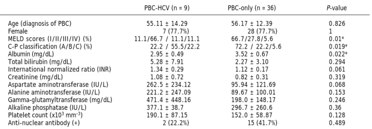
OR 8.13, 95% CI 1.10-60.03, p = 0.014; C-P classifi-cation C vs. C-P classification A: OR 13.0, 95% CI 0.84–200.07, p = 0.017; test for trend, p < 0.01). In the PBC-HCV group, most patients (66.7%) had a MELD score II; PBC-only patients mostly had MELD score I (66.7%). A significant difference was observed in the MELD scores classification (p = 0.01). The PBC-HCV group had poorer laboratory variables (albumin, total bilirubin, international normalized ratio, creatinine, aspartate aminotrans-ferase, alanine aminotransaminotrans-ferase, and gamma-gluta-myltransferase) (Table 1). The clinical and virological details of the PBC-HCV patients are sum-marized in table 2.
Table 3 summarizes the associated major events and autoimmune disorders in the presentation. Between the 2 groups, HCC was noted with a higher
incidence rate in the PBC-HCV group (11.11% vs.
2.8%). Ascites also seemed to be more common in PBC-HCV patients. There was no significant diffe-rence in the association of autoimmune disorders in the 2 groups. The most representative disorder was Sjögren’s syndrome. In the analysis of survival ob-servation time, the PBC-only group showed a slower decline in the curve (Figure 1). Cox regression analysis indicated that high C-P classification sco-res had an unfavorable effect on survival (hazards ratio 2.57; 95% CI 1.15–5.73; p = 0.021).
Clinical characteristics of the HCV-infected pa-tients are summarized in the table 4. Two papa-tients receiving treatment with IFN and ribavirin were noted to have lower viral loads. In the analysis of the survival observation time, the HCV-treated group had a slower decline in the curve (Figure 2).
Table 1. Clinical characteristics of PBC patients.
PBC-HCV (n = 9) PBC-only (n = 36) P-value
Age (diagnosis of PBC) 55.11 ± 14.29 56.17 ± 12.39 0.826
Female 7 (77.7%) 28 (77.7%) 1
MELD scores (I/II/III/IV) (%) 11.1/66.7 / 11.1/11.1 66.7/27.8/5.6 0.01* C-P classification (A/B/C) (%) 22.2 / 55.5/22.2 72.2 / 22.2/5.6 0.019*
Albumin (mg/dL) 2.95 ± 0.49 3.52 ± 0.67 0.022*
Total bilirubin (mg/dL) 5.28 ± 7.91 2.27 ± 3.10 0.294
International normalized ratio (INR) 1.34 ± 0.29 1.12 ± 0.17 0.061
Creatinine (mg/dL) 1.08 ± 0.72 0.82 ± 0.31 0.319
Aspartate aminotransferase (IU/L) 262.5 ± 234.12 95.94 ± 121.69 0.068 Alanine aminotransferase (IU/L) 221.2 ± 247.09 89.67 ± 100.01 0.153 Gamma-glutamyltransferase (mg/dL) 471.4 ± 448.16 198.0 ± 148.17 0.246
Alkaline phosphatase (IU/L) 377.1 ± 38.7 296.7 ± 260.6 0.36
Platelet count (x103 mm-3) 190.1 ± 87.15 152.0 ± 58.87 0.128
Anti-nuclear antibody (+) 2 (22.2%) 15 (41.7%) 0.489
HCV: hepatitis C virus. PBC: primary biliary cirrhosis. MELD scores: Model for End-Stage Liver Disease scores. C-P classification: Child-Pugh (C-P) classifica-tion. *P < 0.05.
Table 2. Clinical characteristics of HCV-infected PBC patients.
No. Gender Inf age of Dx age of Dx age of Dx age of HCV-RNA Source of Outcome HCV (y) HCV (y) AMA PBC (y) IgM titer g /dL (cps/mL) infection
1 F N/A 63 66 1: 160 2.8 2.11 X10⁶ Glass syringe Alive (>156 wks)
2 F N/A 61 63 1: 320 4.7 2.61 X10⁶ Glass syringe Alive (>156 wks)
3 F 28 50 53 1: 80 5.6 3.1 X10⁶ Glass syringe Alive (>156 wks)
4 M 26 42 45 1: 320 2.8 9.33 X10⁶ Glass syringe Alive (>156 wks)
5 F N/A 63 63 1: 640 2.7 1.6 X10⁶ Glass syringe Alive (>156 wks)*
6 F 54 71 71 1: 160 7.6 2.21 X10⁶ Transfusion Sepsis (71 wks)
7 M 8 23 23 1: 320 2.9 2.3 X10⁶ Glass syringe Alive (>156 wks)*
8 F N/A 56 56 1: 640 4.5 2.42 X10⁶ Glass syringe Sepsis (56 wks)
9 F N/A 56 56 1: 160 2.9 4.12 X10⁶ Glass syringe Breast CA (38 wks)
Table 3. Major events and autoimmune diseases of PBC patients (n = 45).
HCV-infected (n=9) PBC only (n=36) P-value
• Major events
Bleeding 1 (11.11%) 7 (19.4%) 0.922
Ascites 3 (33.33%) 6 (16.7%) 0.514
HCC 1 (11.11%) 1 (2.8%) 0.856
Extra-hepatic cancer 1 (11.11%) 0 0.448
Death 3 (33.33%) 7 (19.4%) 0.654
• Autoimmune disorder
Scleroderma 0 0
Sjögren’s syndrome 2 (22.22%) 6 (16.7%) 1
SLE 1 (11.11%) 0 0.448
Raynaud’s disease 0 2 (5.6%) 1
Thyroid disorder 1 (11.11%) 2 (5.6%) 1
Type 2 diabetes mellitus 1 (11.11%) 1 (2.8%) 0.856
HCV: hepatitis C virus. PBC: primary biliary cirrhosis. HCC: hepatocellular carcinoma. SLE: systemic lupus erythematosus.
Figure 1. Kaplan-Meier survival curves in HCV-infected PBC
vs. PBC-only patients. P = 0.322. HCV-treatment Figure 2. Kaplan-Meier survival curves in PBC patients withvs. HCV non-treatment. P = 0.313.
Table 4. Patients of PBC-HCV receiving anti-HCV treatment versus non-treatment.
Tx (n = 2) Non-Tx (n = 7)
HCV-RNA (106 cps/mL) 1.95 ± 0.49 3.7 ± 2.57
Female (n, %) 1, 50% 6, 85.7%
ALP/ALT (initial) 367 ± 9.9/100.5 ± 37.4 380 ± 44/258 ± 271
ALP and ALT (3 years latter) 328 ± 31.1/108.5 ± 78.4 485 ± 77.7/463.5 ± 432.5
HCV: hepatitis C virus. PBC: primary biliary cirrhosis. Dx: diagnosis. Tx: treatment. RNA: ribonucleic acid. ALP: alkaline phosphatase (IU/L). ALT: alanine aminotransferase (IU/L).
1.0
0.8
0.6
0.4
0.2
0.0
Cum survival
0 50 100 150 200
OS weeks
1.0
0.8
0.6
0.4
0.2
0.0
Cum survival
0 50 100 150 200
Time 0.00
1.00
0.00-censored 1.00-censored 0
1
0-censored 1-censored
HCV Treatment
HCV-Tx
P = 0.313
HCV-Non Tx PBC-HCV
PBC-only
Table 5 compares the PBC-HCV patients with the HCV-only controls. The severity of cirrhosis was determined by the C-P classification and MELD scores; there was no significant difference between these 2 groups. However, patients with PBC-HCV had higher total bilirubin levels (p = 0.03).
DISCUSSION
Previous studies on the occurrence of HCV infec-tion in PBC patients reported incidences of 1 in 90 patients and 3 in 55 patients.11,12 A PubMed search
reveals that to date, a total of 23 patients with PBC have been found to have HCV infection.9,13-16 In our
research, we have identified an additional 9 patients with a diagnosis of PBC-HCV, and we further dis-cuss here the effect of HCV infection in PBC pa-tients. Among the 76 patients with PBC, approximately 11.84% (9/76) had HCV infection compared with 1.31% (1/76) patients with concomi-tant HBV infection; however, the prevalence rates of HBV and HCV infections in Taiwan are 15-20% and 1-3%, respectively.17,18 Therefore, there seems to be
a close relationship between HCV infection and PBC. Because HCV can lead to immune disorder6-8
and PBC is an autoimmune hepatic disease,3
immune disturbance may underlie this manifestation. Additionally, a comparison between the HCV infec-tion route (through intravenous injecinfec-tion, sexual behavior, or blood transfusion) and the HBV in-fection route (vertical inin-fection) implicated other infectious agents as the trigger of PBC. Recent stu-dies also support the hypothesis that the develop-ment and progression of PBC hinge on a complex interplay between genetic and environmental risk factors.19-22
Comparisons between groups of HCV, PBC-only and HCV-PBC-only patients, with respect to the analysis of cirrhosis severity, indicate that HCV
infection aggravates the cirrhosis severity of pa-tients with PBC. The mechanism may be due to the synergic effect from PBC and HCV: PBC is a choles-tatic liver disease with the presentation of bile duct destruction and HCV can lead to hepatocyte and pa-renchyma injury. Ramos-Casals, et al. hypothesized that the coexistence of viral and autoimmune fac-tors may accelerate the development of cirrhosis or neoplasia in such patients.16 Although no
signifi-cant difference in the severity of cirrhosis was noted between HCV and HCV-only patients, PBC-HCV patients had higher bilirubin levels (Table 5). We believe that the synergic effect between HCV and PBC may be understood better by studying more ca-ses of PBC-HCV and their respective pathogenesis reported in the future.
In recent studies,23,24 the risk factor for HCC
de-velopment in patients with PBC was thought to be advanced histological stage, and the incidence rate of HCC in PBC cirrhosis was lower than HCV cirrhosis. Therefore, patients with PBC may have better prognosis than patients with HCV. The results of our study show that in patients with PBC, HCV infection seems to be a risk factor for the develop-ment of HCC; this finding is similar to that of a previous study.9 This mechanism may be explained
by the role of HCV infection as an exacerbation factor of cirrhosis.
PBC exhibits a number of autoimmune features; in our study, 1 patient was found to have SLE in the HCV-infected group and PBC developed after the SLE diagnosis. The co-occurrence of SLE and PBC is uncommon. Moreover, cases where PBC developed after SLE are rare; in most cases, PBC emerged before SLE.25,26 Of the
accompan-ying autoimmune disorders, Sjögren’s syndrome (8/ 45, 17.77%) is the most representative disorder, similar to the study of Floreani, et al.9, with a
pre-valence rate of approximately 25%.27 Table 5. PBC-HCV vs. HCV-only in liver profile.
HCV-PBC (n = 9) HCV-only (n = 36) P-value
Age55.11 ±14.29 55.22± 13.1
MELD scores (I/II/III/IV) (%) 11.1/66.7 /11.1/11.1 13.8/61.1 / 22.2/2.7 0.64 C-P classification (A/B/C) (%) 22.2/55.5 /22.2 27.7/52.7 / 19.4 0.94
Albumin (mg/dL) 2.95 ±0.49 2.77 ± 0.49 0.35
Total bilirubin (mg/dL) 5.28 ±7.91 2.02 ± 2.13 0.03*
International normalized ratio (INR) 1.34 ±0.29 1.29 ± 0.38 0.68
Creatinine (mg/dL) 1.08 ±0.72 1.66 ± 1.74 0.24
In the analysis of the survival observation time, we found no significant difference between the PBC-HCV and PBC-only patients (Figure 1). However, there seemed to be a trend that the PBC-only group had a slower decline in survival curve than the PBC-HCV group. Cox regression analysis showed that C-P classification was the influencing factor on survival; patients with high C-P scores had unfavo-rable prognosis on survival. Because both PBC and HCV have a chronic course and HCV can exacerbate the severity of cirrhosis, the difference may be signi-ficant with a longer duration of observation and more patients enrolled.
Currently, pegylated interferon (IFN) plus ribavi-rin become the mainstay of care for HCV infection.28
However, IFN therapy for chronic HCV infection may trigger or exacerbate underlying autoimmune diseases, including PBC,29-31 offering a treatment
challenge in PBC-HCV patients. With antiviral therapy, 2 patients of our PBC-HCV group showed a slower decline in survival curve (Figure 2) vs.
PBC-HCV patients without treament. Moreover, these patients showed a slower progression of liver injury, according to their serum levels of alkaline phosphatase and alanine transaminase at 3 years af-ter therapy (Table 4). Thus, antiviral therapy may be potentially considered in PBC patients with concomi-tant HCV infection, in view of the recently emerged watershed in HCV therapy without IFN.32,33
Our study has several limitations. First, this is a non-randomized and retrospective study; thus, unex-pected bias may exist. Second, because HCV and HBV have higher prevalence rates than PBC, the primary screening is often anti-HCV/HCV-RNA and HBs-Ag in Taiwan. Thus, most PBC patients are only considered after exclusion of endemic viral he-patitis B and C. This often leads to HCV-infected PBC patients identified after HCV diagnosis, others ignored and the others simultaneously noted with HCV infection by experienced doctors. Third, a com-patible liver biopsy can offer more information about the diagnosis and stage of disease. Although liver biopsy has been generally suggested to PBC-HCV pa-tients, most of them refuse this procedure. Thus, the histology severity of PBC cannot be controlled in the analysis of PBC-HCV patients with PBC-only controls. Fourth, the observation time should be longer, which might result in a more significant difference between the 2 groups. Finally and most importantly, only 9 patients are included in the present HCV-infected PBC group; a study with more cases needs to be conducted. In the future, prospective investigations should be conducted to evaluate the
effect of different genotypes of HCV infection on PBC patients and to properly manage this overlap-ping syndrome. Despite these limitations, our study still has clinical implication and is the first stu-dy for the role of HCV infection in Asian Chinese PBC patients.
In conclusion, in PBC patients, concomitant HCV infection is characterized by a biochemical profile with poor values of liver markers, especially albu-min, and is a risk factor for the development of more severe liver damage versus those without infection.
ACKNOWLEDGMENT
This study was supported in parts by National Science Council (NSC94-2314-B-016-047), Tri-Service General Hospital, National Defense Medical Center (TSGH-C97-44, TSGH-C98-48 and TSGH-C99-61) and the C.Y. Foundation for Advancement of Education, Sciences and Medicine, Taiwan.
REFERENCES
1. Kaplan MM, Gershwin ME. Primary biliary cirrhosis. N Engl J Med 2006; 353: 1261-73.
2. Myers RP, Shaheen AA, Fong A, Burak KW, Wan A, Swain MG, Hilsden RJ, et al. Epidemiology and natural history of primary biliary cirrhosis in a Canadian health region: a po-pulation-based study. Hepatology 2009; 50: 1884. 3. Kumagi T, Heathcote EJ. Primary biliary cirrhosis.
Orpha-net J Rare Dis 2008; 3: 1.
4. Poupon RE, Lindor KD, Parés A, Chazouilleres O, Poupon R, Heathcote EJ. Combined analysis of the effect of treat-ment with ursodeoxycholic acid on histologic progression in primary biliary cirrhosis. J Hepatol 2003; 39: 12-16. 5. Davis GL, Albright JE, Cook SF, Rosenberg DM. Projecting
future complications of chronic hepatitis C in the United-States. Liver Transpl 2003; 9: 331-8.
6. Haddad J, Deny P, Munz-Gotheil C, Ambrosini JC, Trinchet JC, Pateron D, Mal F, et al. Lymphocytic sialadenitis of Sjögren’s syndrome associated with chronic hepatitis C virus liver disease. Lancet 1992; 339: 321-3.
7. Marcellin P, Pouteau M, Benhamou JP. Hepatitis C virus in-fection, alpha interferon therapy and thyroid dysfunc-tion. J Hepatol 1995; 22: 364-9.
8. Lunel F, Cacoub P. Treatment of autoimmune and extrahe-patic manifestations of hepatitis C virus infection. J He-patol 1999; 31: 210-6.
9. Floreani A, Baragiotta A, Leone MG, Baldo V, Naccarato R. Primary biliary cirrhosis and hepatitis C virus infection. Am J Gastroenterol 2003; 98: 2757-62.
10. Wiesner R, Edwards E, Freeman R, Harper A, Kim R, Kama-th P, Kremers W, et al. Model for end-stage liver disease (MELD) and allocation of donor livers. Gastroenterology 2003; 124: 91-6.
11. Housset C, Hirschauer C, Degos F. False positive anti-HCV in biliary cirrhosis. Lancet 1991; 114: 252 (Letter). 12. Fusconi M, Lenzi M, Ballardini G, Miniero R, Cassani F,
13. Imada J, Hoshino H, Nishimura D, Morita K, Yoshida N, Ka-tada N, Sano H, et al. Case report: multiple cancers: hepa-tocellular carcinoma and adenocarcinomas of the common bile duct and the gall-bladder in a woman with primary bi-liary cirrhosis. J Gastroenterol Hepatol 1996; 11: 546-50. 14. Hoso M, Nakanuma Y, Kawano M, Oda K, Tsuneyama K, van
de water J, Gershiwin ME. Granulomatous cholangitis in chronic hepatitis C: a new diagnostic problem in liver pa-thology. Pathol Int 1996; 46: 301-5.
15. Mouelhi L, Chaieb M, Sfar I, Debbeche R, Trabelsi S, Gorgi Y, Najjar T. Chronic viral C hepatitis associated with pri-mary biliary cirrhosis. Report of two cases. Rev Med In-terne 2009; 30: 537-9.
16. Ramos-Casals M, Pares A, Jara LJ, Solans R, Viñas O, Váz-quez P, Sánchez-Tapias, et al; HISPAMEC Study Group. An-timitochondrial antibodies in patients with chronic hepatitis C virus infection: description of 18 cases and re-view of the literature. J Viral Hepat 2005; 12: 648-54. 17. Sung JL. Prevention of hepatitis B and C virus infection
for prevention of cirrhosis and hepatocellular carcinoma. J Gastroenterol Hepatol 1997; 12: S370-S376.
18. Wang JT, Wang TH, Sheu JC, Tsai SJ, Hsieh YS, Lin DT, Wang CY, et al. 1993. Hepatitis C virus infection in volun-teer blood donors in Taiwan. Evaluation by hepatitis C an-tibody assays and the polymerase chain reaction. Arch Pathol Lab Med 1993; 117: 152-6.
19. Nakamura M, Yasunami M, Kondo H, Horie H, Aiba Y, Komo-ri A, Migita K, et al; PBC Study Group in NHOSLJ. Analysis of HLA-DRB1 polymorphisms in Japanese patients with pri-mary biliary cirrhosis (PBC): The HLA-DRB1 polymorphism determines the relative risk of antinuclear antibodies for disease progression in PBC. Hepatol Res 2010; 40: 494-504.
20. Gershwin ME, Selmi C, Worman HJ, Gold EB, Watnik M, Utts J, Lindor KD, et al. Risk factors and comorbidities in pri-mary biliary cirrhosis: a controlled interview based study of 1032 patients. Hepatology 2005; 42: 1194-202. 21. Ala A, Stanca CM, Bu-Ghanim M, Ahmado I, Branch AD,
Schiano TD, Odin JA, et al. Increased prevalence of prima-ry biliaprima-ry cirrhosis near superfund toxic waste sites. He-patology 2006; 43: 525-31.
22. Ishibashi H, Komori A, Shimoda S, Ambrosini YM, Gershwin ME, Nakamura M. Risk factors and prediction of long-term
outcome in primary biliary cirrhosis. Intern Med 2011; 50: 1-10.
23. Cavazza A, Caballería L, Floreani A, Farinati F, Bruguera M, Caroli D, Parés A. Incidence, Risk Factors, and Survival of Hepatocellular Carcinoma in Primary Biliary Cirrhosis: Comparative Analysis from Two Centers. Hepatology 2009; 50: 1162-8.
24. Macaron C, Hanouneh IA, Zein NN. Incidence, Risk Fac-tors, and Survival of Hepatocellular Carcinoma in Primary Biliary Cirrhosis. Hepatology 2010; 52: 2239; author reply 2239-40.
25. Michel F, Toussirot E, Wendling D. Primary biliary cirrhosis and systemic lupus erythematosus. A new case report. Rev Rhum Engl Ed 1998; 65: 504-7.
26. Shizuma T, Kuroda H. A case of primary biliary cirrhosis which developed eight years after diagnosis of systemic lupus erythematosus. Intern Med 2011; 50: 321-4. 27. Watt EF, James OF, Jones DE. Patterns of
autoimmuni-ty in primary biliary cirrhosis patients and their fami-lies: a population-based cohort study. QJM 2004; 97: 397-406.
28. Fried MW, Shiffman ML, Reddy KR, Smith C, Marinos G, Gonçales FL Jr, Häussinger D, et al. Peginterferon alfa-2a plus ribavirin for chronic hepatitis C virus infection. N Engl J Med 2002; 26: 975-82.
29. Yoshikawa M, Mimura M, Shiroi A, Kojima H, Fukui H, Sugi-moto Y, Mochi T. Primary biliary cirrhosis exacerbated by a course of acute hepatitis C and subsequent interferon therapy. Am J Gastroenterol 2000; 95: 2396-7.
30. Maeda T, Onishi S, Miura T, Iwamura S, Tomita A, Saibara T, Yamamoto Y. Exacerbation of primary biliary cirrhosis during interferon-alpha 2b therapy for chronic active he-patitis C. Dig Dis Sci 1995; 40: 1226-30.
31. D’Amico E, Paroli M, Fratelli V, Palazzi C, Barnaba V, Callea F, Consoli G. Primary biliary cirrhosis induced by interfe-ron-alpha therapy for hepatitis C virus infection. Dig Dis Sci 1995; 40: 2113-6.
32. Lok AS, Gardiner DF, Lawitz E, Martorell C, Everson GT, Ghalib R, Reindollar R, et al. Preliminary study of two anti-viral agents for hepatitis C genotype 1. N Engl J Med 2012; 366: 216-24.