Hepatic Hydrothorax
Yong Lv,* Guohong Han,* Daiming Fan**
* Department of Liver Diseases and Digestive Interventional Radiology, National Clinical Research Center for Digestive Diseases and Xijing Hospital of Digestive Diseases, Fourth Military Medical University, Xi'an 710032, China. ** State Key Laboratory of Cancer Biology, National Clinical Research Center for Digestive Diseases and Xijing Hospital of Digestive Diseases, Fourth Military Medical University, Xi'an 710032, China.
January-February, Vol. 17 No. 1, 2018: 33-46
INTRODUCTION
Hepatic hydrothorax (HH) is defined as a pleural ef-fusion, typically more than 500 mL, in patients with liv-er cirrhosis without coexisting undliv-erlying cardiac or pulmonary disease.1,2 It is an infrequent complication of
portal hypertension with an estimated prevalence of 5-10% among cirrhotic patients.2-4 Although pleural
effu-sion in association with liver disease was first described in the nineteenth century by Laënnec, HH was first de-fined by Morrow, et al.5 in 1958 while describing a rapid
accumulation of massive right pleural effusion after the diagnosis of cirrhosis. Together with hepatopulmonary syndrome and pulmonary hypertension, HH have been recognized as a major pulmonary manifestation of chronic liver disease and cirrhosis in recent years.1,6 In
most cases, HH develops on the right side (85%), with
13% of cases occurring on the left side and 2% bilater-al.2,3,7 A recent study showed that the frequency of HH
was associated with hepatic function as assessed by Child Pugh scoring system, but not with serum albu-min.8 Although it is commonly seen in conjunction
with ascites, HH can present in the absence of ascites in a small proportion of patients.9 In contrast with ascites
where a significant volume (5 to 8 L) are generally well-tolerated without signicant symptom, a patient with HH will develop dyspnea, shortness of breath, and/or hy-poxia when only 1 to 2 L of fluid accumulates in the pleural space.7,10,11
In this clinical review, the pathophysiology, manifesta-tions, diagnosis, and therapeutic options available for the management of HH will be discussed in order to allow the clinician to better understand these potentially life-threatening complications.
The Official Journal of the Mexican Association of Hepatology, the Latin-American Association for Study of the Liver and
the Canadian Association for the Study of the Liver
Manuscript received: Manuscript received:Manuscript received:
Manuscript received:Manuscript received: November 14, 2017. Manuscript accepted:Manuscript accepted:Manuscript accepted: November 14, 2017.Manuscript accepted:Manuscript accepted: DOI:10.5604/01.3001.0010.7533
A B S T R A C T A B S T R A C T A B S T R A C T A B S T R A C T A B S T R A C T
Hepatic hydrothorax (HH) is a pleural effusion that develops in a patient with cirrhosis and portal hypertension in the absence of car-diopulmonary disease. Although the development of HH remains incompletely understood, the most acceptable explanation is that the pleural effusion is a result of a direct passage of ascitic fluid into the pleural cavity through a defect in the diaphragm due to the raised abdominal pressure and the negative pressure within the pleural space. Patients with HH can be asymptomatic or present with pulmonary symptoms such as shortness of breath, cough, hypoxemia, or respiratory failure associated with large pleural effusions. The diagnosis is established clinically by finding a serous transudate after exclusion of cardiopulmonary disease and is confirmed by radionuclide imaging demonstrating communication between the peritoneal and pleural spaces when necessary. Spontaneous bacteri-al empyema is serious complication of HH, which manifest by increased pleurbacteri-al fluid neutrophils or a positive bacteribacteri-al culture and will require antibiotic therapy. The mainstay of therapy of HH is sodium restriction and administration of diuretics. When medical ther-apy fails, the only definitive treatment is liver transplantation. Therapeutic thoracentesis, indwelling tunneled pleural catheters, tran-sjugular intrahepatic portosystemic shunt and thoracoscopic repair of diaphragmatic defects with pleural sclerosis can provide symptomatic relief, but the morbidity and mortality is high in these extremely ill patients.
Key words. Key words. Key words. Key words.
PATHOPHYSIOLOGY
Although the exact mechanisms involved in the devel-opment of hepatic hydrothorax have not been well-de-fined, several mechanisms have been postulated, including hypoalbuminemia and subsequently decreased colloid os-motic pressure, increased azygos system pressure leading to leakage of plasma into the pleural cavity and transdia-phragmatic migration of peritoneal fluid into the pleural space via lymphatic channels.12-14 However, the most
widely accepted theory is the direct passage of ascitic fluid from peritoneal to the pleural cavity via numerous dia-phragmatic defects.15-17
These defects, which are referred to as pleuroperito-neal communications, are usually < 1 cm and tend to oc-cur on the right side.15,17,18 This right side predominance
could be related to the embryological development of the diaphragm in which the left side of the diaphragm is more muscular and the right side is more tendinous due to the close anatomical relationship with bare areas of the liver.19
On the microscopic examination, these defects were re-vealed as discontinuities in the collagen bundles that make up the tendinous portion of the diaphragm.17
Macroscopi-cally, the diaphragmatic defects associated with the devel-opment of HH have been classified into four morphological types: Type 1, no obvious defect; type 2, blebs lying in the diaphragm; type 3, broken defects (fen-estrations) in the diaphragm; and type 4, multiple gaps in the diaphragm.18
Although diaphragmatic defects occur in the normal population and autopsy series report such defects in up to 20% of cases, they seem to rarely result in pneumothorax following laparoscopic procedures.18,20 In patients with
as-cites, the increasing abdominal pressure and the diaphrag-matic thinning secondary to malnutrition of cirrhotic patients enlarge these defects.1,13,21 Blebs of herniated
peri-toneum can protrude through these defects, and, if a bleb bursts, a communication between peritoneal and pleural space is formed.18,22,23 The movement of fluid from the
abdomen to the pleural space is unidirectional, which is probably due to a permanent gradient pressure as a result of a negative intrathoracic pressure during the respiratory cycle and a positive intra-abdominal pressure.24 If the
vol-ume of accumulation of ascites in the pleural cavity ex-ceeds the absorptive capacity of the pleural membranes, hepatic hydrothorax ensues.
This mechanism has been confirmed by imaging tech-nique demonstrating the communication between the peri-toneal cavity and the pleural space even in the absence of ascites.25-30 Several available methods to evaluate pleural
migration include intraperitoneal injection of blue dye or air,24 contrast-enhanced ultrasonography29-31 and
scinti-graphic studies using intraperitoneal instillation of
99mTc-human serum albumin or 99mTc-sulphor-colloid.16,26,32,33
On the other hand, other theories in that the underlying mechanisms leading to fluid retention in patients with HH are similar to those leading to other forms of fluid accumu-lation in patients with cirrhosis have failed to explain the right predominance of HH.10,12,13,34
CLINICAL MANIFESTATIONS
As HH most frequently occurs in the context of ascites and other features of portal hypertension due to decom-pensated liver disease, the prominent clinical manifesta-tions are nonspecific and related to cirrhosis and ascites in most cases.7,9,35,36 More rarely, HH may be the index
pres-entation for chronic liver disease.9 The respiratory
symp-toms in patients with HH varied, mainly depending on the volume of effusion, rapidity of the effusion accumula-tion in the pleural space and the presence of associated cardiopulmonary disease.1,7,12 Patients may be
asympto-matic in whom pleural effusion is an incidental finding on chest imaging performed for other reasons or they may have pulmonary symptoms of shortness of breath, cough, hypoxemia or respiratory failure associated with large pleural effusions.10,13,21,37 A recent case series including 77
patients with HH indicated that most patients typically had multiple complaints, with the most commonly re-ported symptoms being dyspnea at rest (34%), cough (22%), nausea (11%), and pleuritic chest pain.7 On rare
oc-casions, patients with HH may present with an acute ten-sion hydrothorax, manifesting as severe dyspnoea and hypotension.7
DIAGNOSIS
The diagnosis of HH is based on the presence of hepat-ic cirrhosis with portal hypertension; exclusion of a pri-mary cardiac, pulmonary, or pleural disease; and eventually, confirmation of the passage of ascites into pleu-ral space (Figure 1).2,33,38
Diagnostic thoracentesis
Pleural fluid analysis is mandatory to identify the nature of the fluid, to exclude the presence of infection including spontaneous bacterial empyema (SBEM), and to rule out alternative diagnosis (inflammation or malignancy).39 One
retrospective series40 found that 70% of pleural effusions
Figure 1. Figure 1. Figure 1.
Figure 1. Figure 1. Diagnostic algorithm for hepatic hydrothorax. BPN: Brain natriuretic peptide. CT: Computed tomography. HH: Hepatic hydrothorax. PMN: Polymorphonuclear.
Pleural effusion with liver cirrhosis
Excluded a primary cardiac pulmonary, or pleural disease
by echocardiography, ultrasound, CT, BNP, etc.
Thoracentesis and pleural fluid analysis
Transudative based on Light's criteria
Exudative based on Light's criteria
PMN > 500 cells/mm3 with negative pleural fluid culture
PMN > 250 cells/mm3 with positive pleural fluid culture
PMN < 250 cells/mm3
Consider diaphragmatic evaluation using imaging, scintigraphy or thoracoscopy when the diagnosis of HH is in doubt
Patients on diuretics Patients noton diuretics
Spontaneous bacterial empyema HH Serum/pleuralfluid albumin ratio < 0.6
Serum/pleural fluid albumin ratio ≥ 0.6
Look for causes for exudative pleural
effusion
addition, 80% of right-sided pleural effusions were found to be uncomplicated HH, while only 35% of left-sided pleural effusions were uncomplicated HH.
Pleural fluid analysis should routinely include serum and fluid protein, albumin and lactate dehydrogenase (LDH) levels, cell count, Gram stain and culture in blood culture bottles.2,37,41 Other tests that may be useful
de-pending on clinical suspicion, include triglycerides, pH, adenosine deaminase and polymerase chain reaction (PCR) for mycobacteria, amylase, and cytology to exclude chylothorax, empyema, tuberculosis, pancreatitis, and ma-lignancy, respectively.4,10,36,37,42 The composition of HH is
transudative in nature and therefore similar to the ascetic fluid.42-44 However, total protein and albumin may be
slightly higher in HH compared with levels in the ascitic fluid because of the greater efficacy of water absorption by the pleural surface.12,34,39,40,44,45
In uncomplicated HH, total protein is < 2.5 g/dL in HH with low LDH and glucose levels similar to that in serum.12,42 In addition, it will also have a serum to pleural
fluid albumin gradient > 1.1 as found in ascites secondary
to portal hypertension, although this has not been studied extensively (Table 1).12,42 Although diuresis has widely
been reported to increase the pleural total protein levels, one study found only a single patient had a protein dis-cordant exudate despite 34 patients receiving diuretics.39
However, when HH is an exudate probably because of di-uretics, the serum/pleural fluid albumin ratio should be calculated, and a value <0.6 is classified as transudate.43,44
Exclusion of
Other Causes of Pleural Effusion
To exclude a primary cardiac, pulmonary, or pleural disease, a chest radiograph should be performed in addi-tion to pertinent laboratory tests, such as a brain natriuret-ic peptide (BNP) in the proper clinnatriuret-ical setting.39,46,47 In
the chest will help to exclude mediastinal, pulmonary, or pleural lesions or malignancies.7,42,48 Echocardiography
should be performed to evaluate cardiac function and to rule any cardiac causes of pleural effusions.49,50 In a study
of 41 HH patients, diastolic dysfunction was found in 11 of 21 patients (52%). Contrast echocardiography with agitat-ed saline demonstratagitat-ed an intrapulmonary shunt in 18 of 23 cases (78%).39 However, the study did not mention how
these patients were distinguished from left heart failure. The high prevalence of diastolic dysfunction suggest that heart failure might have contributed to the development of pleural effusions.6,39,49,50 The increased neurohormonal
ac-tivity associated with cirrhosis leading to cardiac hypertro-phy along with impaired relaxation has been speculated as the reason for diastolic dysfunction in cirrhotic pa-tients.10,39 Traditionally, the simplest strategy to reveal the
true transudative nature of heart failure-related effusions, labeled as exudates by Light’s criteria, is to calculate the serum to pleural fluid albumin gradient.44 A recent study
demonstrated that a gradient between the albumin levels in the serum and the pleural fluid > 1.2 g/dL performs sig-nificantly better than a protein gradient > 3.1 g/dL to cor-rectly categorize mislabeled cardiac effusions. On the other hand, the accuracy of a pleural fluid to serum albu-min ratio < 0.6 excelled when compared with albualbu-min and protein gradients in patients with miscategorized HH.43,44
Diaphragmatic evaluation
In cases where the diagnosis of HH is in doubt, in par-ticular when pleural effusion is left sided and/or ascites are absent, diagnosis of HH can be confirmed when a communication is identified between the peritoneal and thoracic cavities.10,13,34,38 Scintigraphic studies using
intra-peritoneal instillation of 99mTc-human serum albumin or 99mTc-sulphor-colloid, is used most frequently be-cause it is simple and safe.24 These radiolabeled particles,
measuring between 3 and 100 μm, are not absorbed by the peritoneum so their intrapleural passage occurs only through an anatomical defect in the diaphragm.24,51 It has
been demonstrated that radiotracers are effective in dem-onstrating peritoneopleural communication even in the absence of ascites.26,27,52 This technique has sensitivity
and specificity rates of 71% and 100%, respectively.52 In
cases with minimal ascites, it has been recommended that intraperitoneal instillation of 300-500 mL of normal saline to favor the pleural passage of radioactivity is help-ful to improve the effectiveness of peritoneal scintigra-phy in the diagnosis of HH.24 The scintigraphic studies
can also provide an estimation of the size of the dia-phragmatic defect(s) by the rapidity with which the radi-oisotope passes from the peritoneum to the pleural space.33 In addition, intraperitoneal injection of
methyl-ene blue can be used intraoperatively to demonstrate and localize defects, and contrast-enhanced ultrasonography has been used to detect flow across the diaphragm in real time.29-31 Although other diagnostic modalities,
includ-ing magnetic resonance imaginclud-ing and CT could also be used to detect the underlying diaphragmatic defects,15,48
direct demonstration of defects with those techniques might be extremely difficult, as the defect itself is usually quite small.39 Video-assisted thoracoscopy, which can
provide a directly visualization of the underlying dia-phragmatic defects,18,20 is an alternative diagnostic option.
However, this modality is invasive and should be consid-ered only when diagnosis is not clear or in case there is a plan to repair the diaphragmatic defects.34
Spontaneous bacterial empyema
Spontaneous bacterial empyema (SBEM) is an infec-tion of a preexisting hydrothorax in the absence of pneu-monia.53,54 It is reported to occur in 2.0% to 2.4% of
patients with cirrhosis and 13% to 16% of patients with HH.55-57 The actual incidence of SBEM may be higher
Table 1. Diagnostic criteria for hepatic hydrothorax.
Diagnostic criteria for uncomplicated hepatic hydrothorax.
Cell count < 500 cells/mm3, PMN count < 250 cells/mm2 and negative culture.
Total protein concentration < 2.5 g/dL or total protein pleural fluid to serum ratio < 0.5. Lactate dehydrogenase pleural fluid to serum ratio < 0.6.
Serum to pleural fluid albumin gradient > 1.1 g/dL.
Pleural fluid amylase concentration < serum amylase concentration. PH 7.40 to 7.55.
Pleural glucose level similar to serum level.
Diagnostic criteria for spontaneous bacterial empyema. Positive pleural culture and PMN count > 250 cell/mm3 or
Negative pleural culture and PMN count > 500 cell/mm3.
No evidence of pneumonia or parapneumonic effusion on chest imaging.
than reported because of underdiagnosis.58-60 The
initia-tion of empirical antibiotics in patients with cirrhosis and fever or hepatic encephalopathy because of a higher suspi-cion for spontaneous bacterial peritonitis masks the diag-nosis of SBEM.58-60 SBEM should be distinguished from
empyema secondary to pneumonia, because there is usual-ly no evidence of pus or abscess in the thoracic cavity in SBEM and it differ in the pathogenesis, clinical course, and treatment strategy with those of empyema secondary to pneumonia.61-63 Therefore, some authors have
pro-posed that it be called spontaneous bacterial pleuri-tis.1,10,12,34
Even-though the exact mechanism of SBEP remains unclear, it is believed that the pathogenesis is similar to spontaneous bacterial peritonitis (SBP) in that transient bacteremia leads to infection of the pleural fluid due to impaired reticuloendothelial phagocytic activity. 3,53,54,56,61-64 In up to 40% of cases, SBEM can occur in the absence of
SBP and even in the absence of ascites, indicating that SBP is not prerequisite for SBEM.3,53,55-57 The risk factors
identified for the development of SBEM in patients with cirrhosis include: low pleural fluid C3 levels, low serum albumin pleural fluid total protein, high Child-Pugh score and concomitant SBP.3,55,56,65 Similar to SBP, the more
fre-quent bacteria involved in SBEP are E. coli, Klebsiella, Strep-tococcus and Enterococcus species.3,55-57, 66,67
The presenting symptoms of SBEM may be those of accompanying SBP (i.e., abdominal pain), may be limited to the thoracic cavity (i.e., dyspnea, thoracic pain), or may be systemic in nature (i.e., fever, shock, new or worsening encephalopathy).54,68 Given the high mortality rate, a high
index of suspicion is essential for the diagnosis of SBEM in cirrhotic patients who develop fever, pleuritic pain, en-cephalopathy, or unexplained deterioration in renal func-tion.3,54,66,68
The diagnostic criteria for spontaneous bacterial em-pyema are similar to those for SBP, requiring a polymor-phonuclear (PMN) cell count > 250 cells/mm3 with a
positive culture or PMN cell count > 500 cells/mm3 in
cases with negative cultures; no evidence of pneumonia and/or contiguous infections process; and a serum/pleural fluid albumin gradient > 1.1 (Table 1).56,65 As with ascites
fluid cultures, pleural fluid should be inoculated immedi-ately into blood culture bottles at the bedside to increase the microbiologic yields.55,56 Bedside inoculation resulted
in positive cultures in 75% of the episodes. The positivity was only 33% when conventional microbiological tech-niques were used.55,56 Detection of pleural neutrophilia
provides an early diagnosis of SBEM when culture results are delayed.54,66
Initial adequate antimicrobial therapy is the corner-stone of the treatment. Given the association with SBP, the initial antibiotic regimen is similar and the
recom-mended treatment is an intravenous third-generation ce-phalosporin given for 7 to 10 days.12,54,58,61 Considering its
proven benefit in SBP, some centers administer albumin similarly in patients with SBEM, although the use of albu-min has not been specifically studied in SBEM.3,68
Place-ment of a chest tube is generally not recommended in SBEM even in culture-positive cases, because it can lead to life-threatening fluid depletion, protein loss, and elec-trolyte imbalance.21,69-71 The only indication for
chest-tube drainage for SBEM is pus in the pleural space. Due to the impaired liver function, frequently with associated re-nal insufficiency in most cirrhotic patients with SBEM, the management of SBEM is a clinical challenge.54 Despite
aggressive therapy, the mortality is high (up to 20%) in these fragile patients.54,56 The independent factors related
to poor outcome are high models for end-stage liver dis-ease (MELD)-Na score, initial ICU admission and initial antibiotic treatment failure.68
MANAGEMENT
Medical management
Because the overwhelming mechanism is transdia-phragmatic ascites flow, the principal treatment should fo-cus on eliminating and preventing the recurrence of ascites. A sodium-restricted diet and judicious use of diu-retics through inducing and maintaining a net negative so-dium balance may provide initial ascites reduction and prevent HH development.72,73 A low sodium diet, with
70-90 mmol per day, and weight loss of 0.5 kg per day in pa-tients without edema, and 1.0 kg per day in those with edema is the goal of therapy.64 Dietary education should be
given to patients at the same time. However, diet therapy is usually not sufficient to achieve such a goal. Therefore, diuretics are required in the vast majority of cases. A distal acting agent (typically spironolactone 100 mg/day) and a loop diuretic (e.g. furosemide 40 mg/day) should be co-administered as the best initial regimen to produce a renal excretion of sodium at least 120 mEq per day.11,74 The
dos-es may be increased in a stepwise fashion every 3-5 days by doubling the doses with furosemide up to 160 mg/day and spironolactone up to 400 mg/day.37,38 Urinary sodium
should be checked before and during therapy to adjust diu-retic dosage as per clinical response.2,13
Refractory hepatic hydrothorax
pre-cipitation of encephalopathy may preclude successful symptomatic control of the pleural effusion. These pa-tients are considered to have refractory hepatic hydrotho-rax.2,12,34 Approximately 21% to 26% of medically treated
patients may fall into this category.73,75 The only definitive
treatment for refractory HH is liver transplantation.14,72 In
patients awaiting liver transplantation and those who are not transplant candidates, the aims of therapy for refractory HH are relief of symptoms and prevention of pulmonary complications (Figure 2).1,10,34 The therapeutic options
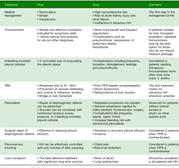
available are therapeutic thoracentesis, chest tube place-ment and indwelling pleural catheter, transjugular intrahe-patic portosystemic shunt (TIPS), and surgical interventions (Table 2).72
Therapeutic thoracentesis
Thoracentesis is a simple and effective procedure indi-cated for relief symptoms of dyspnea in patients with large effusions and those with recurrent or refractory
hydrotho-rax, although the benefits of the procedure are often short lived, and the procedure usually need to be repeated.10,76 In
general, it is recommended that no more than 2 L be re-moved because there is a risk of hypotension or re-expan-sion of pulmonary edema.40,77 Chest X-rays and CT scan of
the chest before therapeutic thoracentesis help to define the size of the effusion. After the procedure, the chest radio-graph is also advisable, not only for detection of pneumoth-orax but also to evaluate pulmonary or pleural pathology that was masked by the effusion.40,77 Coagulopathy of
cir-rhosis is not deemed as contraindication to therapeutic tho-racenthesis unless there is disseminated intravascular coagulation.12,13,21,34,78 Thoracentesis for HH in clinical
practice is usually safe. The risk of pneumothorax after seri-al thoracocentesis increases from 7.7% to 34.7%. Other pos-sible complications include pain at puncture site, pneumothorax, empyema or soft tissues infection, haemop-tysis, air embolism, vasovagal episodes, subcutaneous em-pysema, bleeding (haematoma, haemothorax, or haemoperitoneum), laceration of the liver or spleen.40,77
Figure 2. Figure 2. Figure 2. Figure 2.
Figure 2. Management algorithm for hepatic hydrothorax. ITPC: Indwelling tunneled pleural catheter. TIPS: Transjugular intrahepatic portosystemic shunt.
Hepatic hydrothorax
Evaluation for liver
transplantation Sodium restriction diuretics
Candidate Not a candidate Repeat thoracentesis
Expected waiting list < 3 months
Expected waiting list > 3 months
Recurrent effusion > 2 weeks
Recurrent effusion < 2 weeks
Repeat
thoracentesis TIPS
Resolved Recurrent
effusion Resolved
Recurrent effusion
Chest tube placement and indwelling tunneled pleural catheter
A chest tube should not be placed in this patient popula-tion as it may result in protein loss, secondary infecpopula-tion, pneumothorax, hemothorax and hepatorenal syndrome and electrolyte disturbances.70,71,79,80 It can also be difficult to
re-move the chest tube because there is often a rapid reaccu-mulation of fluid once the chest tube is clamped.70,79
Indwelling tunneled pleural catheter (ITPC, also known as PleurX or Denver catheter) was initially
intend-ed for palliative outpatient therapy of recurrent malignant pleural and ascitic effusions and now it has become a com-mon therapeutic tool in the management of symptomatic malignant effusions.81-83 There are an increasing number of
reports of its usage for benign pleural conditions, includ-ing HH.82,84-88 A recent meta-analysis by Patil, et al.89
re-garding the use of IPTC for nonmalignant pleural effusions demonstrated a spontaneous pleurodesis rate of 51%. In a recent prospective study,81 25 ITPCs were
placed in 24 patients. The mean number of pleural drain-age procedures before ITPC placement was 1.9, with no Table 2. Pros and cons of the different treatment modalities for hepatic hydrothorax.
Treatment Pros Cons Comment
Medical • Noninvasive. • High noncompliance rate The first step in the
management • Simple. • Risk of acute kidney injury and management of HH.
• Inexpensive. renal failure.
• Ineffective in refractory HH.
Thoracentesis • Simple and effective procedure • Short lived benefit and frequent In patients suitable
indicated for symptoms relief. requirement. for liver transplant
• Allows pleural fluid analysis • Complications such as evaluation, repeated to rule out other diagnoses. pneumothorax, reexpansion of thoracentesis
pulmonary edema, may be the best
hemothorax. option for those
who do not require frequent drainage.
Indwelling tunneled • A controlled way of evacuating • Complications including empyema, Considered in pleural catheter the pleural space loculation, dislodgement, leakage, patients needing
and pneumothorax. therapeutic
thoracenteses more often than once every 2 weeks.
TIPS • Response rate of 70 - 80% • Post-TIPS hepatic encephalopathy. The first treatment • Prevention of variceal rebleeding • Shunt dysfunction. choice for and control of refractory ascites. • Deteriorations of liver function. refractory HH
• Bridge to liver transplant. whenever possible.
Pleurodesis • Repair of diaphragmatic defects • Repeated procedures are needed. Reserved for patients can be performed • General anesthesia needed for without clinical • Success can be increased with video-assisted thoracoscopic surgery. ascites in continuous positive airway • Complications like empyema, whom no other pressure, or indwelling tunneled sepsis, septic shock. options exist. pleural catheter. • Increase bleeding risk with
mechanical pleurodesis.
Surgical repair of • Effective in reducing pleural • Residual or recurrent pleural effusion. Considered in patients
diaphragmatic defects effusion. • Invasive. when TIPS is
contraindicated.
Pleurovenous • HH can be effectively controlled • Chest pain. Considered in patients shunting with only minutes of daily pumping. • Risk of air embolism. when TIPS is
contraindicated.
Liver transplant • The best definitive treatment • Short of donor. Should be considered with significant long-term survival. • Long waiting time. in all patients with HH.
further pleural drainages required in any patient after ITPC placement. Spontaneous pleurodesis occurred in 33% patients and pleural fluid infection occurred in 16.7% patients. Even though these results look promising, data are limited and further studies are required to compare the effectiveness with other treatment modalities.72,90,91
Transjugular intrahepatic portosystemic shunt
Transjugular intrahepatic portosystemic shunt (TIPS) is a nonsurgical approach that decompresses the portal system, thereby addressing the mechanism of fluid collec-tion in the abdomen and/or chest.75 In a carefully selected
population, TIPS can lead to significant improvements in the complications related to portal hypertension.75,92-96 It
is now the standard of care in patients with refractory HH. Moreover, TIPS is superior to other treatment modalities in the prevention of rebleeding from varices and its con-trol of refractory ascites which has been well studied in controlled trials.75
The efficacy and safety of TIPS for HH has been in-vestigated in several non-controlled studies and case re-ports.75, 92-98 A recent meta-analysis including six studies
of 198 patients showed that the complete response rate to TIPS was 55.8% (95%CI: 44.7%-66.9%), partial response rate was 17.6% (95%CI: 10.9%-24.2%).99 A recent case
se-ries reported that TIPS was effective in 73.3% of 19 cas-es.100 It should be noted that the stents used in most of
these studies were bare metal stents and, as expected, the rate of shunt dysfunction leading to recurrent hydrotho-rax was high.99 With the use of PTFE-covered stents in
this setting, the shunt patency has been improved great-ly101-103 which will extend the benefit of TIPS for HH.
The incidence of TIPS-related encephalopathy was 11.7% (95%CI: 6.3%-17.2%), most of which is controlla-ble with medical therapy.104,105 Only 5% of cases require
occlusion of TIPS or a reduction in the TIPS caliber to control encephalopathy.
However, TIPS does not improve the overall progno-sis of patients with end-stage liver disease. The average 30-day mortality rate was 18% and the 1-year survival was 52%.92-94,106 Risk factors for mortality after TIPS
place-ment for HH include a Child-Pugh score ≥ 10, MELD score > 15, and an elevated creatinine.92-94 In addition, a
lack of response in the hydrothorax after TIPS placement is associated with an increased mortality rate.92-94 Because
TIPS shunts blood away from the liver and reduces the ef-fective portal perfusion to the liver, it can precipitate liver failure in patients with already significant hepatic dysfunc-tion. Ideally, patients with a high likelihood of decom-pensation after TIPS should also initiate evaluation for liver transplantation, with TIPS serving only as a bridge.93,94,99
Surgical interventions
Three surgical approaches have been used in the man-agement of HH, including chemical pleurodesis (via tube thoracostomy or VATS), repair of diaphragmatic defects or fenestrations with/without pleurodesis, and peritoneov-enous shunts or pleurovperitoneov-enous shunting.10
Pleurodesis
Pleurodesis is a technique that consists of the abla-tion of the space between the parietal and visceral pleu-ra with a sclerosing agent or irritant (such as talc or a tetracycline) that is administered through a tube thora-costomy (chest tube) or by thoracoscopy (VATS).107-111
It is usually reserved for patients without clinical as-cites in whom no other options exist.107,111-113 The
rea-son for this recommendation is that successful pleurodesis requires visceral and parietal pleural appo-sition, which can be difficult to achieve in refractory HH patients due to rapid fluid accumulation.14
Paracen-tesis performed before pleurodesis may also increase the success rate by decreasing ascites and flux of fluid from the peritoneal to the pleural cavity, allowing more time for the pleural spaces to be opposed to each oth-er.114 The ITPC may also be combined with pleurodesis
to avoid and decrease hospitalization.115,116 In addition,
continuous positive airway pressure (CPAP), by in-creasing positive intrathoracic pressure and reversing the peritoneal-pleural pressure gradient, in combina-tion with pleurodesis was reported to improve the suc-cess rate in one study.117
A meta-analysis by Hou, et al.118 comprising 180
refrac-tory HH patients who were subjected to pleurodesis in 13 studies demonstrated an initial mean success rate of 72% with a further symptomatic recurrence in 25% of the cases. The rate off complete response to pleurodesis by chest tube was 78% (95% CI 68-87%), while using a video thora-coscopic (VATS) approach combined with talc poudrage pleurodesis, the rate of complete response rate was up to 84% (95% CI 64-97%).118 According to the drugs used for
pleurodesis, complete response to pleurodesis with talc alone was 71% (95% CI 63-79%), and the complete re-sponse rate with OK-432 alone or in combination with minocycline was 93% (95% CI 78-100%).118
Various complications frequently observed after pleu-rodesis include fever and mild thoracic pain, though em-pyema, pneumothorax, pneumonia, septic shock and hepatic encephalopathy with liver failure have also been reported.107,111-113,118 Persistent high volume ascitic
pleurode-sis carries a high risk of bleeding especially in patients with advanced liver disease and coagulopathy.109
Surgical repair of diaphragm defects
Due to the proposed diaphragmatic defects HH mecha-nism, surgical approaches focused on defects repair with fibrin glue or sutures have been reported.119 Although
clo-sure of transdiaphragmatic defects can be done by open thoracotomy and by VATS with concomitant talc pleurod-esis, open thoracotomy in a cirrhotic patient has a signifi-cant mortality.119
In a small series of eight patients who underwent VATS for refractory HH, demonstrable diaphragmatic defects were founded in six (75%) patients. The pleural effusion did not recur in the six patients with defects that were closed, but the other two patients had recurrent effusion and died 1 and 2 months following the procedure, respec-tively.120 In another series reported by Milanez de
Cam-pos, et al.121 in which 21 thoracoscopies were performed in
18 patients with HH, the overall success rate was 48%. Of those five patients in whom a suture could be performed, three had a good response (60%), one died of postopera-tive pneumonia and liver failure, and the other had empye-ma and drained fluid for 1 month. Ten of the 21 (47.6%) procedures had a good response. However, the high mor-bidity (57.1%) and mortality (38.9%) in this study during a follow-up period of 3 months raised questions about the utility of such an approach.
Although VATS for suturing diaphragmatic defects is effective in reducing pleural effusion in patients with HH, residual or recurrent pleural effusion has been ob-served clinically.122,123 To resolve this problem, the use of
thoracoscopic mesh onlay reinforcement to prevent as-cites leaking from sutured holes in patients with refrac-tory HH has been introduced.124 In a recent surgical
series, Huang, et al.125 reported that HH was controlled in
all 63 patients with refractory HH who underwent thora-coscopic mesh onlay reinforcement to repair diaphrag-matic defects (mesh covering alone was used in 47 patients and mesh with suturing was used in 16 patients). Four patients experienced recurrence after a median 20.5 months of follow-up examinations. The 1-month mortal-ity rate was 9.5% (6 of 63 patients). Underlying impaired renal function and MELD scores were associated with increased 3-month mortality in 16 patients. The main causes of 3-month mortality were septic shock, acute re-nal insufficiency, gastrointestire-nal bleeding, hepatic en-cephalopathy, and ischemic bowel. Although these minimally-invasive approaches appear encouraging, fur-ther evaluation in additional experienced centers is war-ranted to corroborate these results and approaches in these high-risk surgical patients.
Peritoneovenous shunts or pleurovenous shunting
The peritoneovenous shunt is an implantable device that carries the ascites or hydrothorax into the systemic circulation through a surgically placed subcutaneous plas-tic cannula with a one-way pressure valve.126 It has been
reported as an appropriate alternative treatment for manag-ing refractory ascites.126 However, peritoneovenous shunt
for the management of HH has been used in a limited number of patients, with conflicting results.127,128 It has
been noted that the lower pressure in the pleural cavity than in the central vein in case of without ascites usually makes the peritoneovenous shunt ineffective for treat-ment of HH.129 In addition, concerns about serious
com-plications associated with this procedure including infection, diffuse intravascular coagulation, hepatic en-cephalopathy and lack of efficacy due to frequent occlu-sion have led other investigators to conclude that this method has limited effectiveness.128 Therefore,
peritoneo-venous shunt for the treatment of HH was abandoned nearly a decade ago.
The alternative Denver pleurovenous shunt includes a unidirectional pump, which was placed subcutaneously and additionally allowed external manual compression to move fluid.129,130 Use of the Denver shunt is therefore
in-dicated in cases in which the fluid has to be moved against higher pressure.127,131 It can be inserted percutaneously
under local anesthesia, and shunt patency can be main-tained, mechanical occlusion and infection can be man-aged by shunt revision, and HH can be effectively controlled with only minutes of daily pumping.130,132,133
However, complete aspiration of pleural fluid may result in pleuritic chest pain, and shunt insertion must be per-formed with great care to prevent air embolism.134 Several
cases have reported with successful long-term application of a Denver pleurovenous shunt in the treatment of HH, as an “alternative” therapy in selected patients.131-133
Liver transplantation
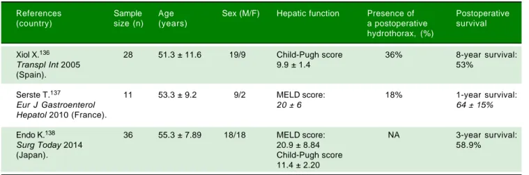
Liver transplantation is the treatment of choice for de-compensated cirrhosis, and thereby provides the best management for HH.1,135 The outcome of HH (refractory
or not) following liver transplantation is very favorable (Table 3).135 Xiol, et al.136 reported 28 HH patients versus a
de-scribed that the postoperative complications and survival were not different in HH (n = 11) compared to those with tense ascites and those with no HH (both groups had n = 11) matched for age, sex, year of transplant, and sever-ity of cirrhosis. No significant differences in the duration of mechanical ventilation, intensive care unit stay, and in-hospital stay, incidence of sepsis and early postoperative death were observed among three groups. One-year sur-vival was also similar (64 ± 15% vs. 91 ± 9% vs. 63 ± 15%). Endo, et al.138 compared the outcomes of patients
with (n = 36) and without (n = 201) uncontrollable HH and massive ascites requiring preoperative drainage who underwent liver transplantation. They found that the inci-dence of postoperative bacteremia was higher (55.6 vs.
46.7%, P = 0.008) and the 1- and 3-year survival rates were lower (1 year: 58.9 vs. 82.9%; 3 years: 58.9 vs. 77.7%; P = 0.003) in patients with uncontrollable HH and massive as-cites than those without. They suggested that postopera-tive infection control may be an important means of improving the outcome for patients with uncontrollable HH and massive ascites undergoing liver transplantation. These findings suggest that liver transplantation provides the best definitive treatment with significant long-term survival and should be considered in all patients.139
SUMMARY
HH is an infrequent complication of portal hyperten-sion in patients with end-stage liver disease. Although the physiopathology of HH is not fully elucidated, transdia-phragmatic passage of ascetic fluid from the peritoneal to the pleural cavity through numerous diaphragmatic de-fects has been shown to be the predominant mechanism in the formation of HH. Although the diagnosis of HH can typically be based on clinical grounds in a patient with
es-tablished cirrhosis and ascites who presents with a right-sided pleural effusion, a diagnostic thoracentesis is manda-tory in all patients with pleural effusions to exclude the presence of infection or an alternate diagnosis. In cases where the diagnosis is uncertain, in particular when as-cites is not detected or the hydrothorax is present on the left side, scintigraphic studies serum albumin can be help-ful. Spontaneous bacterial empyema, the infection of a hy-drothorax, can complicate HH and increase morbidity and mortality. Treatment of HH is primarily medical, with salt restriction and diuretics. However, medical management of this condition often fails and liver transplantation re-mains the ultimate definitive management paradigm. For patients who are not candidates and those who are waiting for a transplant, therapeutic thoracentesis, ITPC, TIPS, pleurodesis, and video-assisted thoracic surgery are useful tools to alleviate symptoms and prevent pulmonary com-plications in selected patients.
FINANCIAL DISCLOSURE
None.
COMPETING INTERESTS
The authors declare that they have no competing inter-ests.
ABBREVIATIONS
• HH: hepatic hydrothorax.
• ITPC: indwelling tunneled pleural catheter. • MELD: models for end:stage liver disease. • SBEM: spontaneous bacterial empyema.
• TIPS: transjugular intrahepatic portosystemic shunt. • VATS: videoassisted thoracoscopic surgery.
Table 3. Results of liver transplantation in refractory hepatic hydrothorax.
References Sample Age Sex (M/F) Hepatic function Presence of Postoperative
(country) size (n) (years) a postoperative survival
hydrothorax, (%)
Xiol X.136 28 51.3 ± 11.6 19/9 Child-Pugh score 36% 8-year survival:
Transpl Int 2005 9.9 ± 1.4 53%
(Spain).
Serste T.137 11 53.3 ± 9.2 9/2 MELD score: 18% 1-year survival:
Eur J Gastroenterol 20 ± 6 64 ± 15%
Hepatol 2010 (France).
Endo K.138 36 55.3 ± 7.89 18/18 MELD score: NA 3-year survival:
Surg Today 2014 20.9 ± 8.84 58.9%
(Japan). Child-Pugh score
11.4 ± 2.20
REFERENCES
1. Machicao VI, Balakrishnan M, Fallon MB. Pulmonary complica-tions in chronic liver disease. Hepatology 2014; 59: 1627-37. 2. Cardenas A, Kelleher T, Chopra S. Review article: hepatic
hydrothorax. Aliment Pharmacol Ther 2004; 20: 271-9. 3. Chen TA, Lo GH, Lai KH. Risk factors for spontaneous
bac-terial empyema in cirrhotic patients with hydrothorax. J Chin Med Assoc2003; 66: 579-86.
4. Liu WL, Kuo PH, Ku SC, Huang PM, Yang PC. Impact of ther-apeutic interventions onsurvival of patients with hepatic hy-drothorax. J Formos Med Assoc2010; 109: 582-8.
5. Morrow CS, Kantor M, Armen RN. Hepatic hydrothorax. Ann Intern Med 1958; 49: 193-203.
6. Singh C, Sager JS. Pulmonary complications of cirrhosis.
Med Clin North Am2009; 93: 871-83.
7. Badillo R, Rockey DC. Hepatic hydrothorax: clinical features, management, and outcomes in 77 patients and review of the literature. Medicine (Baltimore) 2014; 93: 135-42.
8. Abbasi A, Bhutto AR, Alam MT, Aurangzaib M, Masroor M. Frequency of Hepatic Hydrothorax and its Association with Child Pugh Class in Liver Cirrhosis Patients. J Coll Physi-cians Surg Pak 2016; 26: 566-9.
9. Kim JS, Kim CW, Nam HS, Cho JH, Ryu JS, Lee HL. Hepatic hydrothorax without ascites as the first sign of liver cirrho-sis. Respirol Case Rep 2016; 4: 16-8.
10. Al-Zoubi RK, Abu GM, Gohar A, Salzman GA, Yousef O. He-patic hydrothorax: clinical review and update on consensus guidelines. Hosp Pract (1995) 2016; 44: 213-23.
11. Borchardt J, Smirnov A, Metchnik L, Malnick S. Treating he-patic hydrothorax. BMJ 2003; 326: 751-2.
12. Norvell JP, Spivey JR. Hepatic hydrothorax. Clin Liver Dis
2014; 18: 439-49.
13. Gur C, Ilan Y, Shibolet O. Hepatic hydrothorax—pathophysi-ology, diagnosis and treatment—review of the literature. Liv-er Int 2004; 24: 281-4.
14. Kiafar C, Gilani N. Hepatic hydrothorax: current concepts of pathophysiology and treatment options. Ann Hepatol 2008; 7: 313-20.
15. Zenda T, Miyamoto S, Murata S, Mabuchi H. Detection of dia-phragmatic defect as the cause of severe hepatic hydrotho-rax with magnetic resonance imaging. Am J Gastroenterol
1998; 93: 2288-9.
16. Rubinstein D, McInnes IE, Dudley FJ. Hepatic hydrothorax in the absence of clinical ascites: diagnosis and management.
Gastroenterology 1985; 88: 188-91.
17. Gilsanz V, Emons D, Hansmann M, Meradji M, Donaldson JS, Omenaca F, Quero J, et al. Hydrothorax, ascites, and right diaphragmatic hernia. Radiology 1986; 158: 243-6.
18. Huang PM, Chang YL, Yang CY, Lee YC. The morphology of diaphragmatic defects in hepatic hydrothorax: thoracoscop-ic finding. J Thorac Cardiovasc Surg 2005; 130: 141-5. 19. Park CH, Pham CD. Hepatic hydrothorax. Scintigraphic
confir-mation. Clin Nucl Med 1995; 20: 278.
20. Lin DJ, Zhang M, Gao GX, Li B, Wang MF, Zhu L, Xue LF. Thoracoscopy fordiagnosis and management of refractory hepatic hydrothorax. Chin Med J (Engl)2006; 119: 430-4. 21. Krok KL, Cardenas A. Hepatic hydrothorax. Semin Respir
Crit Care Med 2012; 33: 3-10.
22. Kumar S, Kumar R. Hepatic hydrothorax: The shower within.
J Bronchology Interv Pulmonol 2014; 21: 88-9.
23. Dohmen K, Tanaka H, Haruno M, Niho Y. Hepatic hydrotho-rax occurring rapidly after manual abdominal compression.
World J Gastroenterol 2007; 13: 6284-5.
24. Ajmi S, Sfar R, Nouira M, Souguir A, Jmaa A, Golli L, Ben FM, et al. Role of the peritoneopleural pressure gradient in the
genesis of hepatic hydrothorax. An isotopic study. Gastro-enterol Clin Biol 2008; 32: 729-33.
25. Bonacini M, Rammohan G. 99mTc-sulfur colloid studies in he-patic hydrothorax. Gastroenterology 1987; 92: 273. 26. Ajmi S, Hassine H, Guezguez M, Elajmi S, Mrad DK,
Karm-ani M, Zayane A, et al. Isotopic exploration of hepatic hy-drothorax: ten cases. Gastroenterol Clin Biol 2004; 28: 462-6.
27. Benet A, Vidal F, Toda R, Siurana R, De Virgala CM, Richart C. Diagnosis of hepatic hydrothorax in the absence of as-cites by intraperitoneal injection of 99m-Tc-Fluor colloid.
Postgrad Med J1992; 68: 153.
28. Raju EP, Rao PN, Venkateswarlu J, Subhashini S. Ultra-sound documentation of diaphragmatic rent in hepatic hydro-thorax. Indian J Gastroenterol 1989; 8: 111-2.
29. Foschi FG, Piscaglia F, Pompili M, Corbelli C, Marano G, Righi-ni R, Alvisi V, et al. Real-time contrast-enhanced ultra-sound—a new simple tool for detection of peritoneal-pleural communications in hepatic hydrothorax.Ultraschall Med
2008; 29: 538-42.
30. Tamano M, Hashimoto T, Kojima K, Maeda C, Hiraishi H. Diag-nosis of hepatic hydrothorax using contrast-enhanced ultra-sonography with intraperitoneal injection of Sonazoid.J Gastroenterol Hepatol 2010; 25: 383-6.
31. Matono T, Koda M, Murawaki Y. Right diaphragmatic defect in hepatic hydrothorax exposed by contrast-enhanced ultra-sonography after radiofrequency ablation. Hepatology
2012; 56: 784-5.
32. Rajnish A, Sudhakar P. Diagnosis of hepatic hydrothorax by Tc-99m sulfur colloid peritoneal scintigraphy. Clin Nucl Med
2001; 26: 888.
33. Hewett LJ, Bradshaw ML, Gordon LL, Rockey DC. Diagnosis of isolated hepatic hydrothorax using peritoneal scintigra-phy. Hepatology 2016; 64: 1364-6.
34. Baikati K, Le DL, Jabbour II, Singhal S, Anand S. Hepatic hy-drothorax. Am J Ther 2014; 21: 43-51.
35. Jones EG. Hepatic hydrothorax: a retrospective case study.
J Am Acad Nurse Pract 2001; 13: 209-14.
36. Abbasi A, Bhutto AR, Alam MT, Aurangzaib M, Masroor M. Frequency of Hepatic Hydrothorax and its Association with Child Pugh Class in Liver Cirrhosis Patients. J Coll Physi-cians Surg Pak 2016; 26: 566-9.
37. Roussos A, Philippou N, Mantzaris GJ, Gourgouliannis KI. Hepatic hydrothorax: pathophysiology diagnosis and man-agement. J Gastroenterol Hepatol 2007; 22: 1388-93. 38. Cardenas A, Arroyo V. Management of ascites and hepatic
hydrothorax. Best Pract Res Clin Gastroenterol 2007; 21: 55-75.
39. Gurung P, Goldblatt M, Huggins JT, Doelken P, Nietert PJ, Sahn SA. Pleural fluidanalysis and radiographic, sonograph-ic, and echocardiographic characteristics of hepatic hydrot-horax. Chest 2011; 140: 448-53.
40. Xiol X, Castellote J, Cortes-Beut R, Delgado M, Guardiola J, Sese E. Usefulness and complications of thoracentesis in cirrhotic patients. Am J Med 2001; 111: 67-9.
41. Xiol X, Guardiola J. Hepatic hydrothorax. Curr Opin Pulm Med 1998; 4: 239-42.
42. Alonso JC. Pleural effusion in liver disease. Semin Respir Crit Care Med 2010; 31: 698-705.
43. Bielsa S, Porcel JM, Castellote J,Mas E, Esquerda A, Light RW. Solving the Light’s criteria misclassification rate of car-diac and hepatic transudates. Respirology 2012; 17: 721-6. 44. Porcel JM. Identifying transudates misclassified by Light’s
criteria. Curr Opin Pulm Med 2013; 19: 362-7.
45. Light RW. The undiagnosed pleural effusion. Clin Chest Med
46. Bhuniya S. Was it really hepatic hydrothorax? Lancet 2010; 376: 1300, 1300-1301.
47. Ackerman Z, Reynolds TB. Evaluation of pleural fluid in pa-tients with cirrhosis. J Clin Gastroenterol 1997; 25: 619-22. 48. Christoffersen S. CT verified cause of death in hepatic
hy-drothorax without ascites. Forensic Sci Int 2010; 198: e11-e13.
49. Kaplan LM, Epstein SK, Schwartz SL, Cao QL, Pandian NG. Clinical, echocardiographic, and hemodynamic evidence of cardiac tamponade caused by large pleural effusions. Am J Respir Crit Care Med 1995; 151: 904-8.
50. Shah A, Variyam E. Pericardial effusion and left ventricular dysfunction associated with ascites secondary to hepatic cirrhosis. Arch Intern Med 1988; 148: 585-8.
51. Schuster DM, Mukundan SJ, Small W, Fajman WA. The use of the diagnostic radionuclide ascites scan to facilitate treat-ment decisions for hepatic hydrothorax. Clin Nucl Med
1998; 23: 16-8.
52. Kim YK, Kim Y, Shim SS. Thoracic complications of liver cir-rhosis: radiologic findings. Radiographics 2009; 29: 825-37. 53. Flaum MA. Spontaneous bacterial empyema in cirrhosis.
Gastroenterology1976; 70: 416-7.
54. Tu CY, Chen CH. Spontaneous bacterial empyema. Curr Opin Pulm Med2012;18: 355-8.
55. Xiol X, Castellote J, Baliellas C, Ariza J, Gimenez RA, Guardi-ola J, Casais L. Spontaneous bacterial empyema in cirrhotic patients: analysis of eleven cases. Hepatology 1990; 11: 365-70.
56. Xiol X, Castellvi JM, Guardiola J, Sese E, Castellote J, Perello A, Cervantes X, et al. Spontaneous bacterial empyema in cir-rhotic patients: a prospective study. Hepatology 1996; 23: 719-23.
57. Murray HW, Marks SJ. Spontaneous bacterial empyema, pericarditis, and peritonitis in cirrhosis. Gastroenterology
1977; 72: 772-3.
58. Ninan J, Fakhran S. Spontaneous Bacterial Empyema: Its As-sociation With Liver Disease. Mayo Clin Proc 2016; 91: 537-8.
59. B SK, Balajinathan R, Alagavenkatesan AV, Balamurugan, Gurunamasivayam. Spontaneous bacterial empyema in cir-rhosis, an underdiagnosed entity. J Assoc Physicians India
2016; 64: 104-5.
60. Allam NA. Spontaneous bacterial empyema in liver cirrhosis: an underdiagnosed pleural complication. Saudi J Gastroen-terol2008; 14: 43-5.
61. Lam ST, Johnson ML, Kwok RM, Bassett JT. Spontaneous bacterial empyema: not your average empyema. Am J Med2014; 127: e9-e10.
62. Nguyen TA, Liendo C, Owens MW. Counterpoint: does spon-taneous bacterial empyema occur? No. Chest 2015; 147: 1208-10.
63. Lai YK, Eiger G, Fischer RA. Point: doesspontaneous bacte-rial empyema occur? Yes. Chest2015; 147: 1207-8. 64. Kumar S, Sarin SK. Paradigms in the management of hepatic
hydrothorax: past, present, and future. Hepatol Int 2013; 7: 80-7.
65. Sese E, Xiol X, Castellote J, Rodriguez-Farinas E, Tremosa G. Low complement levels and opsonic activity in hepatic hy-drothorax: its relationship with spontaneous bacterial em-pyema. J Clin Gastroenterol 2003; 36: 75-7.
66. Castellote J, Lopez C, Gornals J, Domingo A, Xiol X. Use of reagent strips for the rapiddiagnosis of spontaneous bacte-rial empyema. J Clin Gastroenterol 2005; 39: 278-81. 67. Wang JT, Fang CT, Hsueh PR, Chang SC, Luh KT.
Spontane-ous bacterial empyema caused by Aeromonas veronii bio-type sobria. Diagn Microbiol Infect Dis 2000; 37: 271-3.
68. Chen CH, Shih CM, Chou JW, Liu YH, Hang LW, Hsia TC, Hsu WH, et al. Outcome predictors of cirrhotic patients with spontaneous bacterial empyema. Liver Int 2011; 31: 417-24. 69. Hepokoski M, Makani SS, Lerner AD, Crotty AL.
Spontane-ous Pneumothorax Complicating Chronic Hepatic Hydrotho-rax: Successful Treatment by Small Bore Chest Tube. J Bronchology Interv Pulmonol 2016; 23: e43-e45.
70. Liu LU, Haddadin HA, Bodian CA, Sigal SH, Korman JD, Bod-enheimer HJ, Schiano TD. Outcome analysis of cirrhotic pa-tients undergoing chest tube placement.Chest 2004; 126: 142-8.
71. Runyon BA, Greenblatt M, Ming RH. Hepatic hydrothorax is a relative contraindication to chest tube insertion. Am J Gas-troenterol 1986; 81: 566-7.
72. Porcel JM. Management of refractory hepatic hydrothorax.
Curr Opin Pulm Med 2014; 20: 352-7.
73. Singh A, Bajwa A, Shujaat A. Evidence-based review of the management of hepatic hydrothorax. Respiration 2013; 86: 155-73.
74. Kinasewitz GT, Keddissi JI. Hepatic hydrothorax. Curr Opin Pulm Med 2003; 9: 261-5.
75. Rossle M, Gerbes AL. TIPS for the treatment of refractory ascites, hepatorenal syndrome and hepatic hydrothorax: a critical update. Gut 2010; 59: 988-1000.
76. Argento AC, Murphy TE, Pisani MA, Araujo KL, Puchalski J. Patient-Centered Outcomes Following Thoracentesis. Pleura (Thousand Oaks) 2015; 2.
77. Castellote J, Xiol X, Cortes-Beut R, Tremosa G, Rodriguez E, Vazquez S. Complications of thoracentesis in cirrhotic pa-tients with pleural effusion. Rev Esp Enferm Dig 2001; 93: 566-75.
78. Siddappa PK, Kar P. Hepatic hydrothorax. Trop Gastroen-terol 2009; 30: 135-41.
79. Orman ES, Lok AS. Outcomes of patients with chest tube in-sertion for hepatic hydrothorax. Hepatol Int 2009; 3: 582-6. 80. Pfammatter R, Quattropani C, Reichen J, Goke B, Wagner
AC. Treatment of hepatic hydrothorax and reduction of chest tube output with octreotide. Eur J Gastroenterol Hepatol 2001; 13: 977-80.
81. Chen A, Massoni J, Jung D, Crippin J. Indwelling Tunneled Pleural Catheters for the Management of Hepatic Hydrotho-rax. A Pilot Study. Ann Am Thorac Soc 2016; 13: 862-6. 82. Shah R, Succony L, Gareeboo S. Use of tunneled pleural
catheters for the management of refractory hepatic hydrot-horax. BMJ Case Rep 2011; 2011.
83. Chee A, Tremblay A. The use of tunneled pleural catheters in the treatment of pleural effusions. Curr Opin Pulm Med
2011; 17: 237-41.
84. Bhatnagar R, Reid ED, Corcoran JP, Bagenal JD, Pope S, Clive AO, Zahan-Evans N, et al. Indwelling pleural catheters for non-malignant effusions: a multicentre review of prac-tice. Thorax 2014; 69: 959-61.
85. Mercky P, Sakr L, Heyries L, Lagrange X, Sahel J, Dutau H. Use of a tunnelled pleural catheter for the management of refractory hepatic hydrothorax: a new therapeutic option.
Respiration 2010; 80: 348-52.
86. Shah R, SucconyL, Gareeboo S. Use of tunneled pleural catheters for the management of refractory hepatic hydrot-horax. BMJ Case Rep 2011; 2011.
87. Chee A, Tremblay A. The use of tunneled pleural catheters in the treatment of pleural effusions. Curr Opin Pulm Med
2011; 17: 237-41.
88. Mercky P, Sakr L, Heyries L, Lagrange X, Sahel J, Dutau H. Use of a tunnelled pleural catheter for the management of refractory hepatic hydrothorax: a new therapeutic option.
89. Patil M, Dhillon SS, Attwood K, Saoud M, Alraiyes AH, Harris K. Management of Benign Pleural Effusions Using Indwelling Pleural Catheters: A Systematic Review and Meta-analysis.
Chest 2017; 151: 626-35.
90. Mirrakhimov AE, Ayach T, Gray A. Indwelling Tunneled Pleu-ral Catheters for the Management of Hepatic Hydrothorax: A Word of Caution. Ann Am Thorac Soc 2016; 13: 1432. 91. Haas KP, Chen AC. Indwelling tunneled pleural catheters for
the management of hepatic hydrothorax. Curr Opin Pulm Med2017; 23: 351-6.
92. Gordon FD, Anastopoulos HT, Crenshaw W, Gilchrist B, McEniff N, Falchuk KR, LoCicero JR, et al. The successful treatment of symptomatic, refractory hepatic hydrothorax with transjugular intrahepatic portosystemic shunt. Hepatol-ogy 1997; 25: 1366-9.
93. Parker R, Armstrong MJ, Rowe IA, Houlihan DD. Estimated short-term mortality following TIPS insertion for patients with hepatic hydrothorax. Am J Gastroenterol 2013; 108: 1806-7. 94. Dhanasekaran R, West JK, Gonzales PC, Subramanian R, Parekh S, Spivey JR, Martin LG, et al. Transjugular intrahepatic portosystemic shunt for symptomatic refractory hepatic hydrothorax in patients with cirrhosis. Am J Gastroenterol
2010; 105: 635-41.
95. Haskal ZJ, Zuckerman J. Resolution of hepatic hydrothorax after transjugular intrahepatic portosystemic shunt (TIPS) placement. Chest 1994; 106: 1293-5.
96. Strauss RM, Martin LG, Kaufman SL, Boyer TD. Transjugular intrahepatic portal systemic shunt for the management of symptomatic cirrhotic hydrothorax. Am J Gastroenterol
1994; 89: 1520-2.
97. Conklin LD, Estrera AL, Weiner MA, Reardon PR, Reardon MJ. Transjugular intrahepatic portosystemic shunt for re-current hepatic hydrothorax. Ann Thorac Surg 2000; 69: 609-11.
98. Siegerstetter V, Deibert P, Ochs A, Olschewski M, Blum HE, Rossle M. Treatment of refractory hepatic hydrothorax with transjugular intrahepatic portosystemic shunt: long-term re-sults in 40 patients. Eur J Gastroenterol Hepatol 2001; 13: 529-34.
99. Ditah IC, Al BB, Saberi B, Ditah C, Kamath PS. Transjugular intrahepatic portosystemic stent shunt for medically refrac-tory hepatic hydrothorax: A systematic review and cumula-tive meta-analysis. World J Hepatol 2015; 7: 1797-806. 100.Campos S, Gomes D, Sofia C. Transjugular intrahepatic
por-tosystemic shunt in refractory hydrothorax -a contributionto an unexplored indication. Eur J Gastroenterol Hepatol
2016; 28: 661-6.
101.Holster IL, Tjwa ET, Moelker A, Wils A, Hansen BE, Vermei-jden JR, Scholten P, et al. Covered transjugular intrahepatic portosystemic shunt versus endoscopic therapy + beta-blocker for prevention of variceal rebleeding. Hepatolo-gy2016; 63: 581-9.
102.Bureau C, Pagan JC, Layrargues GP, Metivier S, Bellot P, Perreault P, Otal P, et al. Patency of stents covered with pol-ytetrafluoroethylene in patients treated by transjugular intra-hepatic portosystemic shunts: long-term results of a randomized multicentre study. Liver Int2007; 27: 742-7. 103.Yang Z, Han G, Wu Q, Ye X, Jin Z, Yin Z, Qi X, et al.
Paten-cy and clinical outcomes of transjugular intrahepatic porto-systemic shunt with polytetrafluoroethylene-covered stents versus bare stents: a meta-analysis. J Gastroenterol Hepa-tol 2010; 25: 1718-25.
104.Riggio O, Angeloni S, Salvatori FM, De Santis A, Cerini F, Farcomeni A, Attili AF, et al. Incidence, natural history, and risk factorsof hepatic encephalopathy after transjugular in-trahepatic portosystemic shunt with
polytetrafluoroethyl-ene-covered stent grafts. Am J Gastroenterol 2008; 103: 2738-46.
105.Rossle M. TIPS: 25 years later. J Hepatol2013; 59: 1081-93. 106.Spencer EB, Cohen DT, Darcy MD. Safety and efficacy of
transjugular intrahepatic portosystemic shunt creation for the treatment of hepatic hydrothorax. J Vasc Interv Radiol
2002; 13: 385-90.
107.Cerfolio RJ, Bryant AS. Efficacy of video-assisted thoraco-scopic surgery with talc pleurodesis for porous diaphragm syndrome in patients with refractory hepatic hydrothorax.
Ann Thorac Surg 2006; 82: 457-9.
108.Lin PY, Kuo PH, Yu CJ, Yang PC. Long-term remission of he-patic hydrothorax after OK-432 pleurodesis. J Thorac Car-diovascSurg2008; 136: 1367-9.
109.Northup PG, Harmon RC, Pruett TL, Schenk WR, Daniel TM, Berg CL. Mechanical pleurodesis aided by peritoneal drain-age: procedure for hepatic hydrothorax. Ann Thorac Surg
2009; 87: 245-50.
110.Helmy N, Akl Y, Kaddah S, Hafiz HA, Makhzangy HE. A case series: Egyptian experience in using chemical pleurod-esis as an alternative management in refractory hepatic hy-drothorax. Arch Med Sci 2010; 6: 336-42.
111.Lee WJ, Kim HJ, Park JH, Park DI, Cho YK, Sohn CI, Jeon WK, et al. Chemical pleurodesis for the management of re-fractory hepatic hydrothorax in patients with decompensat-ed liver cirrhosis. Korean J Hepatol 2011; 17: 292-8. 112.Goto T, Oyamada Y, Hamaguchi R, Shimizu K, Kubota M,
Akanabe K, Kato R. Remission of hepatic hydrothoraxafter OK-432 pleurodesis. Ann Thorac Cardiovasc Surg 2011; 17: 208-11.
113.Ferrante D, Arguedas MR, Cerfolio RJ, Collins BG, van Leeuwen DJ. Video-assisted thoracoscopic surgery with talc pleurodesis in the management of symptomatic hepatic hydrothorax. Am J Gastroenterol 2002; 97: 3172-5. 114.Porcel JM. Management of refractory hepatic hydrothorax.
Curr Opin Pulm Med2014; 20: 352-7.
115.Chalhoub M, Harris K, Castellano M, Maroun R, Bourjeily G. The use of the PleurX catheter in the management of non-malignant pleural effusions. Chron Respir Dis2011; 8: 185-91.
116.Harris K, Chalhoub M. The use of a PleurX catheter in the management of recurrent benign pleural effusion: a concise review. Heart Lung Circ 2012; 21: 661-5.
117.Saito R, Rai T, Saito H, Abe K, Takahashi A, Takiguchi J, Ohira H. Two cases of intractable hepatic hydrothorax suc-cessfully treated with nasal CPAP. Nihon Shokakibyo Gakkai Zasshi 2006; 103: 1146-51.
118.Hou F, Qi X, Guo X. Effectiveness and Safety of Pleurodesis for Hepatic Hydrothorax: A Systematic Review and Meta-Analysis. Dig Dis Sci 2016; 61: 3321-34.
119.Porcel JM. Management of refractory hepatic hydrothorax.
Curr Opin Pulm Med 2014; 20: 352-7.
120.Mouroux J, Perrin C, Venissac N, Blaive B, Richelme H. Man-agement of pleural effusion of cirrhotic origin.Chest 1996; 109: 1093-6.
121.Milanez DCJ, Filho LO, de Campos WE, Sette HJ, Fernandez A, Filomeno LT, Jatene FB. Thoracoscopy and talc poudrage in the management of hepatic hydrothorax. Chest 2000; 118: 13-7.
122.NakamuraY, Iwazaki M, Yasui N, Seki H, Matsumoto H, Mas-uda R, Nishiumi N, et al. Diaphragmatic repair of hepatic hy-drothorax with VATS after abdominal insufflation with CO(2). Asian J Endosc Surg 2012; 5: 141-4.
124.Huang PM, Kuo SW, Lee JM. Thoracoscopic diaphragmatic repair for refractory hepatic hydrothorax: application of pleural flap and mesh onlay reinforcement. Thorac Cardio-vasc Surg2006; 54: 47-50.
125.Huang PM, Kuo SW, Chen JS, Lee JM. Thoracoscopic Mesh Repair of Diaphragmatic Defects in Hepatic Hydrothorax: A 10-Year Experience. Ann Thorac Surg 2016; 101: 1921-7. 126.Huang Y, Gloviczki P, Duncan AA, Fleming MD, Driscoll DJ,
Kalra M, Oderich GS, et al. Management of refractory chy-lous ascites with peritoneovenous shunts. J Vasc Surg Ve-nous Lymphat Disord 2017; 5: 538-46.
127.Kim JK, Maynulet R, Goldfarb A. Use of Denver shunt in re-current hepatic hydrothorax. Postgrad Med1982; 71: 236-7, 240-1.
128.Montanari M, Orsi P, Pugliano G. Hepatic hydrothorax with-out diaphragmatic defect. An original surgical treatment.J Cardiovasc Surg (Torino) 1996; 37: 425-7.
129.Ghandour E, Carter J, Feola M, Nugent KM. Management of hepatic hydrothorax with a peritoneovenous (Denver) shunt. South Med J 1990; 83: 718-9.
130.Bayram AS, Koprucuoglu M, Aygun M, Gebitekin C. Pleurov-enous shunt for treating refractory benign pleural effusion.
Eur J Cardiothorac Surg 2008; 33: 942-3.
131.Park SZ, Shrager JB, Allen MS, Nagorney DM. Treatment of refractory, nonmalignant hydrothorax with a pleurovenous shunt. Ann Thorac Surg 1997; 63: 1777-9.
132.Artemiou O, Marta GM, Klepetko W, Wolner E, Muller MR. Pleurovenous shunting in the treatment of nonmalignant pleural effusion. Ann Thorac Surg 2003; 76: 231-3. 133.Hadsaitong D, Suttithawil W. Pleurovenous shunt in treating
refractory nonmalignant hepatic hydrothorax: a case report.
Respir Med 2005; 99: 1603-05.
134.Perera E, Bhatt S, Dogra VS. Complications of Denver shunt. J Clin Imaging Sci 2011; 1: 6.
135.Krowka MJ, Wiesner RH, Heimbach JK. Pulmonary contrain-dications, indications and MELD exceptions for liver trans-plantation: a contemporary view and look forward. J Hepatol 2013; 59: 367-74.
136.Xiol X, Tremosa G, Castellote J, Gornals J, Lama C, Lopez C, Figueras J. Liver transplantation in patients with hepatic hy-drothorax. Transpl Int 2005; 18: 672-5.
137.Serste T, Moreno C, Francoz C, Razek WA, Paugham C, Belghitti J, Valla D, et al. The impact of preoperative hepatic hydrothorax on the outcome of adult liver transplantation.
Eur J Gastroenterol Hepatol 2010; 22: 207-12.
138.Endo K, Iida T, Yagi S, Yoshizawa A, Fujimoto Y, Ogawa K, Ogura Y, et al. Impact of preoperative uncontrollable hepatic hydrothorax and massive ascites in adult liver transplanta-tion. Surg Today 2014; 44: 2293-9.
139.Bozbas SS, Eyuboglu F. Evaluation of liver transplant candi-dates: A pulmonary perspective. Ann Thorac Med 2011; 6: 109-14.
Correspondence and reprint request: Prof. Daiming Fan
State Key Laboratory of Cancer Biology,
National Clinical Research Center for Digestive Diseases and Xijing Hospital of Digestive Diseases,
Fourth Military Medical University, No. 127 Changle West Road, Xi'an 710032, China.