Annals of Hepatology 6(3) 2007: 174-180
174
medigraphic.com
Annals of Hepatology 2007; 6(3): July-September: 174-180Annals of Hepatology
Original Article
Antiviral therapy: Inhibition of Hepatitis C Virus
expression by RNA interference directed against the
NS5B region of the Viral Genome
Laura Trejo-Ávila;1 Regina Elizondo-González;1 Karina del C. Trujillo-Murillo;2 Pablo Zapata-Benavides;1
Cristina Rodríguez-Padilla;1 Ana María Rivas-Estilla2
1Laboratorio de Inmunología y Virología, Facultad de Ciencias
Biológicas, Universidad Autónoma de Nuevo León, Monterrey, Nuevo León, México.
2Departamento de Bioquímica, Facultad de Medicina,
Universidad Autónoma de Nuevo León, Monterrey, Nuevo León, México.
Address for correspondence: Dra. Ana María Rivas Estilla
Departamento de Bioquímica, Facultad de Medicina Universidad Autónoma de Nuevo León
Av. Madero y Av. Aguirre Pequeño s/n Col. Mitras Centro 64460 Monterrey, NL, México.
Teléfono: (81) 8329-4174; Fax: (81) 8333-7747 E-mail: [email protected]
Manuscript received and accepted: 15 June and 19 July 2007
Abstract
Background: Hepatitis C virus (HCV) is a major public health problem with 170 million chronically infected people throughout the world. Currently, the only treat-ment available consists of a combination of pegylated interferon (INF-ααααα) and ribavirin, but only half of the patients treated show a sufficient antiviral response. Thus there is a great need for the development of new treatments for HCV infections. RNA interference (RNAi) represents a new promising approach to devel-op effective antiviral drugs and has been extremely ef-fective against HCV gene expression in short-term cell culture. Our aim was to determine the effect of RNAi directed against the NS5B-HCV region on HCV ex-pression in a human hepatoma cell line that expresses HCV-subgenomic replicon (Huh7 HCV replicon cells). Methods: We transfected Huh7 HCV replicon cells with different concentrations of RNAi (100-200 nM) targeting the NS5B region of the viral genome. 2-6 days post-transfection HCV-RNA was quantified by semiquantitative and real-time RT-PCR, and HCV NS5B protein levels were assayed by western blot. Cell viability was also quantified by MTT assay. Results: Our results indicate that the NS5B-siRNAs used in this
study can specifically inhibit HCV-RNA replicationand protein expression (more than 90%) compared to con-trol cells. Conclusions: Synthetic siRNA against NS5B-HCV inhibited NS5B-HCV replication and viral proteins lev-els and thereby becomes a powerful strategy to combat hepatitis C virus.
Key words: Hepatitis C virus (HCV), RNA interference (RNAi), small interfering RNA (siRNA), non-structural 5B HCV protein (NS5B), HCV ribonucleic acid (HCV-RNA).
Introduction
Infection with hepatitis C virus (HCV) is a global pub-lic health issue and is a significant cause of cirrhosis, liv-er failure, hepatocellular carcinoma and livliv-er trans-plant.1,2 HCV is a member of the Hepacivirus genus in the
virus family, Flaviviridae.3The HCV genome is a
single-stranded RNA molecule of positive polarity.4 It contains
a single open reading frame (ORF) encoding a polypro-tein of about 3,000 amino acids which is cleaved by both cellular and viral proteases to generate 10 functional viral proteins. The structural proteins include the core (C), en-velope glycoproteins E1 and E2, and p7. The nonstruc-tural (NS) proteins NS2 to NS5B are involved in polypro-tein processing and viral replication.5-7 Particularly,
NS5B is the RNA-dependent RNA polymerase (RdRp) that is crucial for viral genome replication.6,7 This viral
enzyme has been extensively characterized at the bio-chemical8 and structural levels9 and has emerged as a
ma-jor target for antiviral intervention.10,11
Current standard therapy consists of the use of pegy-lated interferon and the nucleoside analogue
ribavi-rin.12,13 However, this therapy is only effective in 50-60%
of infected individuals and is associated with serious side effects,11,13 therefore alternative antiviral drugs that
effi-ciently block virus production are needed.
The search for selective inhibitors of HCV replication has long been hindered by the lack of appropriate cell culture models.14 A major progress was made in 1999 with
the generation of HCV replicon-expressing systems,
Artemisa
medigraphic.com
which replicate in human hepatoma cell lines and pro-vide a valuable model to investigate viral expression and for characterizing those factors that regulate HCV
expres-sion.15-17 In addition, replicons provide an excellent
sys-tem to evaluate HCV antiviral agentsin cell culture.18
RNAi interference (RNAi) is a cellular process that
in-duces gene silencing.19 RNAi is initiated by the RNase
III-like nuclease Dicer, which promotes progressive cleavage of long dsRNAs into 21 to 27 nucleotide (nt) short interfering RNAs (siRNAs) with two nt 3'-over-hangs. Subsequently, the siRNAs are incorporated into an RNA-induced silencing complex (RISC), identified in Drosophila, and the protein-RNA effector nuclease
com-plex recognizes and destroys the target mRNAs.20,21 RNAi
has become a powerful and widely used tool for the anal-ysis of gene function and represents a promising new ap-proach to developing effective antiviral drugs.
The HCV genome is a single-stranded RNA that functions as both viral messenger RNA and a template for RNA replication via negative-strand
intermedi-ate.4,14 This situation suggests that HCV-RNA
mole-cules could be a particularly attractive target for RNAi therapy with the potential to cure patients infected with HCV. A number of research groups have demon-strated that strong RNAi activity can be induced against HCV using synthetic small interference RNAs (siRNA) duplexes as triggers, or by expressing short
hairpin RNAs from plasmid or viral vectors.22-24
How-ever, much work remains to improve delivery, main-tain specificity and limit the development of virus re-sistance. In this study we evaluated the effect of siRNA directed against the RdRp on HCV replicon expres-sion at both RNA and protein level.
Material and methods
Cell culture and siRNA transfection
The original wild-type pFKI389-NS3-3’ replicon
DNA from genotype 1b and the generation of Huh7
HCV replicon cells have been described earlier.15
Sta-ble HCV subgenomic replicon cell line was main-tained in Dulbecco’s Modified Eagle Medium (DMEM; GIBCO-BRL, Grand Island, NY, USA) sup-plemented with 10% (v/v) heat-inactivated fetal bo-vine serum (GIBCO-BRL), 1% (v/v) nonessential ami-no acids, 100 U of penicillin G per mL and 100 µg of streptomycin per mL. The flasks were maintained in a
humidified atmosphere with 5% CO2 at 37°C. The
cul-ture medium contained 500 µg/mL of G418 (Geneti-cin; GIBCO-BRL), which was removed before each ex-periment. Huh7 HCV replicon cells were incubated with an increasing amount of SIPORT Lipid transfec-tion reagent (Ambion Inc, Austin, TX, USA) (0.3 - 0.6 µL) for up to 48 hours in order to evaluate cell cyto-toxicity by tetrazolium salt reduction assay.
Synthetic siRNA
To perform RNAi assays two chemically synthesized RNA oligonucleotides (siRNA-NS5B) a directed against the NS5B region of the HCV genome and a siRNA con-trol (non-sense sequence) were designed (Ambion, Inc., Austin, TX, USA). The position and sequence of the
syn-thetic siRNA-NS5B are illustrated in Figure 1. The
siR-NA control and siRsiR-NA-NS5B (100 and 200 nM) were transfected into Huh7 HCV Replicon cells using SIPORT Lipid reagent for 2, 4 and 6 days. After each experiment total cellular RNA and proteins were isolated from the cell cultures and subjected to RT-PCR semiquantitative and real time RT-PCR to detect HCV-RNA, and immuno-blot analysis to identify NS5B viral protein.
RNA Extraction
Total RNA was extracted from Huh7 HCV replicon cells transfected with siRNA-NS5B (100 and 200 nM) or siRNA control using Trizol (Invitrogen, Carlsbad, CA, USA) according to the manufacturer’s specification. RNA was then washed once in 75% alcohol and resuspended in 30 µL of RNase- free water.
RT-PCR for HCV-RNA semiquantification
The RNA samples were subjected to reverse transcrip-tion (RT) using Moloney Murine Leukemia Virus (MMLV) reverse transcriptase and random primers, and the resultant complementary DNAs (cDNAs; 1 µg) were subsequently amplified with Taq DNA polymerase (Promega, Madison, WI, USA) by appropriate PCR cycles (26, 28, 30 and 32 cycles), each one consisting of 2 min at 94°C followed by cycles of 94°C for 1 min, 58°C for 1min and 72°C for 1 min, and finally at 72°C for 10 min. A set of primers, HCV1 (sense; 5'-CTGTGAGGAAC-TACTGTCTTC-3') and HCV2 (antisense; 5'-CAACAC-TACTCGGCTAGCAGT-3') were used to amplify a por-tion of 5’UTR of the HCV genome (221 bp). RT-PCR for Glyceraldehide-3-phosphate dehydrogenase (GAPDH) mRNA was performed in parallel to show an equal amount of total RNA in each sample. The set of primers, gapdh5 (sense; 5‘CCATCACCATCTTCCAGGAGCG -3’) and gapdh3 (antisense; 5‘-AAGGCCATGCCAGT-GAGCTTC -3’) were used for amplify a portion of GAP-DH gene (483 bp). PCR products were visualized in UV light by using a 2% agarose gel.
Real Time RT-PCR for HCV RNA quantification
Amplifica-medigraphic.com
ESTE DOCUMENTO ES ELABORADO POR MEDI-GRAPHIC
tions were conducted in triplicate using the following primers labeled with 6-carboxyfluorescein (6-FAM) and probes labeled with tetrachloro-6-carboxyfluorescein (TAMRA) (Applied Biosystems): HCV replicon TaqMan probe, 5'-6FAM-CTGCACGACACTCATAC-TAMRA-3'; HCV replicon RNA Forward, 5'-GCGTCTAGCCATG-GCGTTA-3'; HCV replicon RNA Reverse, 5'-GGTTCCG-CAGACCACTATGG-3'. Thermal cycling conditions were designed as follows: initial setup at 50°C for 2 min, then 95°C for 10 min, followed by 40 cycles of 95°C for 15 seconds and 60°C for 60 seconds. For each PCR reac-tion, 25 µl of TaqMan Universal PCR Master Mix, 2.5 µL of 20X Assay Mix, and 22.5 µL of cDNA diluted in RNase-free water were added. Fluorescence was moni-tored during every PCR cycle at the annealing step and the amplification plots were generated. GAPDH mRNA expression was used to normalize the RNA concentration in each sample tested. For GAPDH-RNA quantification we used GAPDH (20X) assay (Applied Biosystems: Part number 4326317E) according to the manufacturer’s specification.
Protein extraction and immunoblot assay
Total cell lysates were prepared from Huh7 HCV rep-licon cells transfected with siRNA-NS5B (100 and 200 nM) or control cells (cells transfected with siRNA con-trol) for 2-6 days. Proteins were extracted with 1X lysis buffer containing 10 mM Tris-HCl pH 7.5, 50 mM KCl,
2 mM MgCl2, 1% Triton X-100, 1 mM dithiothreitol
(DTT), 1 mM phenylmethylsulfonyl fluoride (PMSF) and complete EDTA-free protease inhibitors. After incu-bation on ice for 20 minutes, the lysates were centri-fuged at 13,000 x g for 10 minutes at 4°C. The superna-tant was transferred to a fresh tube and protein concen-tration was measured by the Bradford assay (Bio-Rad). Equal amounts of protein (40 µg) were separated by 10% SDS-PAGE and transferred onto Nitrocellulose membrane (Hybond-ECL Amersham Biosciences). The membranes were blocked with phosphate-buffered sa-line (PBS)/Triton (0.1% Triton X-100 in PBS, pH 7.2) supplemented with 5% of BSA (w/v) for 1 hour and then incubated overnight at 4°C with one of the following antibodies: goat polyclonal anti-HCV NS5B antibody (dilution 1:500, Santa Cruz Biotechnology, Inc.); goat polyclonal anti-actin antibody (dilution 1:500, Santa
Cruz Biotechnology, Inc. Santa Cruz, California, USA). After washing with PBS/Triton, the membranes were in-cubated with HRP-Rabbit Anti-Goat IgG Conjugate (di-lution 1:1000, ZYMED Laboratories, San Francisco, CA, USA) for 2 hours at room temperature. Detection of peroxidase-coupled antibodies was performed with the enhanced chemiluminescence detection system (Roche).
Statistical analysis
All variables were tested in triplicate and experiments were repeated at least three times. One-way analysis of variance was used to test for differences in means and t-test was used for comparisons. The differences were
con-sidered significant if P < 0.05.
Results
siRNA-NS5B reduces HCV-RNA levels in Huh7 cells expressing HCV-replicon
In order to determine the right amount of transfec-tion reagent (SIPORT) and siRNA that do not induce cell cytotoxicity, we performed cell viability
determi-nations (MTT assay) using 0.3-0.6μL of the SIPORT
reagent and 100-400 nM of siRNA control. We
select-ed the amount of 0.5 μL of transfection reagent (Figure
2A) and 100-200 nM of siRNA which showed no
cyto-toxic or growth inhibitory effect in culture cells
(Fig-ure 2B). To evaluate the effect of siRNA-NS5B (direct-ed against the NS5B region of the viral genome) on HCV-RNA levels, the Huh7 HCV Replicon cells were transfected with 100 and 200 nM of siRNA-NS5B for 2 and 4 days. At the end of each time cells were har-vested and HCV-RNA levels were measured by semi-quantitative RT-PCR. Semi-semi-quantitative PCR analysis showed that 100 nM of siRNA-NS5B did not appear to have significant effect on HCV-RNA levels (data not shown). However, at a concentration of 200 nM, siR-NA-NS5B dramatically decreased HCV-RNA levels (Figure 3A, lanes 3-4) compared with control cells (cells transfected with siRNA control). The ratio of HCV/GAPDH mRNA from RT-PCR detection was
quantified with Phoretix 1D v2003.02 software
(Fig-ure 3B).
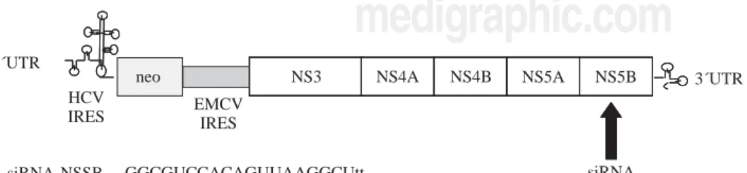
Figure 1. Schematic
representa-tion of the HCV subgenomic plicon and the siRNA targeted re-gion. The arrow indicates that the siRNA is directed against the NS5B region of the viral genome and is showed the sequence of synthetic siRNA.
GGCGUCCACAGUUAAGGCUtt AGCCUUAACUGUGGACGCCtt siRNA-NSSB
NS5B HCV IRES
neo
EMCV IRES
NS3 NS4A NS4B NS5A 3´UTR
siRNA Target 5´UTR
medigraphic.com
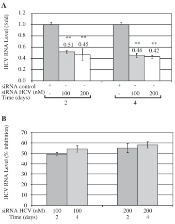
These results were further confirmed by Quantitative Real Time RT-PCR, which is a more sensitive tech-nique. Quantitative analysis showed that both concen-trations of siRNA-NS5B (100 and 200 nM) decreased HCV-RNA levels, reaching a maximum inhibition value at 4 days post-transfection (0.46 and 0.42-fold times
compared with control cells) (Figure 4A). Figure 4B
shows that siRNA-NS5B significantly inhibited HCV RNA level at 2 and 4 days postransfection with both concentrations assayed, reaching a maximum inhibition 4 days postransfection (54% and 58% for 100 and 200 nM, respectively).
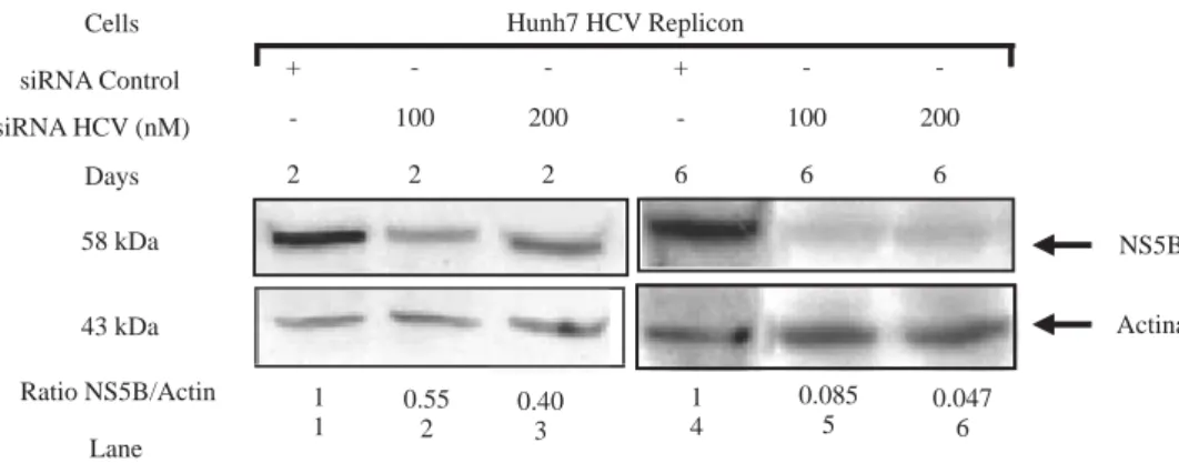
siRNA-NS5B inhibited HCV protein expression
To determine whether siRNAs-NS5B could inhibit the synthesis of viral proteins, NS5B protein levels were de-termined by Western blot. Replicon cells were transfect-ed with 100 and 200 nM of siRNA-NS5B for up to 6 days and then harvested to perform immunoblot analysis. We observed that siRNA-NS5B at both concentrations used
decreased NS5B protein levels several times starting from
day 2 until day 6 post-transfection (Figure 5). These data
suggest that synthetic siRNA-NS5B not only has a nega-tive effect on HCV-RNA levels, but also it could de-crease viral protein production. These results indicate that effect of the siRNA on HCV replicon protein and
RNAlevels were specific. Theeffects of siRNA on HCV
protein and RNA levels in this study could be theresult
of siRNA-directed degradation of the HCV replicon RNA
byRISC, but at this moment we can not exclude other
RNA degradation or decreasing stability mechanisms in-volved.
Figure 3. Effect of siRNA on HCV-RNA levels in Huh7 HCV
re-plicon cells. (A) Huh7 HCV rere-plicon cells (2 x 10⁵ cells) plated in
6-well plates were transfected with 200 nM siRNA directed against the NS5B region of HCV genome for 2 and 4 days. At the end of incubation total cellular RNA was extracted at each time point and analyzed by semi-quantitative RT-PCR. PCR was performed at 26 cycles. RT-PCR for glyceraldehyde-3-phosphate dehydrogenase (GAPDH) mRNA was performed in parallel to show an equal amount of total RNA in each sample. (B) The ratio of HCV/GAP-DH mRNA from RT-PCR detection was quantified with Phoretix 1D v2003.02 software. Data are expressed as relative band intensi-ties to control (transfected cells with siRNA control) which is defi-ned as 1.0. The data shown are the mean ± SD of three separate ex-periments, each one performed in triplicate (**P < 0.01).
Lane 1 2 3 4 0.003 0.006 1
483 pb 221 pb 2
-+
Huh7 HCV Replicon
siRNA HCV 200 nM
Ratio HCV/GAPDH
4 -+
2 +
-4 + -Cells
siRNA control
Days HCV GAPDH
- +
2
- +
4 0.003 0.006
** **
siRNA Control siRNA HCV 200 nM
Relative
band
intensities
1.2 1.0 0.8 0.6 0.4 0.2 0.0
Days
+
-+
-A
B
Figure 2. Assessing cell viability. (A) The Huh7 HCV replicon
ce-lls (5 x 10³ cells) plated in a 96-well plate were treated with
SI-PORT Lipid transfection reagent at indicated concentrations for 48 hours. (B) The Huh7 HCV replicon cells (5 x 10³ cells) plated
in a 96-well plate were transfected with 100-400 nM of siRNA control (non-sense sequence) for 48 hours. The cell viability was then determined in triplicates by MTT assay and compared with untreated cells (100% viability).
A
120
100
80
60
40
20
0
0.3 0.4 0.5 0.6
SIPORT Lipid (µl)
Huh7 HCV Replicon
% Cel viability
B
104 102 100 98 96 94 92 90 88
% Cel viability
Huh7 HCV Replicon
100 200 300 400
medigraphic.com
DiscussionHCV is an RNA virus and, in many cases, is difficult to eradicate from infected individuals even with an inten-sive antiviral therapy that utilizes pegylated interferon
alfa and ribavirin.25,26 Although a number of other
antivi-ral compounds, including inhibitors against the NS3-4A
protease12,27,28 and NS5B RNA dependent RNA
poly-merase,11,27,29,30 are currently being tested for their
thera-peutic applicability, such attempts have not always been promising.
Synthetic siRNAs have been used both in vitro and
in vivo to cause gene silencing.31,32 The potential of
us-ing RNAi activity for treatment of viral diseases and
cancer has aroused a great deal of interest in the
scien-tific community. The mechanisms of gene silencing by RNA interference does not induce non-specific intrac-ellular interferon defense mechanisms and therefore represents a promising approach to elucidate gene function or to inhibit certain RNA viruses such as
HCV.33
The HCV genome is a positive-sense
single-strand-ed RNA that functions as botha messenger RNA and
replication template via a negative-strand
intermedi-ate, making it an attractivetarget for the study of RNA
interference. A number of groups have demonstrated that siRNAs interfere with HCV gene expression and
replication.22,23 Other laboratories have reported the
useof siRNA against HIV-1, HPVand poliovirus in
culture cells.34,35 We have demonstrated that
introduc-tion of siRNA-NS5B into target cells that contained HCV replicon caused a dramatic decrease of viral RNA
and protein levels (Figures 4 and 5). This effect was
likely due to the degradationof HCV messenger RNA
by the RISC endonuclease. We do not know the effect
of RNAi on HCV immediately aftervirus entry into
cells because an efficient cell-culture system for
growth of HCV is not available at this time. Because of the great variability in RNA sequences between dif-ferent quasispecies and genotypes of HCV, for thera-peutic applications it may be necessary to include sev-eral different combinations of siRNA to target a partic-ular region of the genome. In addition, the high mutation rate of HCV that is apparent during replica-tion makes the appearance of escape mutant from siR-NA. The utility of siRNA as a therapy against HCV in-fection will depend on the development of efficient
delivery systems thatinduce long-lasting RNAi
activi-ty. HCV is an attractive targetbecause of its
localiza-tion in the infected liver, an organ that can be readily targeted by nucleic acid molecules and viral vectors.
In the future,chemically modified synthetic siRNAs,
with improved resistanceto nucleases coupled with
enhanced duration of RNAi, may becomea possibility
for therapeutic applications. On the other hand,gene
therapy offers another possibility to express siRNAs
thattarget HCV in a patient’s liver. The data in this
study suggest that siRNAs targeting NS5B viral poly-merase can elicit an anti-HCV response in cell culture. It represents a promising therapy that could eliminate viral RNA from the infected cell and potentially cure patients with hepatitis C.
Acknowledgments
This work was supported by grants from the SEP, Mexico Grant Number: PROMEP/103.5/04/2590 to AMR and by the Autonomous University of Nuevo Leon Grant Number: PAICYT SA1010-04 to AMR and SA1436-06 to LTA.
Figure 4. Quantitative real time RT-PCR of HCV RNA levels from
Huh7 HCV replicon cells transfected with 100 and 200nM of siR-NA-NS5B. (A) 2 x 10⁵ cells plated in 6-well plates were
transfec-ted at indicatransfec-ted concentrations of siRNA-NS5B for 2 and 4 days. Viral RNA levels were normalized based on the ratio of HCV/GA-PDH mRNA that was amplified in the same plate by the real-time RT-PCR. Data are expressed as HCV RNA levels relative (fold) to control (transfected cells with siRNA control), which is defined as 1.0. (B) The data are expressed as HCV RNA levels (% inhibition) relative to control that is defined as 0. The data shown are the mean ± SD of triplicate cultures, and the experiment was repeated three times (**P<0.01).
** 0.51
**
0.45 **
0.46 ** 0.42
-100 A
1.2
1.0
0.8
0.6
0.4
0.2
0.0
HCV
RNA
Level
(f
old
)
+
-200
2
-100 +
-200
4 siRNA control
siRNA HCV (nM) Time (days)
B
HCV
RNA
Level
(%
inhibition
)
100 2
100 4
200 2
200 4 siRNA HCV (nM)
medigraphic.com
Figure 5. HCV NS5B protein levels
in Huh7 HCV replicon cells trans-fected with siRNA-NS5B. 2 x 10⁵
Huh7 HCV replicon cells were transfected with 100 nM of siRNA-NS5B (lanes 2 and 5) and 200nM of siRNA-NS5B (lanes 3 and 6) for 2 and 6 days. Cell lysates were pre-pared and equal amounts of pro-tein extracts (40 μg) were subjected to immunoblot analysis to detect NS5B (top panel) and actin levels (bottom panel). The ratio of NS5B/ actin proteins from immunoblot detection was quantified with Pho-retix 1D v2003.02 software.
1 1
0.55 2
0.40 3
1 4
0.085
5 0.0476
Actina NS5B Hunh7 HCV Replicon
-2 +
100 2
-200 2
-6 +
100 6
-200 6
-Lane Ratio NS5B/Actin
43 kDa 58 kDa Days siRNA HCV (nM)
siRNA Control Cells
References
1. Lieber CS. Alcohol and hepatitis C. Alcohol Res Health 2001; 25: 245-54.
2. Poynard T, Yuen MF, Ratziu V, Lai CL. Viral hepatitis C. Lancet
2003; 362: 2095-100.
3. Simmonds P, Bukh J, Combet C, Deleage G, Enomoto N, Feinstone S, Halfon P, Inchauspe G, Kuiken C, Maertens G, Mizokami M, Murphy DG, Okamoto H, Pawlotsky JM, Penin F, Sablon E, Shin IT, Stuyver LJ, Thiel HJ, Viazov S, Weiner AJ, Widell A. Consen-sus proposals for a unified system of nomenclature of hepatitis C virus genotypes. Hepatology 2005; 42: 962-73.
4. Penin F, Dubuisson J, Rey FA, Moradpour D, Pawlotsky JM. Struc-tural biology of hepatitis C virus. Hepatology 2004; 39: 5-19. 5. Chisari FV. Unscrambling hepatitis C virus-host interactions.
Nature 2005; 436: 930-2.
6. Tellinghuisen TL, Rice CM. Interaction between hepatitis C virus proteins and host cell factors. Curr Opin Microbiol 2002; 5: 419-27. 7. Bartenschlager R, Lohmann V. Novel cell culture systems for the
hepatitis C virus. Antiviral Res 2001; 52: 1-17.
8. Behrens SE, Tomei L, De Francesco R. Identification and prop-erties of the RNA-dependent RNA polymerase of hepatitis C virus. Embo J 1996; 15: 12-22.
9. Bressanelli S, Tomei L, Rey FA, De Francesco R. Structural analysis of the hepatitis C virus RNA polymerase in complex with ribo-nucleotides. J Virol 2002; 76: 3482-92.
10. Sookoian SC. New therapies on the horizon for hepatitis C. Ann Hepatol 2003; 2: 164-70.
11. Neyts J. Selective inhibitors of hepatitis C virus replication. Anti-viral Res 2006; 71: 363-71.
12. Zeuzem S, Heathcote JE, Martin N, Nieforth K, Modi M. Peginterferon alfa-2a (40 kDa) monotherapy: a novel agent for chronic hepatitis C therapy. Expert Opin Investig Drugs 2001; 10: 2201-13.
13. Feld JJ, Hoofnagle JH. Mechanism of action of interferon and ribavirin in treatment of hepatitis C. Nature 2005; 436: 967-72. 14. Barth H, Liang TJ, Baumert TF. Hepatitis C virus entry: molecular biology and clinical implications. Hepatology 2006; 44: 527-35. 15. Lohmann V, Korner F, Koch J, Herian U, Theilmann L, Bartenschlager R. Replication of subgenomic hepatitis C virus RNAs in a hepatoma cell line. Science 1999; 285: 110-3. 16. Bartenschlager R. Hepatitis C virus replicons: potential role for
drug development. Nat Rev Drug Discov 2002; 1: 911-6. 17. Bartenschlager R, Pietschmann T. Efficient hepatitis C virus cell
culture system: what a difference the host cell makes. Proc Natl Acad Sci USA 2005; 102: 9739-40.
18. Escuret V, Martin A, Durantel D, Parent R, Hantz O, Trepo C, Menguy T, Bottius E, Dardy J, Maral J, Escary JL, Zoulim F. Novel alpha interferon (IFN-alpha) variant with improved in-hibitory activity against hepatitis C virus genotype 1 replication
compared to IFN-alpha2b therapy in a subgenomic replicon sys-tem. Antimicrob Agents Chemother 2006; 50: 3984-91. 19. Bumcrot D, Manoharan M, Koteliansky V, Sah DW. RNAi
thera-peutics: a potential new class of pharmaceutical drugs. Nat Chem Biol 2006; 2: 711-9.
20. Scherer LJ, Rossi JJ. Approaches for the sequence-specific knock-down of mRNA. Nat Biotechnol 2003; 21: 1457-65.
21. Schwarz DS, Hutvagner G, Haley B, Zamore PD. Evidence that siRNAs function as guides, not primers, in the Drosophila and human RNAi pathways. Mol Cell 2002; 10: 537-48.
22. Wilson JA, Jayasena S, Khvorova A, Sabatinos S, Rodrigue-Gervais IG, Arya S, Sarangi F, Harris-Brandts M, Beaulieu S, Richardson CD. RNA interference blocks gene expression and RNA synthesis from hepatitis C replicons propagated in human liver cells. Proc Natl Acad Sci USA 2003; 100: 2783-8. 23. Takigawa Y, Nagano-Fujii M, Deng L, Hidajat R, Tanaka M,
Mizuta H, Hotta H. Suppression of hepatitis C virus replicon by RNA interference directed against the NS3 and NS5B regions of the viral genome. Microbiol Immunol 2004; 48: 591-8. 24. Hamazaki H, Ujino S, Miyano-Kurosaki N, Shimotohno K,
Takaku H. Inhibition of hepatitis C virus RNA replication by short hairpin RNA synthesized by T7 RNA polymerase in hepa-titis C virus subgenomic replicons. Biochem Biophys Res Commun
2006; 343: 988-94.
25. Lindsay KL, Trepo C, Heintges T, Shiffman ML, Gordon SC, Hoefs JC, Schiff ER, Goodman ZD, Laughlin M, Yao R, Albrecht JK. A randomized, double-blind trial comparing pegylated inter-feron alfa-2b to interinter-feron alfa-2b as initial treatment for chronic hepatitis C. Hepatology 2001; 34: 395-403.
26. Fried MW, Shiffman ML, Reddy KR, Smith C, Marinos G, Goncales FL, Jr., Haussinger D, Diago M, Carosi G, Dhumeaux D, Craxi A, Lin A, Hoffman J, Yu J. Peginterferon alfa-2a plus ribavirin for chronic hepatitis C virus infection. N Engl J Med
2002; 347: 975-82.
27. Pause A, Kukolj G, Bailey M, Brault M, Do F, Halmos T, Lagace L, Maurice R, Marquis M, McKercher G, Pellerin C, Pilote L, Thibeault D, Lamarre D. An NS3 serine protease inhibitor abro-gates replication of subgenomic hepatitis C virus RNA. J Biol Chem 2003; 278: 20374-80.
28. Perni RB, Almquist SJ, Byrn RA, Chandorkar G, Chaturvedi PR, Courtney LF, Decker CJ, Dinehart K, Gates CA, Harbeson SL, Heiser A, Kalkeri G, Kolaczkowski E, Lin K, Luong YP, Rao BG, Taylor WP, Thomson JA, Tung RD, Wei Y, Kwong AD, Lin C. Preclinical profile of VX-950, a potent, selective, and orally bioavailable inhibitor of hepatitis C virus NS3-4A serine pro-tease. Antimicrob Agents Chemother 2006; 50: 899-909. 29. Nakagawa M, Sakamoto N, Enomoto N, Tanabe Y, Kanazawa N,
medigraphic.com
30. Paeshuyse J, Kaul A, De Clercq E, Rosenwirth B, Dumont JM, Scalfaro P, Bartenschlager R, Neyts J. The non-immunosup-pressive cyclosporin DEBIO-025 is a potent inhibitor of hepa-titis C virus replication in vitro. Hepatology 2006; 43: 761-70. 31. Xia H, Mao Q, Paulson HL, Davidson BL. siRNA-mediated gene silencing in vitro and in vivo. Nat Biotechnol 2002; 20: 1006-10.
32. McCaffrey AP, Meuse L, Pham TT, Conklin DS, Hannon GJ, Kay MA. RNA interference in adult mice. Nature 2002; 418: 38-9.
33. Elbashir SM, Harborth J, Lendeckel W, Yalcin A, Weber K, Tuschl T. Duplexes of 21-nucleotide RNAs mediate RNA interference in cultured mammalian cells. Nature 2001; 411: 494-8.
34. Gu W, Putral L, Hengst K, Minto K, Saunders NA, Leggatt G, McMillan NA. Inhibition of cervical cancer cell growth in vitro and in vivo with lentiviral-vector delivered short hairpin RNA targeting human papillomavirus E6 and E7 oncogenes. Cancer Gene Ther 2006; 13: 1023-32.