HA Baptista González et al.Association of HFE mutations (C282Y and H63D) with iron overload in blood donors from Mexico City 55
edigraphic.com
Annals of Hepatology 2007; 6(1): January-March: 55-60Annals of Hepatology
Original Article
Association of HFE mutations (C282Y and H63D)
with iron overload in blood donors from Mexico City
Héctor A. Baptista-González;1 Fany Rosenfeld-Mann;1 Rocío Trueba-Gómez;1 Luisa Bermejo-Martínez;1
Nahum Méndez-Sánchez2
1Hematología Perinatal. Instituto Nacional de Perinatología. Secretaría de Salud.
2Departments of Biomedical Research, Gastroenterology & Liver Unit, Médica Sur Clinic & Foundation. Mexico City, México.
Address for correspondence:
Dr. Héctor A. Baptista González, Hematología Perinatal. 1st floor, Research building, Instituto Nacional de Perinatología. Montes Urales Núm. 800, Lomas Virreyes, Delegación Miguel Hidalgo, 1100, phone 55209900-323. E-mail [email protected]
Manuscript received and accepted: 23 November and 8 December 2006.
Abstract
Background and objective: Iron overload has been as-sociated with HFE mutations (C282Y and H63D). We investigated the association between these mutations and high serum ferritin in a sample of healthy adult men. Design and methods: We enrolled unrelated blood donors from three hospitals in Mexico City in a cross-sectional study. Serum ferritin (SF) was determined to define iron overload, and HFE gene mutations were identified by PCR–RFLP. Results: We evaluated 2524 male blood donors and included 246 individuals for each group. We identified 108 individuals with HFE gene mutation, 20.5 % were heterozygote (wt/H63D or wt/C282Y) and the remaining homozygote (H63D/ H63D). The genotype wt/C282Y was observed in two cases, none cases with C282Y/C282Y. The allelic fquency of H63D and C282Y was 0.115 and 0.002, re-spectively. We observed different association for H63D allele with iron overload (OR 1.54, CI 95 %1.16-2.03) and none in allele C282Y. Although values averages were different, the extreme dispersion of serum ferritin not showed statistically significant differences between H63D and C282Y alleles and ferritin concentrations.
Conclusions: The male unrelated blood donors from Mexico City with iron overload prevalence of 13.8% hold similarities with other populations from Europe o America continent, respecting the allele frequency H63D. Nevertheless, allele frequency C282Y is lower than that observed in descendents from northern Eu-rope. We have not observed statistic difference of SF or iron overload frequency by effect of both alleles.
Key words: HFE gene, hereditary hemochromatosis, iron overload, serum ferritin.
Introduction
The gene involved in hereditary hemochromatosis is located at 6p21.3 locus and was described in 1996 by Feder.1 The normal product is a protein of 343 amino ac-ids located on the cell surface, with a sequence similar to MHC class I molecules. The HFE protein extends through the cell membrane to form a heterodimer with
β2-microglobulin.
2 The best-known variants of HFE gene are the C282Y and H63D mutants. A no directional G→A mutation occurs at nucleotide 845 (G845A), which changes a cysteine to tyrosine at residue 282 (C282Y). This cysteine is highly conserved and forms an intermo-lecular disulfide bridge. This loss impedes the interac-tion of HFE with β2-microglobulin and prevents the ex-pression of the protein on cell surface, with the protein instead being retained within the Golgi complex.2 The H63D mutation is located in the alpha 1 chain of the pro-tein and does not affect the expression of the propro-tein or its interaction with β2-microglobulin, C→G change oc-curs at nucleotide 187 (C187G), which causes the substi-tution of aspartate for histidine at residue 63 (H63D) that alters the interaction between HFE protein and the trans-ferrin receptor on cell surface.3
A large number of diseases related to defects in the ab-sorption, distribution, or storage of iron have been iden-tified,2 which in all cases result in a pathological in-crease in iron stores. Iron overload has attracted the atten-tion of clinical researchers because it is associated with different chronic degenerative diseases, such as diabetes mellitus, hepatic cirrhosis, cardiomyopathy, cancer, re-productive failure, vascular damage, and other less spe-cific conditions, such as chronic fatigue syndrome.9
The most common cause of hereditary iron overload is the presence of mutations in HFE gene. The prevalence of these mutations is influenced by ethnic origin.4 The C282Y mutation has been reported in up to 26% of a no selected population of individuals of Celtic ancestry, whereas it is practically absent from individuals of orien-tal or African origin.4,5 Conversely, the H63D mutation has a multicentric origin, with no ethnic associations.
Ini-Artemisa
edigraphic.com
SUSTRAÍDODE-M.E.D.I.G.R.A.P.H.I.C
:ROP ODAROBALE FDP
VC ED AS, CIDEMIHPARG
ARAP
ACIDÉMOIB ARUTARETIL :CIHPARGIDEM
tial evidence indicates that the prevalence of mutations of the HFE gene in the population of mixed Mexican ori-gin (mestizos) is similar to that observed in Spanish peo-ple.6 The impact of mutations of HFE gene on iron over-load is also determined by other environmental vari-ables, such as the life style,7 regular blood donation, alcohol intake, and gender.8
The most common test used in the evaluation of iron store in clinical practice or epidemiological setting in-cluded the determination of serum ferritin (SF) and trans-ferrin saturation (Ts), with different results in external or internal validation. The cut-off to define iron overload is SF > 300 ug/L or Ts > 55% form men and > 200 ug/L or Ts > 50% for women. The international literature reports that the prevalence of iron overload varies from 9.3 to 22.5%,4 whereas in Mexico, our group has identified a prevalence of up to 29% in selected healthy popula-tion.10 The occurrence of iron overload is variable due to the complex interaction of ethnic origin, life style and mutations of HFE gen. This antecedent forces to evaluate association of these three variables in each particular clinical setting.
With this perspective, the aim of this study was to in-vestigate the association between HFE mutations and iron overload in a sample of male blood donors residing in Mexico City.
Design and methods
We enrolled unrelated volunteer blood donors from three hospitals in Mexico City in a cross-sectional study, case-control design. We included male individuals, 18– 64 years of age, who agreed to participate as blood do-nors were included in this study. Written informed con-sent was obtained from all participants before entry. All subjects were asked to complete a questionnaire that ad-dressed demographic and medical variables. A physical examination was made, and erythrocyte index were de-termined according to established criteria for acceptation of blood donors in our country.11 The subjects diagnosed with post-transfusion iron overload, sideroblastic anemia, iron deficiency (SF < 30 ug/L), who had been transfused in the past five years, were excluded.
Analytical procedures
A blood sample was taken to determine the blood cell count and serum ferritin (SF) level, and for the extraction of DNA. Blood samples with hemolysis, lipemia, or in-sufficient or inadequate leukocyte DNA were eliminated. The values for hemoglobin, hematocrit, mean corpuscu-lar volume, mean corpuscucorpuscu-lar hemoglobin, mean corpus-cular hemoglobin concentration, and red distribution width, were determined in an AcT 5 Diff Hematology An-alyzer (Beckman Coulter Electronics, Miami, FL, USA). SF was determined in duplicate samples and expressed in
µg/L, which allowed us to stratify the donors with iron overload or «Group I» (SF ≥ 300 µg/L), according to population values above the 90th percentile, as proposed by Koziol and Beutler,12 and Group II with normal SF (30–299 µg/L). A commercial reagent was used (Vidas® Ferritin, bioMerieux®, Lyon, France), based on an immu-noenzymatic method with a final reading in fluorescence (enzyme-linked fluorescent assay), according to proce-dures validated in our laboratory.
The genomic DNA was extracted and the amplifica-tion protocol was based on the original proposals of Fed-er1 and Adams,13 with oligonucleotide sequence de-signed to allow the exploration of two mutations in HFE gene. The oligonucleotide sequences used in this experi-ment to amplify codon 282 were 5´-TGGCAAGGG-TAAACAGATCC-3´ (forward) and CTCAGGCACTC-CTCTCAACC-3´ (reverse); and for codon 63, 5´-ACATGGTTAAGGCCTGTTGC-3´ (forward) and 5´GCCACATCTGGCTTGAAATT-3´ (reverse). The ampl-icons were digested with RsaI (codon 282, exon 4) and BclI (codon 63, exon 2).
Statistical analysis
To establish the statistically significant risk of cases with iron overload and controls, with an exposed portion of 0.10, an alpha value of 0.05 (two tailed), and a beta value of 0.20, we estimated that an OR ≥ 2 was required. We compare the frequency of each HFE mutations in both groups with non parametric procedure of Mann-Whitney test. Descriptive statistics for the whole sample were obtained.
This protocol was approved by the Research and Bio-ethical Committees of Instituto Nacional de Perina-tología. The confidentiality of both the information and the identity of the participants were maintained. To guar-antee the anonymity of the individuals evaluated, the se-rum and DNA samples were identified by unique progres-sive numbers that allowed us to file, track, or repeat the necessary tests. This project was supported financially with public funds from the Instituto Nacional de Perina-tología and Fundación Clínica Médica Sur.
Results
inde-edigraphic.com
pendent distributions, but not statistical differences be-tween two groups. The SF values were 466 (CI 95% 443-490 ug/L) and 138 (129-147 ug/L) respectively.
Mutations in HFE gene were determined in the 492 individuals included in the study (246 for each group). Using molecular analysis, we identified 384 individuals (78.0%) with the wild-type (wt) gene presentation, pro-portion 0.47 and 0.53 for each group (Table II). In 108 individuals with HFE gene mutation, 101 (20.5%) were identified as heterozygote (wt/H63D or wt/C282Y) and the remaining seven as homozygotes (1.4%, H63D/ H63D). We do not observed statistical association be-tween two situations, OR 1.48 (CI 95%, 0.95-2.31) and 2.75 (CI 95% 0.53-14.33), respectively.
The mutation wt/H63D were present in 99 subjects (20.1%), with proportion of 57 and 42 cases for each
group, without statistical association (OR 1.49, CI 95% 0.55-2.33) and H63D/H63D in seven cases (proportion 0.71 and 0.29, for Group I and II, respectively), without statistical differences (OR 2.75, CI 95% 0.53-14.33). The variant wt/C282Y was observed in two cases (one for each group, OR 1.10, CI 95% 0.07-17.69). We do not identified cases with variant C282Y/C282Y.
The allelic frequency of wt was 0.883, H63D and C282Y was 0.115 and 0.002, respectively. The distribu-tion of allelic frequency showed different associadistribu-tion for H63D allele (OR 1.54, CI 95%1.16-2.03), with frequency 0.136 and 0.093 for Group I and II, respectively. There was not difference in allelic frequency for C282Y (OR 1.05, CI 95% 0.15-7.47).
To evaluate the global relationship between HFE gene mutations and iron overload, we grouped all cases with iron overload and all subjects with HFE mutation (either H63D or C282Y); in a «mutated» group with versus none mutated or wild-type group (Table III). There were 63 (58.3%) and 45 mutated subjects (41.7%) for group I and II, respectively, with border line statistical association (OR 1.54 CI 95%, 1.00-2.37). Although a greater (OR 1.54) association existed between individuals with iron overload and the presence of a mutations in the HFE gene, the value observed did not exceed the value calcu-lated for OR in the design of the study (OR ≥ 2).
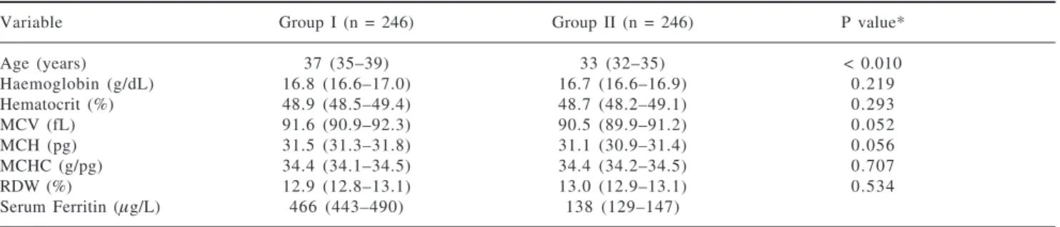
Table I. Distribution of erythrocyte indices among groups of the study.
Variable Group I (n = 246) Group II (n = 246) P value*
Age (years) 37 (35–39) 33 (32–35) < 0.010
Haemoglobin (g/dL) 16.8 (16.6–17.0) 16.7 (16.6–16.9) 0.219 Hematocrit (%) 48.9 (48.5–49.4) 48.7 (48.2–49.1) 0.293
MCV (fL) 91.6 (90.9–92.3) 90.5 (89.9–91.2) 0.052
MCH (pg) 31.5 (31.3–31.8) 31.1 (30.9–31.4) 0.056
MCHC (g/pg) 34.4 (34.1–34.5) 34.4 (34.2–34.5) 0.707
RDW (%) 12.9 (12.8–13.1) 13.0 (12.9–13.1) 0.534
Serum Ferritin (µg/L) 466 (443–490) 138 (129–147)
Average (95% confidence interval). MCV, mean corpuscular volume; MCH, mean concentration haemoglobin; RDW, red cell distribution width; MCHC, mean corpuscular haemoglobin concentration.
* Mann-Whitney test (P < 0.01)
Table III. Distribution of the mutant HFE gene between study groups.
HFE Gene Group I (n = 246) Group II (n = 246)
Mutated (108/22.0) 63 (58.3) 45 (41.7) Non mutated (384/78.0) 183 (47.6) 201 (52.4)
OR 1.54 (95% CI 1.00–2.37)
Table II. Distribution of the zygosity of the HFE alleles.
Variants (n) Group I (n = 246) Group II (n = 246) OR
wt/wt (384/78.0) 183 (0.470) 201 (0.530)
wt/H63D (99/20.1) 57 (0.570) 42 (0.430) 1.49 (0.55-2.33) wt/C282Y (2/0.40) 1 (0.500) 1 (0.500) 1.10 (0.07-17.69) H63D/H63D (7/1.4) 5 (0.710) 2 (0.290) 2.75 (0.53-14.33)
C282Y/C282Y (0) 0 0
Heterozygote (101/20.5) 58 (0.580) 43 (0.420) 1.48 (0.95-2.31) Homozygote (7/1.4) 5 (0.710) 2 (0.290) 2.75 (0.53-14.33) Allelic frequency
WT (0.883) 0.861 0.904
H63D (0.115) 0.136 0.093 1.54 (1.16-2.03)
C282Y (0.002) 0.002 0.002 1.05 (0.15-7.47)
edigraphic.com
SUSTRAÍDODE-M.E.D.I.G.R.A.P.H.I.C
:ROP ODAROBALE FDP
VC ED AS, CIDEMIHPARG
ARAP
ACIDÉMOIB ARUTARETIL :CIHPARGIDEM
We evaluated the differences on the distribution of se-rum ferritin values between the individuals with the natu-ral form of the gene (wild type), as well as with the muta-tions wt/H63D, H63D/H63D and wt/C282Y. Although values averages were different, the extreme dispersion of such concentrations not generates statistically signifi-cant differences. The same situation was documented with the allele’s wt, H63D and C282Y (data no showed).
Discussion
The mutations of C282Y and H63D of HFE gene are documented widely in international literature.13-15 The se-lection bias in the type of target population is a deter-mining factor to establish the frequency of these muta-tions and their correlation with the increase of iron store observed. Different reports have included newborns
dur-ing neonatal screen,16 general population,8 subjects in healthy appraisal clinic or blood donors6,17 and relatives of patients with clinical hereditary haemochromatosis.5,18 The study of gene HFE mutations has been used in blood donors, although the frequent donation can be an impor-tant factor to modify the iron store, as to prevent its es-tablishment the direct relation with mutations C282Y and H63D.17,19 However, the effect of frequent blood do-nations diminishes serum ferritin values and increases iron deficiency prevalence, without the protective effect from the HFE gene mutations.
The genotype and allele frequencies for C282Y and H63D in blood donors vary around the world (Table IV). The allele frequency C282Y is more prevalent in Europe with 0.011,13,15 particularly in Northern Ireland with 0.020 and South Wales with 0.012. In Spain, it varies widely from 0.020 to 0.007.20,21 The allele frequency of C282Y, compared between diverse population groups from North America, shows that white people from USA is more prevalent (0.115-0.196) than Afro-Americans (0.025)18,22 and practically null in Brazil,23 and native Amerindians.6,13,24,25 Among the Mexican American pop-ulation, resident in the USA, it is three to five times great-er,13,18,22 than reported in the present study for blood do-nors living in Mexico City, with 0.001, but less than Spanish blood donors.20,21 These results could be ex-plained by the differences in the origin of the diverse American native populations, in addition to the possibil-ity of the Mestizo Mexican population having a different proportion of mixed genes from European-Spanish ori-gin,25 independently of the selection bias effect.
The H63D allele is highly prevalent in the world pop-ulation, varying from 0 to 0.22513 for individuals not ethnically related to Celtic ancestors,27 which is
consis-Table IV. Genotype and allele frequencies for HFE gene mutations in diverse populations of blood donors.*
Geographic area Genotype frequency Allele frequency
Population C282Y/C282Y wt/C282Y H63D/H63D C282Y/H63D wt/H63D wt/wt C282Y H63D
Europe 0.4 9.2 2 1.8 21.6 65.1 0.011 0.027
Denmark 1.4 1 1 1.5 2.5 2 0 6 5 0.016 0.026
Germany 0 3.3 2 2 1 9 73.9 0.005 0.025
Italy 0.8 8.2 6.2 3.7 28.6 50.4 0.068 0.224
Northern Ireland 1.2 14.8 1.5 2.5 22.8 57.2 0.02 0.028
Norway 0 12.8 2.1 0 18.1 6 7 0.013 0.022
Spain 0-0.2 4.1-4.5 4.1-4.3 1.4-1.7 32.3-33.8 57.1-66.5 0.02-0.007 0.16-0.044
South Wales 1 5.9 3 4 21.8 64.3 0.012 0.032
England 0.68 12.7 2.4 2.4 23.6 58.3 0.016 0.031
America 8.1-10.0?? 0.3-7.8 2.1-5.0 1.7-5.3 21.5-24.2 55.7-65.3 0.020-0.046 0.051-0.069 EUA White 0.12– 0.82 8.08–11.3 1.45–3.08 1.63–3.34 21.3–25.9 59.5–62 0.115-0.196 0.067-0.121
San Diego, Cal. 1 11.4 2.9 3.2 20.9 60.6 0.017 0.03
USA (Blacks) 0.06 2.33 0.32 0.06 5.55 91.69 0.025 0.062
Brazil. 0.001 0.001 0.001 0.001 0.001 99.99 0 0
Chile Indian 0 1.28 0 0 5.13 93.59 0.001 0.005
Chile White ethnicity 0 2.56 1.28 0 18.59 77.57 0.01 0.087
Caracas, Venezuela. 0 3.7 0 0.41 18.2 77.69 0.004 0.019
Mexican American 0.02-0.4 1.6-3.9 0.6-2.0 0.02-0.9 17.4-23.3 71.1-76.0 0.002-0.006 0.09-0.028
México City ** 0 0.4 1.4 0 20.2 77.9 0.001 0.023
* References 6, 13-15, 17-25 **Presen report.
Figure 1. Ferritin concentrations in blood donors with HFE muta-tions. (number on the box are median values).
1000
800
600
400
200
0
N- 384
wt/wt 269
315
99 wt/H63D
7 H63D/H63D
2 wt/C282Y
Gen HFE mutations
Serum ferritin (
g/L)
m
444
edigraphic.com
tent with the frequency of 0.023 reported in this issue. The fact that allele frequency of H63D in Mexican popu-lation is similar to that reported for other groups can be explained in two ways. The first is that this allele fre-quency was inherited during the Spanish racial intermix-ing and the second is that mutation H63D,26 contrary to the C282Y mutation, rose in more than one geographic area.14 This means that genetic frequency of C282Y is de-termined by the ethnic origin,13,15 whereas mutation H63D has a uniform world-wide distribution.13 Likewise, allele C282Y had a multicentric origin and did not de-pend initially on the flow of genes or genetic mixing.14 Finally, based on differences between allelic frequencies, it is assumed that H63D is more ancient than C282Y al-lele.27
The serum ferritin concentrations are related to bone marrow hemosiderin and modifiable iron. In the adult, 1
µg/L of ferritin corresponds to 7–8 mg of iron stored.28 The physiological variables related to changes in iron concentrations differ between men and women,10 due to the interactions of diverse biological and social factors. Physiological changes in the content of iron store occur slowly, varying with age.12 The balance in iron content, and the change from one state to another, occurs over a long period of 7–10 years.10,28 The prevalence of iron overload is similar in our report (12%) to that reported in other publications12,27,28 for blood donors (8.3%) or no se-lected populations (8.7–16.5%). We observed that indi-viduals with alleles C282Y or H63D held higher median SF concentrations, lacking statistical differences (Figure 1).
Why is not there absolute statistical association be-tween iron overload and HFE gene mutations? It is clear that iron-overload individuals tend to accumulate iron store during their course of life, but there is also an inevi-table interaction with individual, environmental, and bi-ological factors,10,18 including the gender,22 iron con-sumption on the diet,7 iron supplementation,8 and other social variables, such as consumption of alcohol28 or fre-quent blood donations.17,20 Our group has shown that there is an increase of over 20% in the average serum fer-ritin values between the 18–29 and 30–49 year age stra-ta.10 The specific effect of each differs according to the context of each individual or population stud-ied.8,10,13,12,18
In conclusion, male blood donors from Mexico City with iron overload prevalence of 13.8%, hold similarities with other populations from Europe or America conti-nent, respecting the allele frequency H63D. Neverthe-less, allele frequency C282Y is lower than that observed in descendents from northern Europe. We have not ob-served, however, statistic difference of SF nor iron over-load frequency by effect of both alleles. The global prev-alence of HFE gene mutations is high (22%), suggesting a need to extend the study and interactions of these mu-tations in Amerindian Mexican population.
References
1. Feder JN, Penny DM, Irrinki A, Lee VK, Lebron JA, Watson N, et al. The hemochromatosis gene product complexes with the trans-ferrin receptor and lowers its affinity for ligand binding. Proc
Natl Acad Sci USA 1998; 95: 1472–7.
2. Andrews CN. Molecular control of iron metabolism. Best Pract
Res Clin Haematol 2005; 18: 159–69.
3. Ganz T. Hepcidin-a regulator of intestinal iron absorption and iron recycling by macrophages. Best Pract Res Clin Haematol
2005; 18: 171–82.
4. Waalen J, Nordestgaard GB, Beutler E. The penetrance of heredi-tary hemochromatosis. Best Pract Res Clin Haematol 2005; 18: 203–20.
5. DuBois S, Kowdley KV. Review article: targeted screening for hereditary haemochromatosis in high-risk groups. Aliment
Pharmacol Ther 2004; 20: 1–14.
6. Ruiz AG. Analysis of HFE codon 63/282 (H63D/C282Y) gene variants in Mexican mestizos, blood donors and patients with hereditary haemochromatosis. Arch Med Res 2000; 31: 422–4. 7. Hunt JR, Zeng H. Iron absorption by heterozygous carriers of the
HFE C282Y mutation associated with hemochromatosis. Am J
Clin Nutr 2004; 80: 924–31.
8. Rossi E, Bulsara MK, Olynk JK, Cullen DJ, Summerville L, Powell LW. Effect of hemochromatosis genotype and lifestyle factors on iron and red cell indices in a community population. Clin Chem
2001; 47: 202–8.
9. Yen AW, Fancher TL, Bowlus CL. Revisiting hereditary hemo-chromatosis: current concepts and progress. Am J Med 2006; 119: 391–9.
10. Baptista GH, Rosenfeld MF, Trueba GR, Méndez SN, Uribe EM. Evaluation of iron overload in healthy adult residents of Mexico City. Arch Med Res 2005; 36: 142–7.
11. Norma Oficial Mexicana para la disposición de sangre humana
con fines terapéuticos. NOM SSA003. Diario Oficial de la
Federación, 1993.
12. Koziol JA, Ho NJ, Felitti VJ, Beutler E. Reference centiles for serum ferritin and percentage of transferrin saturation, with ap-plication to mutations of the HFE gene. Clin Chem 2001; 47: 1804–10.
13. Adams PC, Reboussin DM, Barton JC, McLaren CE, Eckfeldt JH; Hemochromatosis and Iron Overload Screening (HEIRS) Study Research Investigators. Hemochromatosis and iron-overload screening in a racially diverse population. N Engl J Med 2005; 352: 1769–78.
14. Rochette J, Pointon JJ, Fisher CA, Perera G, Arambepola M, Arichchi DS, et al. Multicentric origin of hemochromatosis gene (HFE) mutations. Am J Hum Genet 1999; 64: 1056–62. 15. Hanson EH, Imperatore G, Burke E. HFE gene and hereditary
Hemochromatosis: A HuGe review. Am J Epidemiol 2001; 154: 193-206.
16. Hoppe C, Watson RM, Long CM, Lorey F, Robles L, Klitz W, et al. Prevalence of HFE mutations in California newborns. Pediatr
Hematol Oncol 2006; 23: 507-16.
17. Jackson HA, Carter K, Darke C, Guttridge MG, Ravine D, Hutton RD, et al. HFE mutations, iron deficiency and overload in 10,500 blood donors. Br J Haematol 2001; 114: 474–84.
18. Schmitt B, Golub RM, Green R. Screening primary care patients for hereditary hemochromatosis with transferrin saturation and serum ferritin level: systematic review for the American College of Physicians. An Intern Med 2006; 143: 522-536.
19. De Gobbi M, D’Antico S. Castagno S, Testa D, Merlini R, Bondo A, Camaschella C. Screening selected blood donors with bio-chemical iron overload for hemochromatosis: a regional experi-ence. Haematologica 2004; 89: 1161-67.
edigraphic.com
SUSTRAÍDODE-M.E.D.I.G.R.A.P.H.I.C
:ROP ODAROBALE FDP
VC ED AS, CIDEMIHPARG
ARAP
ACIDÉMOIB ARUTARETIL :CIHPARGIDEM
21. Alvarez S, Mesa MS, Bandres F, Arroyo E. C282Y and H63D mutation frequencies in a population from central Spain. Dis
Markers 2001; 17(2): 111-4.
22. Steinberg KK, Cogswell ME, Chang CJ, Caudill PS, McQuillan MG, Bowman AB, et al. Prevalence of C282 and H63D mutations in the hemochromatosis (HFE) gene in the United States. JAMA
2001; 285: 2216–22.
23. Dias NKV, Meirelles de souza AF, Fonseca CJM, Proietti FA, Portes MRS, Leite de Souza J. Hereditary Hemochromatosis. Population screening based on phenotype in Brazilian blood donors. J Clin Gastroenterol 2005; 39: 430-34.
24. Vizzi E, Loureriro CL, Geder M, de las Nieves GCM, Rodriguez LA, Gerace L, et al. Mutation analysis of the HFE gene associated with Hereditary Hemochromatosis in a Venezuelan sample. Ann
Hematol 2005; 84: 802-6.
25. Wohllk N, Zapata R, Acuna M, Reyes H, Navarro A, Roa I, Roa JC. HFE gene mutations in Chile. Ann Intern Med 2003; 139: 708-9.
26. Fragoso JM, Juarez-Cedillo T, Hernandez-Pacheco G, Ramirez E, Zuniga J, Izaguirre R, de la Pena A, Granados J, Vargas-Alarcon G. Cytochrome P4501A1 polymorphisms in the Amerindian and Mestizo populations of Mexico. Cell Biochem
Funct 2005; 23: 189-93.
27. Lucotte G, Dieterlen F. A European allele map of the C282Y mutation of hemochromatosis: Celtic versus Viking origin of the mutation? Blood Cells Mol Dis 2003; 31: 262–7.
28. Milman N, Byg KE, Ovesen L, Kirchhoff M, Jurgensen KS. Iron status in Danish men 1984–94: a cohort comparison of changes in iron stores and the prevalence of iron deficiency and iron overload. Eur J Haematol 2002; 68: 332–40.