775
Intoxication with Amanita phalloides. , 2016; 15 (5): 775-787
Early initiation of MARS
®
dialysis in
Amanita phalloides-induced acute liver injury
prevents liver transplantation
Mike Hendrik Pillukat,* Tina Schomacher,* Peter Baier,* Gert Gabriëls,** Hermann Pavenstädt,** Hartmut H. J. Schmidt*
* Department for Transplant Medicine, University Hospital Münster, Münster, Germany. ** Department for Internal Medicine D, General Internal Medicine and Nephrology, University Hospital Münster, Münster, Germany.
A B S T R A C T A B S T R A C T A B S T R A C T A B S T R A C T A B S T R A C T
Amanita phalloides is the most relevant mushroom intoxication leading to acute liver failure. The two principal groups of toxins, the amatoxins and the phallotoxins, are small oligopeptides highly resistant to chemical and physical influences. The amatoxins inhibit eukaryotic RNA polymerase II causing transcription arrest affecting mainly metabolically highly active cells like hepatocytes and re-nal cells. The clinically most characteristic symptom is a 6-40 h lag phase before onset of gastrointestire-nal symptoms and the rapid progression of acute liver failure leading to multi-organ failure and death within a week if left untreated. Extracorporeal albumin dialy-sis (ECAD) was reported to improve patient’s outcome or facilitate bridging to transplantation. In our tertiary center, out of nine in-toxicated individuals from five non-related families six patients presented with acute liver injury; all of them were treated with ECAD using the MARS® system. Four of them were listed on admission for high urgency liver transplantation. In addition to standard
medi-cal treatment for Amanita intoxication we initiated ECAD once patients were admitted to our center. Overall 16 dialysis sessions were performed. All patients survived with full native liver recovery without the need for transplantation. ECAD was well tolerated; no severe adverse events were reported during treatment. Coagulopathy resolved within days in all patients, and acute kidney injury in all but one individual. In conclusion, ECAD is highly effective in treating intoxication with Amanita phalloides. Based on these ex-periences we suggest early initiation and repeated sessions depending on response to ECAD with the chance of avoiding liver trans-plantation.
Key words. Key words.Key words. Key words.
Key words. Mushroom intoxication. Albumin dialysis. Amatoxin. Silibinin. Death cap.
September-October, Vol. 15 No. 5, 2016: 775-787
CASE REPORT
INTRODUCTION
Acute liver failure (ALF) may be caused by a wide range of conditions such as toxins, medications, ischemia, venous and bile obstruction, and genetic disorders.1 The
most common mushroom intoxication leading to acute liver failure is caused by ingestion of Amanita phalloides or some other species of the genus Amanita, as well as a few other, closely related genera of basidiomycetes, which all can sometimes be mistaken for wild champignons or oth-er edible mushrooms.2,3
The Amanita mushroom contain two principal toxin groups, the heptapeptide phallotoxins and the octapeptide amatoxins, both cyclic peptide toxins synthesized as 35 aa-long proproteins.2,3 The three most important amatoxins α-,
β- and γ-amanitin have molecular masses between 0.88 and
0.92 kDa, are resistant to heat, low pH, proteases and are soluble in alcohol and water. While the phallotoxins are not taken up by the enteral mucosa resulting only in early gas-trointestinal symptoms, the amatoxins are readily taken up into the enterohepatic circulation, cross cytoplasmic mem-branes and inactivate RNA polymerase II, leading to inhibi-tion of transcripinhibi-tion especially in metabolically very active cells like liver and kidney cells, which leads to cell death and in case of the liver, to rapid, progressive organ failure.3
Intoxication provokes four distinct clinical phases: a lag phase devoid of any symptoms, in contrast to many other mushroom intoxications, occurring for 6 to 40 h before on-set of the gastrointestinal phase, lasting 12 to 24 h character-ized by nausea, crampy abdominal pain, vomiting, diarrhea as well as consequent electrolyte imbalances, dehydration, hypoglycemia, and hypotension. After a phase of apparent
The Official Journal of the Mexican Association of Hepatology, the Latin-American Association for Study of the Liver and
the Canadian Association for the Study of the Liver
Manuscript received: Manuscript received:Manuscript received:
convalescence (36-48 h after ingestion) with intermittent clinical improvement and characterized by deterioration of liver enzyme tests with raising transaminase levels culmi-nating in jaundice the intoxication progresses into the phase of acute liver failure (4-9 d) characterized by dramatic raises in transaminase levels and deterioration of liver and kidney function. These conditions then lead to hyperbilirubine-mia, coagulopathy, hypoglycehyperbilirubine-mia, acidosis, hepatic encepha-lopathy, and hepatorenal syndrome. Ultimately, multi organ failure and death may occur after 1-3 weeks.4 Therefore,
OLT is the treatment of choice in cases with ALF.
ECAD methods such as the Molecular Adsorbent Re-circulating System (MARS®)5 or the Fractionated Plasma
Separation and Adsorption System (FPSA, Prometh-eus®),6,7 have repeatedly shown to improve patient
out-come in acute liver failure caused by intoxications. ECAD may create a time bridge until an appropriate donor organ becomes available for OLT or for avoiding the need for transplantation at all.8-11 The governing principle of these
albumin dialysis methods is the selective removal of albu-min-bound liver toxins in addition to removal of hy-drophilic small molecules via a conventional dialysis loop.10 Rapid urinary and fecal clearance of amatoxins and
the importance of sustained aggressive IV hydration therapy have been reported in amanitin intoxication.12,13,31 It is also
sug-gested that the amatoxins themselves do not bind to albumin.14
MARS® employs an albumin-impermeable membrane with a pore size cut-off at 60 kDa against a continuously circulating loop with 20% human serum albumin passing through col-umns of charcoal, an anion exchange resin and a low-flux dia-lyzer connected to a secondary circuit.8,10 FPSA on the other
hand uses a larger filter pore size (cut-off at 250 kDa) which allows the patient’s own albumin to get in contact with two fil-ter columns, while wafil-ter-soluble substances are removed di-rectly from blood by a separate high-flux dialysis loop.9,10 The
availability of using MARS is very limited due to various rea-sons including deficits in evidence-based outcome data, regu-latory aspects and reimbursement issues in many countries. Therefore, this system may not be available for many patients suffering from amanitin intoxication.
There are several reports on MARS® dialysis in ALF.
Camus, et al. reported on a transplant-free recovery apply-ing the MARS® system in acute liver failure.15,16 In
addi-tion, the Helsinki group published their experience for using MARS® in amatoxin intoxication. The concept of
initiating this dialysis treatment before signs of ALF devel-oped resulted also in less need for liver transplantation.17
Additional data were published by Cisneros-Garza, et al.
confirming these experiences in native liver recovery in ALF patients in a multicentric study in several Mexican hospitals.18
Here we report on our experience of employing ECAD in treatment of severe mushroom poisonings at our center from October 2010 to August 2014. Early initiation of MARS® dialysis resulted in survival without any need for
OLT. The treatment was tolerated well by the patients. No clinical relevant side effects were observed.
MATERIAL AND METHODS
Patients
Between late summer 2010 and August 2014 nine in-toxications were reported within five independent fami-lies leading to admission to a local hospital fulfilling the criteria of i) suspected amanitin intoxication and ii) pre-dicted acute liver failure. In all cases, Amanita intoxica-tion was confirmed by the patients, in some cases with the help of a mycologist. Out of these nine cases, three female and three male patients were transferred thereaf-ter to our university hospital due to rapid increase of INR > 1.5. The total mean time intervals between inges-tion of the mushrooms and onset of gastrointestinal (GI) symptoms were 7 h (range 3-10 h), to local hospital ad-mission 27.83 h (range 21-43 h) and to commencement of ECAD 55.5 h (range 35-76 h). The mean admission-to-MARS® time for our center was 3.5 h (range 1-6 h) and
for admission-to-HU listing the interval was 10.5 h (0-23 h) (Table 1). Mean age was 56.8 years (range 34-78 years). Mean number of ECAD treatments were 2.67 (range 2-5),
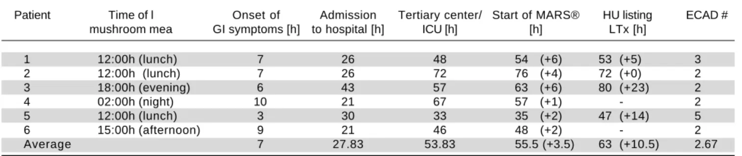
Table 1. Total time intervals between ingestion, hospital and ICU admissions, HU listing and initiation of ECAD.
Patient Time of l Onset of Admission Tertiary center/ Start of MARS® HU listing ECAD #
mushroom mea GI symptoms [h] to hospital [h] ICU [h] [h] LTx [h]
1 12:00h (lunch) 7 26 48 54 (+6) 53 (+5) 3
2 12:00h (lunch) 7 26 72 76 (+4) 72 (+0) 2
3 18:00h (evening) 6 43 57 63 (+6) 80 (+23) 2
4 02:00h (night) 10 21 67 57 (+1) - 2
5 12:00h (lunch) 3 30 33 35 (+2) 47 (+14) 5
6 15:00h (afternoon) 9 21 46 48 (+2) - 2
Average 7 27.83 53.83 55.5 (+3.5) 63 (+10.5) 2.67
777
Intoxication with Amanita phalloides. , 2016; 15 (5): 775-787
mean duration was 6.63 h (range 1.08-13.84 h) and mean length of total university hospital stay was 13.38 d (range 4-36 d). Tables 2A-2F illustrate fluid balances, blood gas analysis and laboratory parameters of the presented patients. Our standard medical treatment for amanitin intoxications for all six individuals during their ICU stay and at the day of discharge is outlined in table 3. Four of the six patients were initially listed for high urgent liver transplantation, but all could be de-listed afterwards without fatalities (Fig-ure 1). Due to comorbidity, age or rapid clinical improve-ment two patients were not listed for liver transplantation despite initial presentation with acute liver injury.
ECAD
For MARS® treatment we used a Fresenius 5008 or a
Genius dialysis machine (Fresenius Medical Care Deut-schland GmbH, 61346 Bad Homburg v. d. H.) and a MARS® Monitor (Gambro Lundia AB, Sweden). A
con-ventional hemodialysis catheter access either through the jugulary or the subclavian vein was established. To prevent clotting in the albumin-impregnated, highly permeable dia-lyzer system we used a continuous infusion of unfractionat-ed low molecular weight heparin (Ratiopharm GmbH, Ulm, Germany). The closed loop contained 500 ml of 20% commercial human serum albumin. The flow in the blood loop, the albumin dialysate circuit and the low-flow albu-min-regeneration dialyzer were kept at 250 mL/min.
Safety monitoring
The most frequently reported serious adverse events due to ECAD in medical literature are bleeding due to an-ticoagulation and/or thrombocytopenia.19 Platelets may
drop during and after ECAD in individual cases.19
Blood pressure and heart rate were continuously moni-tored during the ECAD sessions, while laboratory meas-urements were taken at least daily. Routine creatinine measurements were taken photometrically, as there are currently no official recommendations to use the enzy-matical method in MELD calculations.20
RESULTS
Case 1
A 56-year-old woman was admitted to a local hospital after having ingested a mushroom meal at lunch together with her family the day before. Her medical history in-cluded arterial hypertension and one historic episode of pyelonephritis. In the evening, about 6 h after the meal, she started to suffer from diarrhea, nausea, vomiting and leg cramps. Her husband and two of her children
devel-oped similar, but milder symptoms. Together, the family was admitted to a hospital. All except her daughter (see case 2) developed transaminases below 1000 U/l and re-mained clinically stable. A mycologist examined remain-ing samples from the rest of the mushroom meal and discovered several mildly gastrointestinal-toxic fungi in-cluding Amanita species, but could not detect A. phalloides
anymore. After initiation of treatment for Amanita intoxica-tion with silibinin, charcoal and fluid resuscitaintoxica-tion the pa-tient developed fever, raising transaminase values and falling liver synthesis parameters. The patient was then transferred to the ICU of our tertiary care center for high-ly urgent (HU) liver transplantation listing. Sonography revealed hepatomegaly with parenchymal damage without cholestasis, while CT scanning showed no necrotic foci within the liver at that early stage.
In addition to standard medical treatment (Table 3) the patient received three ECAD therapies on three consecu-tive days, which resulted in reversal of transaminases and normalization of parameters for liver synthesis (Figure 1A, Table 2A). The clinical condition of the patient im-proved markedly, she was taken off the high urgency OLT list. Acute kidney injury occurred accompanied by anuria, which resolved within days (Table 2A). She was hospital-ized for 4.3 days.
Case 2
The 34-year-old daughter of patient #1 developed a similar rise in transaminases, albeit several hours later than her mother. This patient had no relevant medical history. She had ingested the mushroom meal together with her mother (see case 1 for details), and was transferred from the local hospital to our tertiary center one day later, suf-fering from the same clinical symptoms as her mother with laboratory findings consistent with hyperacute liver injury. Onset of clinical symptoms in her case was about 11 h after the meal. She received the same supportive med-ical treatment (Table 3) as her mother and was also listed for HU OLT, but after two sessions of ECAD her lab val-ues had already improved markedly and she could be taken off the waiting list (Figure 1B, Table 2B). Without any signs of acute kidney injury and in markedly improved clinical condition she was transferred back to her local hospital (Figure 1B). Hospitalization was overall 3.95 days.
Case 3
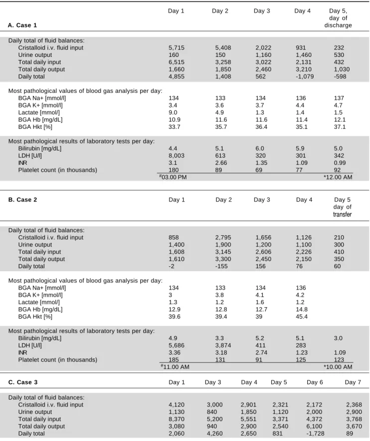
Table 2. A-F. Fluid balances (*per 24 h, at 6.00 a.m.; in millilitres), blood gas analysis and laboratory parameters (most pathological values for each respective day) for all six patients at admission and during ICU treatment for the first seven days, or until transfer to the normal ward.
Day 1 Day 2 Day 3 Day 4 Day 5,
day of
A. Case 1 discharge
Daily total of fluid balances:
Cristalloid i.v. fluid input 5,715 5,408 2,022 931 232
Urine output 160 150 1,160 1,460 530
Total daily input 6,515 3,258 3,022 2,131 432
Total daily output 1,660 1,850 2,460 3,210 1,030
Daily total 4,855 1,408 562 -1,079 -598
Most pathological values of blood gas analysis per day:
BGA Na+ [mmol/l] 134 133 134 136 137
BGA K+ [mmol/l] 3.4 3.6 3.7 4.4 4.7
Lactate [mmol/] 9.0 4.9 1.3 1.4 1.5
BGA Hb [mg/dL] 10.9 11.6 11.6 11.4 12.1
BGA Hkt [%] 33.7 35.7 36.4 35.1 37.1
Most pathological results of laboratory tests per day:
Bilirubin [mg/dL] 4.4 5.1 6.0 5.9 5.0
LDH [U/l] 8,003 613 320 301 342
INR 3.1 2.66 1.35 1.09 0.99
Platelet count (in thousands) 180 89 69 77 92
#03.00 PM *12.00 AM
B. Case 2 Day 1 Day 2 Day 3 Day 4 Day 5 day of transfer
Daily total of fluid balances:
Cristalloid i.v. fluid input 858 2,795 1,656 1,126 210
Urine output 1,400 1,900 1,200 1,100 300
Total daily input 1,608 3,145 2,606 2,226 410
Total daily output 1,610 3,300 2,450 2,150 350
Daily total -2 -155 156 76 60
Most pathological values of blood gas analysis per day:
BGA Na+ [mmol/l] 134 133 134 136
BGA K+ [mmol/l] 3 3.8 4.1 4.2
Lactate [mmol/] 1.3 1.2 1.6 1.2
BGA Hb [mg/dL] 12.9 12.8 12.7 14.8
BGA Hkt [%] 39.6 39.4 39 45.4
Most pathological results of laboratory tests per day:
Bilirubin [mg/dL] 4.9 3.3 5.2 5.1 3.0
LDH [U/l] 5,686 3,874 411 283
INR 3.36 3.18 2.74 1.23 1.09
Platelet count (in thousands) 185 131 91 125 123
#11.00 AM *10.00 AM
C. Case 3 Day 1 Day 3 Day 4 Day 5 Day 6 Day 7 Daily total of fluid balances:
Cristalloid i.v. fluid input 4,120 3,000 2,901 2,321 2,172 2,368
Urine output 1,130 840 1,850 1,120 2,000 2,900
Total daily input 8,370 5,200 5,551 3,371 4,372 3,768
Total daily output 3,080 940 2,900 2,540 6,100 3,670
779
Intoxication with Amanita phalloides. , 2016; 15 (5): 775-787
Most pathological values of blood gas analysis per day:
BGA Na+ [mmol/l] 133 132 134 137 138
BGA K+ [mmol/l] 3.6 2.7 4.0 4.2 3.8
Lactate [mmol/] 3.8 3.2 3.0 2.9 2.5
BGA Hb [mg/dL] 10.6 10.1 10.7 11.3 10.3
BGA Hkt [%] 32.8 33.5 33 34.7 32.4
Most pathological results of laboratory tests per day:
Bilirubin [mg/dL] 3.9 5.3 12.3 14.7 23 22.1
LDH [U/l] 2,491 2,003 406 348 353
INR 4.38 6.14 2.3 1.53 1.4 1.3
Platelet count (in thousands) 107 20 6 11 19 17
#01.00 AM *24.00 AM
D. Case 4 Day 1 Day 2 Day 3 Day 4 Day 5 Daily total of fluid balances:
Cristalloid i.v. fluid input 824 1,453 542
Urine output 350 2,150 350
Total daily input 1,474 3,853 1,392
Total daily output 650 3,300 650
Daily total 824 553 742
Most pathological values of blood gas analysis per day:
BGA Na+ [mmol/l] 136 134 135
BGA K+ [mmol/l] 3.6 3.4 3.4
Lactate [mmol/] 1.4 1.8 1.2
BGA Hb [mg/dL] 11.1 10.8 11.2
BGA Hkt [%] 34.2 33.4 34.5
Most pathological results of laboratory tests per day:
Bilirubin [mg/dL] 1.1 2.1 2.8 2.7 2.6
LDH [U/l] 259 624 4,790 785 345
INR 1.05 1.35 2.14 2.15 1.54
Platelet count (in thousands) 253 229 97 82 54
#07.00 AM *10.00 PM
E. Case 5 Day 1 Day 2 Day 3 Day 4 Day 5 Daily total of fluid balances:
Cristalloid i.v. fluid input 3,704 2,769 2,716 2,806 495
Urine output 500 570 3,350 3,900 500
Total daily input 4,101 4,519 4,016 5,356 695
Total daily output 1,900 2,820 5,100 5,800 500
Daily total
Most pathological values of blood gas analysis per day:
BGA Na+ [mmol/l] 139 134 133 133 133
BGA K+ [mmol/l] 3.0 2.5 3.4 3.5 3.6
Lactate [mmol/] 2.9 2.4 1.7 0.8 0.8
BGA Hb [mg/dL] 16.4 12.4 9.8 9.0 9.8
BGA Hkt [%] 50.1 38.2 30.3 28 30.3
Most pathological results of laboratory tests per day:
Bilirubin [mg/dL] 0.7 1.4 2.8 3.3 3
LDH [U/l] 1,924 8,398 543 273 258
INR 1.01 1.64 1.35 0.94 0.91
Platelet count (in thousands) 327 277 70 65 75
#08.00 PM *07.00 PM
F. Case 6 Day 1 Day 2 Day 3 Day 4 Day 5 Daily total of fluid balances:
Cristalloid i.v. fluid input 1963 617 282
Total daily input 2,683 3,067 2,782
Total daily output 500 1,300 2,150
Daily total 2,183 1,767 632
Most pathological values of blood gas analysis per day:
BGA Na+ [mmol/l] 134 133 132 133
BGA K+ [mmol/l] 3.8 3.6 3.6 3.8
Lactate [mmol/] 1.4 1.6 1.2 0.8
BGA Hb [mg/dL] 11.4 10.1 9.9 10.1
BGA Hkt [%] 35 31.4 30.5 30.5
Most pathological results of laboratory tests per day:
Bilirubin [mg/dL] 2.1 1.7 2.0 1.7 1.3
LDH [U/l] 2,196 3,513
INR 1.62 1.87 1.43 1.27 1.08
Platelet count (in thousands) 240 197 162 153
#13.00 PM *05.00 AM
#Monitoring time until midnight. *Monitoring time from midnight.
of rising transaminase values to initiate treatment for amanitin intoxication with silibinin and N-acetylcysteine (Table 3). Due to further deteriorating lab tests and addi-tional onset of acute kidney injury with edema of the low-er extremities the patient was quickly transflow-erred to our university hospital to initiate ECAD therapy and possible HU listing for OLT. Ultrasound revealed signs of hepatic parenchymal swelling without signs of obstructions. After two MARS® sessions ECAD was suspended because of
suspected heparin induced thrombocythaemia type II (HIT II) and HU OLT listing was carried out. At time of a donor offer, the patient had already improved liver labo-ratory values and therefore no OLT was required any-more. Subsequently, his renal function also returned to normal values (Figure 1C, Table 2C). The patient was dis-charged after 15.35 days from our hospital in good general condition.
Case 4
A 67-year-old man with a medical history of arterial hy-pertension, hepatitis B, hyperthyroidism and cachexia was admitted to our emergency department and then trans-ferred directly to our hospital´s ICU because of an Amani-ta mushroom poisoning with subsequent acute liver injury. Approximately 10 h after completely ingesting a meal of self-collected mushrooms (his description of one of them resembled a white death cap), he developed nau-sea, emesis, watery diarrhea and gastric pain. After admis-sion we began intensive care treatment with standard medical therapy for Amanita intoxication with charcoal, silibinin, N-acetylcysteine (Table 3) and carried out two supporting ECAD sessions. He required continuous anti-hypertensive medication with urapidil and sublingual va-sodilatators. His transaminase values decreased rapidly; therefore HU OLT listing was not carried out (Figure
1D, Table 2D). Overall the patient was 10.63 days hospi-talized.
Case 5
A 58-year-old woman with a medical history of several episodes of cholelithiasis, breast cancer and arterial hyper-tension was transferred from a local hospital to our Inten-sive Care Unit due to acute liver and kidney injury 33 hours after Amanita intoxication. She had developed nau-sea, emesis and diarrhea after eating the larger part of a meal of mushrooms which all had been collected by rela-tives, while her husband took the smaller portion. He was only briefly hospitalized with the same but milder symp-toms. She presented with extensively elevated liver and kidney retention parameters. After admission to our ICU a therapy with silibinin, charcoal and lactulose was initiat-ed, together with antihypertensive intravenous urapidil treatment (Table 3). She was promptly listed for HU OLT. ECAD was administered five times in four days un-til liver and coagulation parameters had improved. After al-bumin dialysis was discontinued, hemodialysis was reintroduced since renal parameters increased again. Dial-ysis was performed three times until discharge, 15 days af-ter intoxication.
Two weeks later, serum creatinine levels had fallen to about 1.3 mg/dl under constant ambulatory surveillance (Figure 1E, Table 2E). About eight months after discharge her creatinine levels had returned to almost normal values. Hospital length of stay was 36 days.
Case 6
781
Intoxication with
Amanita phalloides
.
, 2016; 15 (5): 775-787
Table 3. Standard medical treatment: intoxication-specific and general medication orders for all six cases during ICU stay and at day of discharge.
Case 1 Case 2 Case 3 Case 4 Case 5 Case 6
Anti-intox. charcoal (p.o.), charcoal (p.o.), charcoal (p.o.), charcoal (p.o.), charcoal (p.o.), charcoal (p.o.), (all i.v., except silibinine (Legalon®), silibinine (Legalon®), silibinine (Legalon®), silibinine (Legalon®), silibinine (Legalon®), silibinine (Legalon®),
noted otherwise) vitamin K, vitamin K, vitamin K, vitamin K, vitamin K, vitamin K,
acetylcysteine acetylcysteine acetylcysteine acetylcysteine acetylcysteine acetylcysteine thiamine
Gastroenterological ranitidine, lactulose, pantoprazole, metoclopramide, dimenhydrinate, (all p.o., except noted metoclopramide, metoclopramide, metoclopramide butylscopolamine, lactulose,
otherwise) magaldrate dimeticone alizapride, pantoprazole
(Riopan® Gel) (Sab simplex®), lactulose,
sodium sulfate ranitidine
decahydrate (Glauber’s salt), butylscopolamine
Cardiological (all i.v., urapidile, glyceryl trinitrate, Enalaprile, urapidile, urapidile, glyceryl trinitrate, except noted noradrenaline urapidile, glyceryl trinitrate amlodipine (p.o.), ramiprile (p.o.),
otherwise) lercandipine (p.o.), ramiprile (p.o.), urapidile, clonidine,
clonidine glyceryl trinitrate lercandipine (p.o.)
Nephrological (all i.v., potassium chloride, potassium chloride, potassium chloride, potassium chloride, Sterofundin ISO®, Sterofundin ISO®, except noted otherwise) sodium chloride, sodium chloride, sodium chloride, sodium chloride, potassium chloride, potassium chloride,
Sterofundin ISO® Sterofundin ISO®, Sterofundin ISO®, Sterofundin ISO® sodium chloride, sodium chloride Potassium chloride Potassium chloride torasemide
Pain (all i.v., except metamizole metamizole piritramide, metamizole, metamizole metamizole
noted otherwise) morphine
Metabolic (all i.v., glucose/sodium glucose/sodium glucose/sodium glucose/sodium insulin, insulin, glucose/sodium except noted chloride 40% chloride 40% chloride 40%, chloride 40% glucose/sodium chloride 40%
otherwise) Olimel emulsion chloride 40%
Antibiotic (all i.v., ceftriaxone piperacilline- meropenem
except noted otherwise) tazobactam,
ceftriaxone
Other (all i.v., except Aminoplasmal Hepa®, Aminoplasmal freshly frozen plasma,
noted otherwise) thiamine, nicotine patch Hepa® human albumin
Day of discharge heparin, ranitidine, metoprolole, enalapril, amlodipine, ramiprile, ramiprile, lercandipine,
(all p.o.) metamizole amlodipine amlodipine, ranitidine, lactulose pantoprazole
the afternoon of the day before. During the night he had developed severe gastroenteritic symptoms with severe emesis and diarrhea. After initiation of standard medical treatment (penicillin G, activated charcoal, silibinin) in the local hospital the patient developed a dramatic in-crease in transaminases and dein-crease in coagulation
param-eters. He was rapidly transferred to our hospital’s ICU and received two ECAD treatments in addition to our standard of care (Table 3). His liver parameters started to decrease, the prothrombin time began to rise slowly and the renal parameters remained stable (Figure 1F, Table 2F). One week later, the patient was discharged from our Figure 1.
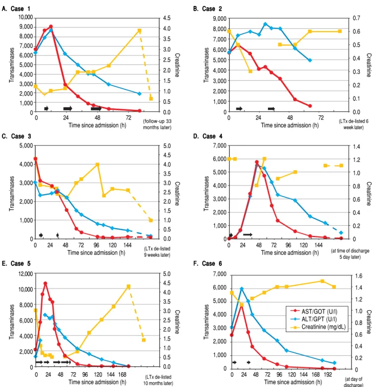
Figure 1. Figure 1. Figure 1.
Figure 1. Liver parameters over time after admission to our hospital are illustrated. On each graph the left y-axis depicts transaminase enzyme activities in units per liter (ALT/GPT, alanine aminotransferase; AST/GOT, aspartate aminotransferase). The right y-axis displays serum creatinine in milligrams per deciliter. The horizontal black arrows denote each ECAD treatment and its duration.
A. Case 1 A. Case 1 A. Case 1 A. Case 1 A. Case 1 10,000 9,000 8,000 7,000 6,000 5,000 4,000 3,000 2,000 1,000 0 4.5 4.0 3.5 3.0 2.5 2.0 1.5 1.0 0.5 0.0
0 24 48 72
Time since admission (h)
Transaminases
Creatinine
(follow-up 33 months later)
B. Case 2 B. Case 2B. Case 2 B. Case 2B. Case 2 9,000 8,000 7,000 6,000 5,000 4,000 3,000 2,000 1,000 0 0.7 0.6 0.5 0.4 0.3 0.2 0.1 0.0
0 24 48 72
Time since admission (h)
Transaminases
Creatinine
(LTx de-listed 6 week later)
C. Case 3 C. Case 3 C. Case 3 C. Case 3 C. Case 3 5,000 4,000 3,000 2,000 1,000 0 5.0 4.5 4.0 3.5 3.0 2.5 2.0 1.5 1.0 0.5 0.0
0 24 48 72 96 120 144
Time since admission (h)
Transaminases
Creatinine
(LTx de-listed 9 weeks later)
D. Case 4 D. Case 4D. Case 4 D. Case 4D. Case 4 7,000 6,000 5,000 4,000 3,000 2,000 1,000 0 1.4 1.2 1.0 0.8 0.6 0.4 0.2 0
0 24 48 72 96 120 144
Time since admission (h)
Transaminases
Creatinine
(at time of discharge 5 day later)
E. Case 5 E. Case 5 E. Case 5 E. Case 5 E. Case 5 12,000 10,000 8,000 6,000 4,000 2,000 0 5.0 4.5 4.0 3.5 3.0 2.5 2.0 1.5 1.0 0.5 0.0 0 24 48 72 96 120 144 168
Time since admission (h)
Transaminases
Creatinine
(LTx de-listed 10 months later)
F. Case 6 F. Case 6F. Case 6 F. Case 6F. Case 6 7,000 6,000 5,000 4,000 3,000 2,000 1,000 0 1.6 1.4 1.2 1.0 0.8 0.6 0.4 0.2 0 0 24 48 72 96 120 144 168 192
Time since admission (h)
Transaminases
Creatinine
(at day of discharge)
783
Intoxication with Amanita phalloides. , 2016; 15 (5): 775-787
internal medicine ward in good condition with only creat-inine levels still elevated above normal values. His serum creatinine levels already had been at 1.3 mg/dL eight months before the intoxication and remained elevated at 1.4 mg/dL six months later. Total length of stay in our hos-pital was 10 days.
Outcome
Among nine intoxicated individuals, six were admitted to our center for OLT due to acute liver injury. All of these patients fulfilled the criteria for amanitin intoxica-tion and predicted acute liver failure. Four of these six pa-tients developed acute kidney injury, one patient (case 6) already had elevated serum creatinine values before the in-toxication and developed only a mild decrease in renal function, and another never showed any increase in kidney retention parameters. Oliguria was detectable before any changes in serum creatinine were noticed. Volume expan-sion was used in all patients to treat or prevent pre-renal acute kidney failure (Tables 2A-2F, Table 3). Both liver and kidney injury parameters recovered in all patients to their initial values, except for case 5, who retained slightly elevated serum creatinine values afterwards. Transaminas-es always recovered within the very first days, whereas bi-lirubin and renal function recovered usually within days to a few weeks after intoxication. Since all patients had no history of both liver and renal disease, transaminases seemed to be the best parameter to predict the time course of the disease. Nevertheless, MARS® dialysis may
be even continued until bilirubin is decreasing as long as the treatment is tolerated. We use a drop of platelets be-low 50.000/μL as a trigger to stop albumin dialysis. Any de-crease in bilirubin may enhance regeneration of the liver after ECAD has been started. Therefore, MARS® may be
continued even longer to increase patient benefit. MARS®
therapy was well tolerated in all six patients; however, drop of platelets, bleeding events or infections as well as the logistics and general availability of this form of dialysis may limit their usage. No patient required OLT although four out of six patients were listed HU. All organ offers were rejected due to fast recovery of medical status and pa-tients were delisted subsequently.
Safety and adverse events during treatment
To assess the patient’s condition we at least daily moni-tored serum electrolytes, blood urea nitrogen, serum creatinine, total bilirubin, transaminases (AST/GOT, ALT/GPT), lactate dehydrogenase, prothrombin time, In-ternational Normalized Ratio (INR), partial prothrombin time (PTT), albumin, and blood cell counts. Only platelet
counts significantly deteriorated during the ECAD treat-ment in each patient. All other determined laboratory pa-rameters benefited from ECAD. The most important parameters are shown in figures 1A-1F and tables 2A-2F.
During each MARS® treatment we continuously
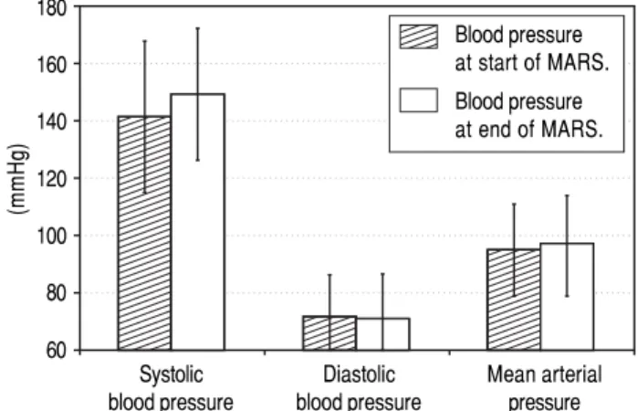
moni-tored arterial blood pressure (Figure 2). In this analysis we included all patients with at least two or more blood pressure measurements taken and without any antihyper-tensive medication (12 out of 16 sessions fulfilled these criteria). The first measured mean systolic blood pressure value was 141.42 ± 26.32, diastolic pressure was 71.67 ± 14.46 (MAP 94.92 ± 15.97) and the last measured mean systolic value was 149.08 ± 22.98 with a diastolic value of 71.08 ± 15.28 (MAP 97.08 ± 16.84). Therefore, blood pressure during ECAD remained stable in contrast to oth-er intoth-ermittent dialysis methods reported.
DISCUSSION
In this work we present our tertiary care center’s expe-rience in treatment of Amanita intoxication with extracor-poreal albumin dialysis from October 2010 till August 2014. Out of nine individuals from five families exposed to varying amounts of poisonous mushrooms, six patients were predicted to develop symptoms of acute liver failure and had to be transferred from their respective local hos-pitals to our liver transplant center. In all six cases liver support therapy was started almost immediately on admis-sion (Table 1). The liver parameters of all patients im-proved during the next 2-4 days of treatment. Four patients were eligible for HU listing, whereas two patients were not listed due to age, comorbidity or clinical improve-ment. In all patients, coagulopathy resolved itself during
Figure 2. Figure 2.Figure 2. Figure 2.
Figure 2. Mean arterial blood pressure values during 12 out of a total of 16 ECAD treatments in six patients with Amanita phalloides intoxication taken during routine monitoring of vital parameters at the beginning and at the end of MARS® sessions. Data from sessions with less than two blood
pressure measurements or under continuous antihypertensive medication du-ring the sessions were omitted from analysis (4 out of 16 treatments).
Systolic Diastolic Mean arterial blood pressure blood pressure pressure 180
160
140 120 100 80
60
(mmHg)
two to three days of ECAD treatment. Since patients seemed to be stable under MARS® therapy, organ offers
from ET were declined and later on patients were delisted due to full recovery. HU listing in all four cases was con-firmed by an audit committee of Eurotransplant (Leiden, The Netherlands).
Kidney retention parameters stayed stable in two pa-tients or returned to the normal range in one case within weeks. First renal symptom was oliguria (Tables 2A-2F). Two patients developed acute renal failure during their ICU stay requiring continuous veno-venous hemodialysis (CVVH), necessitating temporary ambulatory renal re-placement therapy until kidney function recovered. One patient already had a slightly impaired renal function eight months before the intoxication as well as six months after-wards. Transaminases returned to normal values within a few days in all patients, as did total bilirubin in three indi-viduals before their discharge. In the other three patients the bilirubin values had almost reached normal values at discharge or at the time of a further blood test during a subsequent ambulatory counseling in our outpatient clinic (Figures 1A-1F, Tables 2A-2F).
In our experience, MARS® was generally tolerated very
well by all patients without major adverse events. Tran-sient thrombocytopenia was also present in our patients, but recovered quickly as was described previously for MARS®19,21 and Prometheus®.22 In addition, severe
out-comes of amanitin intoxications like death, often de-scribed in earlier studies,23 as well as emergency liver
transplantation associated with its higher intrinsic risk of peri- and post-operative mortality, could be avoided in our cohort.24
ECAD treatment for mushroom-induced acute liver failure has already been described by several groups. In 2003, Faybik, et al. published their experience with MARS® in six patients with a mean time interval between
exposure and first ECAD session of 76 h, with 1-3 ses-sions per patient lasting between 10-24 h. No ECAD-relat-ed adverse events were reportECAD-relat-ed.25 Two of their patients
regenerated spontaneously, two were bridged successfully to liver transplantation (one adult died in septic shock a few days after transplantation, while one pediatric patient survived without complications). In one patient with graft dysfunction after emergency OLT of a marginal organ ECAD helped to avoid re-transplantation. One patient de-veloped a fatal cerebral herniation shortly before trans-plantation was to be carried out. The authors concluded that ECAD seemed to be a successful treatment perspec-tive for support of liver regeneration either to avoid trans-plantation, for bridging until Tx becomes possible, or for treatment of graft dysfunction.25
Covic, et al. published in 2003 an early series of six pediatric patients aged 7-16 years from Romania accidently
intoxicated with mushrooms.26 They performed two
MARS® sessions per patient lasting 6 h each without any
complications. Four out of six patients survived with complete recovery of liver function. The authors conclud-ed that ECAD was a safe and effective depurative therapy in children in acute liver failure and that survival was pre-dicted in their study only by the impact of the initial MARS® sessions and not by baseline laboratory
parame-ters.26
Kantola, et al. in 2009 emphasized the urgency of com-mencing ECAD treatment in mushroom intoxications while the patients are still in a relatively good clinical condition.21 They recorded a mean time interval of 18 h
from ingestion to start of medical treatment and of 48 h until start of MARS® therapy in their series of ten adult patients from Finland. They performed a median of three ECAD sessions per patient lasting for 15.4 h on average, without any MARS®-related adverse events. Five out of
six patients with laboratory signs of acute liver failure ex-perienced native liver recovery, only one was bridged to successful OLT. The other four patients never developed any degree of liver failure prompting the authors to specu-late if in retrospect ECAD was indicated at all. They con-cluded that early aggressive MARS® therapy in combination with optimum supportive measures seemed to be beneficial in cases of Amanita intoxication. Addition-ally, they also acknowledged the fact that the optimal in-tensity, duration and initiation criteria for ECAD in these cases have still not been developed.21
Sorodoc, et al. published in 2010 a retrospective case se-ries of six Romanian patients of whom the first three had received ECAD plus optimal intensive care and the last three had received optimal intensive care without MARS®.27 The former group received three six-hour
ses-sions per patient, with the first as late as 89-121 h after in-gestion of the mushroom meal, without any significant adverse reactions. In the non-ECAD group, all patients died within 10 days post-intoxicationem and in the ECAD group, only one out of three individuals survived, attribut-ed by the authors to the almost complete lack of options for emergency liver transplantations caused by severe do-nor organ shortage in their country. They concluded that the time point of initiation of ECAD therapy, its initial impact as well as the amatoxin inoculum size predicted the outcome of the patients in their small study.27
Recently, Cisneros-Garza, et al. reported on 38 Mexican patients with ALF (of whom 54.3% received MARS®), 15
patients with AoCLF (24.2% with MARS®) and 17
choles-tatic patients with intractable pruritus (21.5% MARS®)
785
Intoxication with Amanita phalloides. , 2016; 15 (5): 775-787
other 10.5% were successfully bridged until a suitable do-nor organ became available. Cisneros-Garza, et al. there-fore concluded that MARS® is a safe and effective
procedure especially for patients with acute liver failure with the potential to contribute to native liver recovery.18
Concerning other modalities of albumin dialysis, Ber-gis, et al. were the first group to report on amanitin intoxi-cations treated with the Fractionated Plasma Separation and Adsorption (FPSA, Prometheus®) system.22 They
compared a group of nine patients with 1-2 FPSA cycles lasting six hours each to eleven matched patients which re-ceived standard medical therapy only. Mean time between mushroom ingestion and onset of diarrhea was 16.2 ± 4.63 h and from ingestion to hospital admission 35.7 ± 6.37 h. All patients in the FPSA group tolerated the procedures well and recovered fully without need for liver transplan-tation, while in the control group one patient died from shock and cardiac arrest, one patient developed renal in-sufficiency and one retained elevated kidney retention pa-rameters after discharge. The authors concluded that liver support therapy by FPSA was a safe method with the po-tential of reducing the need for emergency liver transplan-tations. They also acknowledged the current lack of large controlled studies addressing the questions of the optimal detoxification system, the optimum number of treatment cycles and the identification criteria of patients that would benefit the most from liver support therapy.22
In our case series, the mean time of onset of gastrointes-tinal symptoms was 7 h, admission to local hospitals oc-curred about 28 h after ingestion. After initial treatment for on average 26 h at the local hospital, the patients were trans-ferred to our center where ECAD was commenced with a delay of only 3 h 30 min after admission. HU listing was es-tablished on average in less than 11 h (Table 1). These time intervals of about one day to hospital admission and another one to two days until start of MARS® are comparable to the
intervals reported by previous ECAD studies.21,22,25
The aforementioned reports on ECAD therapies in amatoxin-induced acute liver failure including our here-in reported patients may have had a similar outcome without interventional ECAD treatments. However, based on the experience in acute or hyperacute liver failure the survival is surprisingly very impressive despite the low numbers of patients studied.
Concerning endogenous mediators of liver toxicity, re-cent in vitro studies utilizing an albumin dialysis model system indicated removal of water-soluble substances, proinflammatory mediators like IL-6 and TNFα using the MARS Flux® membrane. An even better removal was
achieved using membranes with larger pores.28,29
Howev-er, the authors point out that it is still unclear whether in-creased or dein-creased levels of cytokines are important for the survival of patients.28 Concerning in vivo data from a
clinical setting, Donati, et al. reported in 2013 on their ex-perience with 64 patients and 269 MARS sessions showing that small molecules like bilirubin, ammonia and bile ac-ids were significantly reduced and hepatocyte growth fac-tor (HGF) concentrations improved, while cytokines were not affected by MARS.30
Our clinical experience with our strategy of ‘the earlier initiation of albumin dialysis, the better the outcome in ALF’, needs further evaluation and a better understanding of the initial pathophysiological mechanisms resulting in liver failure. Early markers for both liver and renal failure to bet-ter define timing and modes of ECAD in this setting are missing. Experience in management of using ECAD may also influence outcome. A prospective randomized study is desired. However, with our experience at our center it would be of some ethical concern to have a controlled study including a control arm offering no ECAD therapy in amatoxin induced liver failure. In addition, we provide evi-dence that, in contrast to the previously reported experience in other studies that dialysis tends to lower blood pressure, the MARS® system resulted in stable hemodynamics. In
patients with low blood pressure, this is of interest espe-cially in the setting of liver diseases, since blood pressure impacts renal perfusion and therefore renal excretion as well as volume balance. We observed that renal function re-covered fully in all our patients to their pre-intoxication levels. While there seems to be general consensus on the early initiation of ECAD treatment, fewer consensuses ex-ists on the duration of and the time intervals between the respective sessions.15,19,21,22,25,31
CONCLUSION
This study highlights the efficacy of the MARS® system
in treating intoxications with Amanita phalloides. Based on these experiences we suggest early initiation, e.g. once transaminases reach 1,000 U/l or parameters such as INR increase. Repeated sessions depending on response of ECAD are required offering the chance of avoiding OLT. Platelets are critical for deciding timing and duration of MARS®. Further studies are required to determine
dos-age, intervals and the optimum characteristics for ECAD treatment of patients suffering from acute liver failure due to mushroom intoxication. These experiences are also rel-evant for other forms of liver failure, reconsidering ECAD as an optional tool in the multimodal treatment pattern in liver failure or intoxications caused by substances with or without affinity to albumin.
ABBREVIATIONS
• ALF: acute liver failure.
• ECAD: extracorporeal albumin dialysis. • HU: high urgency.
• ICU: Intensive Care Unit.
• MARS®: molecular adsorbent recirculating system.
• OLT: orthotopic liver transplantation. • Tx: transplantation.
CONFLICT OF INTEREST
The authors declare that there are no conflicts of interest.
INFORMED CONSENT
All procedures followed were in accordance with the ethical standards of the responsible committee on human experimentation (institutional and national) and with the Helsinki Declaration of 1975, as revised in 2008. Informed consent was obtained from all patients for being included in the study.
CONTRIBUTIONS
MHP collected the data, prepared the figures, reviewed the literature and prepared the manuscript. TS contribut-ed in data collection and helpcontribut-ed in preparing the manu-script. PB gave valuable advice on the clinical management of acute liver failure, and reviewed the manuscript togeth-er with GG and HP, who additionally offtogeth-ered invaluable insights into the clinical as well as technical aspects of di-alysis methods. HS was responsible for all cases present-ed, supervised the project and helped to prepare the final draft of the manuscript.
REFERENCES
1. Lee WM, Squires RH, Jr., Nyberg SL, Doo E, Hoofnagle JH.
Acute liver failure: Summary of a workshop. Hepatology
2008;47:1401-1415.
2. Vetter J. Toxins of Amanita phalloides. Toxicon 1998; 36:
13-24.
3. Walton JD, Hallen-Adams HE, Luo H. Ribosomal biosynthesis
of the cyclic peptide toxins of Amanita mushrooms.
Biopoly-mers 2010; 94: 659-64.
4. Santi L, Maggioli C, Mastroroberto M, Tufoni M, Napoli L,
Ca-raceni P. Acute Liver Failure Caused by Amanita phalloides
Poisoning. Int J Hepatol 2012; 2012: 480-7.
5. Stange J, Mitzner S, Ramlow W, Gliesche T, Hickstein H,
Schmidt R. A new procedure for the removal of protein
bound drugs and toxins. Asaio J 1993; 39: M621-M625.
6. Falkenhagen D, Strobl W, Vogt G, Schrefl A, Linsberger I,
Gerner FJ, Schoenhofen M. Fractionated plasma separation and adsorption system: a novel system for blood purification
to remove albumin bound substances. Artif Organs 1999;
23: 81-6.
7. Rifai K, Ernst T, Kretschmer U, Bahr MJ, Schneider A, Hafer
C, Haller H, et al. Prometheus—a new extracorporeal system
for the treatment of liver failure. J Hepatol 2003; 39: 984-90.
8. Wauters J, Wilmer A. Albumin dialysis: current practice and
future options. Liver Int 2011; 31(Suppl. 3): 9-12.
9. Rifai K. Fractionated plasma separation and adsorption: current
practice and future options. Liver Int 2011; 31(Suppl. 3): 13-5.
10. Krisper P, Stadlbauer V, Stauber RE. Clearing of toxic sub-stances: are there differences between the available liver
support devices? Liver Int 2011; 31(Suppl. 3): 5-8.
11. Mitzner SR, Stange J, Klammt S, Koball S, Hickstein H, Reis-inger EC. Albumin dialysis MARS: knowledge from 10 years
of clinical investigation. Asaio J 2009; 55: 498-502.
12. Jaeger A, Jehl F, Flesch F, Sauder P, Kopferschmitt J. Kinet-ics of amatoxins in human poisoning: therapeutic
implica-tions. J Toxicol Clin Toxicol 1993; 31: 63-80.
13. Faulstich H, Talas A, Wellhoner HH. Toxicokinetics of labeled
amatoxins in the dog. ArchToxicol 1985; 56: 190-4.
14. Fiume L, Sperti S, Montanaro L, Busi C, Costantino D.
Aman-itins do not bind to serum albumin. Lancet 1977; 1: 1111.
15. Camus C, Lavoue S, Gacouin A, Compagnon P, Boudjema K, Jacquelinet C, Thomas R, et al. Liver transplantation avoided in patients with fulminant hepatic failure who received albu-min dialysis with the molecular adsorbent recirculating sys-tem while on the waiting list: impact of the duration of
therapy. Ther Apher Dial 2009; 13: 549-55.
16. Camus C, Lavoue S, Gacouin A, Le Tulzo Y, Lorho R, Boud-jema K, Jacquelinet C, et al. Molecular adsorbent recirculat-ing system dialysis in patients with acute liver failure who
are assessed for liver transplantation. Intensive Care Med
2006;32: 1817-25.
17. Kantola T, Ilmakunnas M, Koivusalo AM, Isoniemi H. Bridging therapies and liver transplantation in acute liver failure, 10
years of MARS experience from Finland. Scand J Surg
2011; 100: 8-13.
18. Cisneros-Garza LE, Munoz-Ramirez M del R, Munoz-Espino-za LE, Ruiz Velasco JA, Moreno-Alcantar R, Marin-Lopez E, Mendez-Sanchez N. The molecular adsorbent recirculating system as a liver support system: summary of Mexican
ex-perience. Ann Hepatol 2014; 13: 240-7.
19. Stange J. Extracorporeal liver support. Organogenesis
2011; 7: 64-73.
20. Kaiser T, Kinny-Koster B, Bartels M, Parthaune T, Schmidt M, Thiery J. Impact of different creatinine measurement
meth-ods on liver transplant allocation. PLoS One 2014; 9:
e90015.
21. Kantola T, Kantola T, Koivusalo AM, Hockerstedt K, Isoniemi H. Early molecular adsorbents recirculating system treatment
of Amanita mushroom poisoning. Ther Apher Dial 2009; 13:
399-403.
22. Bergis D, Friedrich-Rust M, Zeuzem S, Betz C, Sarrazin C, Bojunga J. Treatment of Amanita phalloides intoxication by fractionated plasma separation and adsorption
[Prometheus(R)]. J Gastrointestin Liver Dis 2012; 21: 171-6.
23. Enjalbert F, Rapior S, Nouguier-Soule J, Guillon S, Amour-oux N, Cabot C. Treatment of amatoxin poisoning: 20-year
retrospective analysis. J Toxicol Clin Toxicol 2002; 40:
715-57.
24. Willars C. Update in intensive care medicine: acute liver fail-ure. Initial management, supportive treatment and who to
transplant. Curr Opin Crit Care 2014; 20: 202-9.
25. Faybik P, Hetz H, Baker A, Bittermann C, Berlakovich G, Wer-ba A, Krenn CG, et al. Extracorporeal albumin dialysis in
pa-tients with Amanita phalloides poisoning. Liver Int 2003;23
(Suppl. 3): 28-33.
787
Intoxication with Amanita phalloides. , 2016; 15 (5): 775-787
poisoned by toxic mushroom ingestion. Liver Int 2003;23
(Suppl. 3): 21-7.
27. Sorodoc L, Lionte C, Sorodoc V, Petris O, Jaba I. Is MARS system enough for A. phalloides-induced liver failure
treat-ment? Hum Exp Toxicol 2010; 29: 823-32.
28. Dominik A, Stange J, Pfensig C, Borufka L, Weiss-Reining H, Eggert M. Reduction of elevated cytokine levels in acute/ acute-on-chronic liver failure using super-large pore albumin
dialysis treatment: an in vitro study. Ther Apher Dial 2014;
18: 347-52.
29. Pfensig C, Dominik A, Borufka L, Hinz M, Stange J, Eggert M. A New Application for Albumin Dialysis in Extracorporeal Or-gan Support: Characterization of a Putative Interaction Be-tween Human Albumin and Proinflammatory Cytokines IL-6
and TNFalpha. Artif Organs 2015.
30. Donati G, La Manna G, Cianciolo G, Grandinetti V, Carretta E, Cappuccilli M, Panicali L, et al. Extracorporeal detoxification for hepatic failure using molecular adsorbent recirculating system: depurative efficiency and clinical results in a
long-term follow-up. Artif Organs 2014; 38: 125-34.
31. Banares R, Nevens F, Larsen FS, Jalan R, Albillos A, Doll-inger M, Saliba F, et al. Extracorporeal albumin dialysis with the molecular adsorbent recirculating system in
acute-on-chronic liver failure: the RELIEF trial. Hepatology 2013; 57:
1153-62.
32. Vesconi S, Langer M, Iapichino G, Costantino D, Busi C,
Fiume L. Therapy of cytotoxic mushroom intoxication. Crit
Care Med 1985; 13: 402-6.
Correspondence and reprint request:
Hartmut H.J. Schmidt, Univ. Prof. Dr. med. Klinik für Transplantationsmedizin
Universitätsklinikum Münster Albert-Schweitzer-Campus 1, Building A14
48149 Münster, Germany