ABSTRACT. The effect produced on Vero cell monolayers by toxins derived from Staphylococcus strains was characterized. 210 milk samples taken from dairy cows suffering from sub-clinical mastitis were ana-lyzed. Strains belonging to the Staphylococcus genus were isolated from 73 of these milk samples. The production of toxins was then stimulated from these strains when they were cultured in Dolman’s me-dium. The study of cell cultures showed that 53 toxin samples induced marked and irreversible cellular changes. This is compared to 42 samples (57.5%) which were strongly cytotoxic. The remaining 11 sam-ples were shown to be slowly cytotoxic. 16% of the total toxins did not induce cell damage and 11% of the toxins produced cellular damage that was reversible in less than 24 hrs, and were designated as cytotonic. Haemolytic actively in vitro, using sheep red blood cells, was assessed using toxins that caused alteration in the monolayers. The results indicate that 46.51% of the toxins showed ββ haemolytic activity, 2.32% αα haemolytic activity, and 51.16% showed neither αα nor ββ haemolytic activity. The later type of activity did however cause damage to cultured cells, which suggests that the causative agent could be δδ toxin. This study reveals a strong predominance of ββ haemolytic strains in the dairy farm studied. These strains in-duced in vitro cell damage, and it is possible to speculate that mammary gland tissue damage is similarly produced, which may be attributed to both ββ and/or δδ haemolytic toxins.
Key words: Staphylococcus aureus, Cytolysins, Vero cell cultures, Haemolytic activity, Bovine mastitis.
RESUMEN. El efecto de toxinas de cepas de Staphylococcus sobre monocapas de células Vero fue caracte-rizado. 210 muestras de leche tomadas de vacas afectadas de mastitis subclínica fueron analizadas. De esas muestras fueron aisladas 73 cepas pertenecientes al género Staphylococcus. La producción de toxinas por las cepas fue estimulada cuando se cultivaron en medio Dolman. El estudio realizado en cultivos celulares demostró que 53 muestras de toxina indujeron cambios celulares marcados e irreversibles. Esto es compa-rado a 42 muestras (57.5%) que fueron fuertemente citotóxicas. Las 11 muestras restantes mostraron ser ligeramente citotóxicas. El 16% del total de las toxinas no indujo daño a las células y 11% de las toxinas produjeron un daño celular, designado como citotónico, que fue reversible dentro de 24 h. La actividad hemolítica in vitro sobre eritrocitos de borrego fue probada usando toxinas que causaron alguna altera-ción sobre las monocapas. Los resultados indican que el 46.51% de las toxinas presentaron una actividad ββ hemolítica, 2.32% actividad αα hemolítica, y 51.16% no mostraron actividades tipo αα o ββ. Esta última ac-tividad dañó las células posiblemente por una toxina δδ. Este estudio revela una fuerte predominancia de cepas ββ hemolíticas en la granja lechera estudiada. Esas cepas indujeron daño in vitro, y es posible especu-lar que el daño en el tejido de la glándula mamaria puede ser atribuido a las toxinas hemolíticas ββ y δδ. Palabras clave. Staphylococcus aureus, Citolisinas, Células Vero, Actividad Hemolítica, Mastitis Bovina.
Effect of
Staphylococcus
Toxins Isolated from Dairy Cow Milk
on Vero Cell Monolayers
L
ILIANAS
ABINI,
1*
C
RISTINAT
ORRES,
2M
IRTAD
EMO,
1S
ONIAS
UTIL,
2ANDL
ORENZAL
ARA2Departamento de Microbiología e Inmunología. Facultad de Ciencias Exactas Físico-Químicas y
Naturales. Universidad Nacional de Río Cuarto. Ruta Nacional Nº 36, km 601 (5800) Río Cuarto,
Córdoba, Argentina
*Corresponding author: E-mail [email protected] Phone (058) 676113 Fax (058) 680280
INTRODUCTION
Studies on the etiology of intra mammary bacterial in-fections have shown that 95% of such inin-fections are caused by the Staphylococcus and Streptococcus genera.5 The most isolated bacterial species being Staphylococcus aureus.6,7
In an attempt to evaluate and characterize the
Staphylo-coccus genus it is necessary to be aware of certain impor-tant epidemiological indicators, such as the frequency of presentation and prevalence of the microorganism, risk factors, pathogenicity and virulence factors.6 These indica-tors may then be applied to the analysis of bovine intra mammary infection dynamics.
contribute to the development of the clinical picture. Amongst these toxins are found the haemolysins, which have been denominated as α, β, γ and δ.2,17 The haemoly-sins are differentiated by their different activities mani-fested on erythrocytes from different animal species, al-though they can also produce toxic effects on other cell types such as, fibroblasts, macrophages and white blood cells.5
The haemolysins can be detected by immunological tests or in certain animal models, and when released into culture medium may be analyzed according to the effects they induce on cell cultures. The work presented here fcuses on the characterization of Staphylococcus strains is o-lated from milk taken from cows with sub-clinical mastitis. Their toxigenicity on Vero cell cultures has been assessed, and in parallel this phenotypic expression has been com-pared with in vitro haemolytic activity. It is suggested that this in vitro model could then be used to simulate tissue damage found the in vivo situation.
MATERIAL AND METHODS
Samples. During September 1995 to August 1996, milk samples were taken from a dairy farm situated 12 Km from Río Cuarto city, Córdoba, Argentina. A total of 210 milk samples were taken manually from diffe rent cow teats, and were collected into sterile containers. Care was taken to maintain the greatest aseptic conditions possible, and the samples were kept cold until they were later proc-essed in the laboratory.
Bacteriological examination. Each milk sample was placed in Hotis medium (0.5 ml 0.5% purple bromocresol + 9.5 ml milk).14 The cultures were then incubated at 37ºC
for 24 h. Following this initial incubation, colonies were isolated by streaking drops onto Petri dishes containing manitol agar salt (Merck) and were incubated for 24 h at 37ºC.
The colonies were then stained with Gram stain, and those cultures whose cocci were both Gram positive and positive for the catalase test were selected.3 To select strains belonging to the Staphylococcus genus, the follow-ing tests were carried out: Resistance to bacitracin (at 0.04 U/ml),8 fermentation of glucose and motility.19 In addition,
the rapid coagulase binding test was carried out.10 In order to select for haemolytic strains, those strains identified as belonging to the Staphylococcus genus were seeded into Petri dishes containing sheep tryptose blood agar (5%). Toxin production. The selected strains were seeded in brain heart broth and incubated for 12 h at 37ºC. In order to induce production of the Staphylococcus toxins 1 ml was taken from these cultures and placed in semisolid Do l-man medium. The cultures were then incubated at 37ºC for 24 h with 20% C02.21 Cultures were later filtered through
Whatman Nº2 paper, and centrifuged at 8,000 rpm. for 15 min at 4ºC.9 The supernatants were then sterilized by filtra-tion though 0.22 µm Millipore filters. The control for
ste-rility was carried out in thioglycolate medium.
Toxigenicity test in cell cultures. The Vero cell line used for this assay, was obtained from the Argentine Cell Bank Association (ABAC), and had undergone 40-72 pas-sages. Cell monolayers were grown in 96 well micro-plates with Eagle’s minimal essential medium (MEM), plus Earle’s salts supple mented with 8% fetal bovine serum.13 To these cell cultures either the undiluted supernatants were added, or supernatants diluted by a factor of 2 or 5, in maintenance medium (MM) consisting of MEM supple-mented with 2% fetal bovine serum. The assays were car-ried out in triplicate and were incubated for no more than 7 days at 37ºC. The control consisted of cell monolayers containing MM alone.9,11
Microhaemagglutination test. Bacterial culture super-natants that were found to induce cellular change in the monolayers, were selected and submitted to analysis of haemolytic activ ity. This was carried out by adding a 1% suspension of sheep red blood cells in sterile physiological solution to each supernatant diluted by a factor of 2. This mixture was then incubated in 96 well “U” bottomed mi-cro-plates for 1 hour at 37ºC. At this temperature it was possible to determine α-haemolysis activity. The micro-plates were later incubated at 4ºC over night (hot-cold shock), which revealed the β-haemolysis activity.1
RESULTS
Bacteriological analysis. Of the total 210 milk sam-ples collected and cultured on manitol salt agar medium, it was possible to select 73 strains which had clusters of Gram positive cocci. 100% (73) of these cultures were also found to be positive to the catalase test.
All strains tested were resistant to bacitracin, fermented glucose and were non-motile. Based on these criteria they were therefore considered to be members of the Staphylo-coccus genus.18
Toxicity test in Vero cell cultures. Cell damage in-duced by the supernatants was analyzed by inverted light microscopy. The effect of each supernatant was character-ized according to the following four deffinitions:
a) Strongly cytotoxic toxins (SCT): This category included those samples that caused strong cellular destruction, which was evident and irreversible during 24 h post inocu-lation (p.i.), (Fig. 1a). The number of strains that induced toxins with these characteristics was 42 (Fig. 7).
b) Slowly cytoto xic toxins (SCTb): This category includes those samples that caused irreversible cellular death, in a progressive manner over time, not exceeding 7 days p.i., (Fig. 2, 3, 4 and 5). The number of strains that induced to x-ins with these characteristics was 11 (Fig. 7).
c) Cytotonic toxins (CT): This category includes those samples that induce rounding up of cells, with reversion to the normal state in no less than 24 h p.i., (Fig. 1b). The number of strains that induced toxins with these character-istics was 8 (Fig. 7).
d) Negative toxins (NT): This category includes those sam-ples that did not induce changes in the cell monolayers, and which were found to be identical to the untreated con-trols, (Fig. 6). The number of strains with these character-istics was 12 (Fig. 7).
Microhaemagglutination test. The analysis of haemo-lytic activity was carried out on the 43 strains that were found to produce SCT and SCTb effects on Vero cells.
The results of t he haemolytic activity on sheep red blood cells are shown in Table 1. It was possible to detect one strain with a pure α-haemolysis effect (2.32%). This is compared with the high activity of β-haemolytic strains (18) (41.86%), and two strains with α-β haemolysis activ-Fig. 2. Cytotoxic effect of Staphylococcus toxins, induced
on Vero cell monolayers at 10-12 hours p.i. (X 40). These toxins were characterized as being slowly cytotoxic toxins or SCTb.
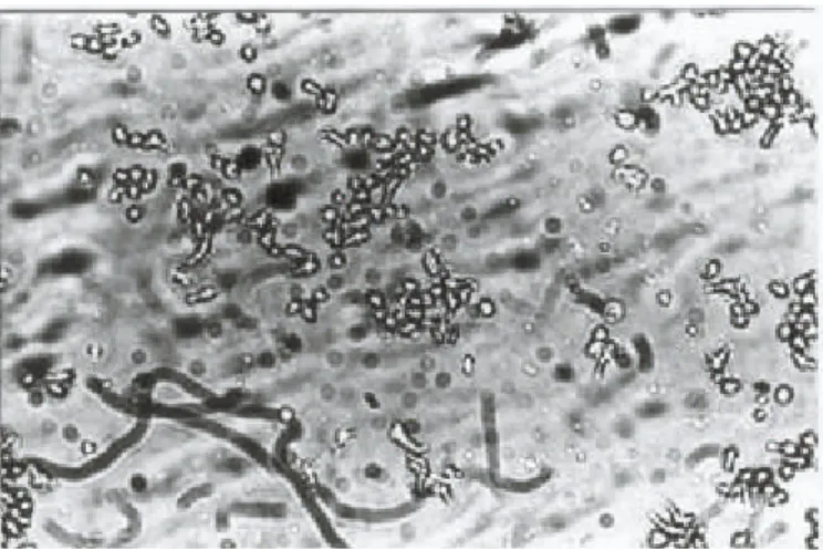
Fig. 3. Cytotoxic effect of Staphylococcus toxins, induced on Vero cell monolayers at 24 hours p.i. These toxins were characterized as slowly cytotoxic toxins SCTb (X 40).
Fig. 4. Cytotoxic effect of Staphylococcus toxins on Vero cell monolayers at 36 hours p.i. These toxins were charac-terized as slowly cytotoxic toxins or SCTb (X 40).
ity (4.85%), whose joint activity added to the activity of both toxins (α and β) increased the value to 48.83% (21). As an example, Fig. 8 shows the results of the haemolytic activities produced by the same toxins when the test was developed at 37ºC and then submitted to hot-cold shock (Fig. 9). The supernatants of the remaining 51.56% (22) strains did not show α and/or β haemolysis activity.
DISCUSSION
The 73 strains belonging to the Staphylococcus genus were analyzed according to their capacity to generate
changes in in vitro cell culture systems. The Vero cells, set up as a cell line, are mammalian cells, which implies that the results obtained may reflect the activity of toxins in the mammary gland in vivo.
In support of this there is information in the literature on Fig. 6. Control Vero cell monolayer cultured without
tox-ins (X 20).
Strongly cytotoxic 57.5%
Not cytotoxic
16.4% Cytotonic 11.0%
Slowly cytotoxic 15.1% 42
12 8 11
Strongly cytotoxic Not cytotoxic Cytotonic Slowly cytotoxic
Type of
haemoly-sin
Positive
Haemolytic titre (range)
Percent-age
Character-ized in
CC
••
α 1 1/2-1/4 2.32 SCT
β 18 1/2-1/32 41.86 SCT
α and β 2 1/2-1/32 4.65 SCT
No α-No
β
11 SCT
No α-No
β
11 51.16 SCTb
TOTAL 43 - 100 SCT
CC•: Cell culture; SCT: Strongly Cytotoxic; SCTb: Slowly cytotoxic (there were 3 in total).
Table 1. Classification of the haemolytic activity of staphy-lococcus toxins by the micro-haemoagglutination test using sheep red blood cells.
Fig. 8. Haemolytic effect of staphylococcus toxins on sheep red blood cells, carried out at 37ºC.
toxins derived from both the Staphylococcus genus,5,20,21 and from other bacteria.11,9,15,16 Based on the definition given by t hese authors it was also possible to define toxins within the samples studied, as cytotoxic or cytolytic. Ac-cording to their criteria it was also possible to identify to x-ins that induced progressive and irreversible rounding up of cells, ending in lysis or death with total detachment of the cell monolayer. According to our observations it is also possible to distinguish two sub -categories of toxins within this category, as based on the speed at which the cells died. Such that, when cell death occurred between 18 and 24 h p. i. the cells were characterized as being highly cytotoxic, and those designated as slowly cytotoxic, totally destroyed the monolayers at 7 days p.i. Thes e two sub-categories suggest that the strains that produced them are different with respect to this virulence factor, since the character of the strain producing the toxin is an intrinsic factor for each species.
It is necessary to point out that although the production of toxins in all the mono-microbial cultures is induced in Dolman’s medium under identical laboratory conditions, the level of toxin production would not be the same for each of the isolated species or perhaps the mechanisms of action would differ. Amongst he toxin s produced by the staphylococci the haemolysins are the most prevalent. Hence, in the light of the fact that these toxins produce a marked lytic action on erythrocytes from different animal species as well as on other cell types, we infer that the ac-tivity described in our assays as cytotoxic, may be due to said haemolysins. This hypothesis was supported by the correlated study of the haemolytic activity on sheep red blood cells.
It is know that β toxin is a phospholipase that provokes hydrolysis of the sphingomielin component of cell me m-brane phospholipid bilayers. This toxin does not induce cell lysis but does generate instability of cell membranes, making cells susceptible to the action of certain effects such as, marked temperature variation, and changes in pH
and ionic strength.17 Taking this into account the action of
temperature is a relevant parameter that permits the identi-fication of β toxin when erythrocytes are submitted to hot-cold shock.
When the haemolytic assay was carried out at 37ºC, the sheep red blood cells were altered by the effect of α and β toxins. It is known that at this temperature α toxin acts to induce lysis of the cells, such that it is possible for it to d e-tected and titred (Fig. 7). However, at this temperature, although β toxin makes the erythrocytes extremely fragile, its haemolytic activity is only evident following exposure to 4ºC (Fig. 8).
With these findings it was possible to analyze the results obtained from the development of the haemoagglutination technique, which are summarized in Table 1. It can be seen that a greater percentage (51.16%) did not show haemo-lytic activity. In contrast, amongst those that did reveal this activity, there is a large percentage (41.86%) that could be characterized as having β haemolytic activity, and when the joint effect of α and β haemolytic activity is accounted for there is an increase to 48.83%. A low percentage (2.32%) were characterized as being due to pure α hae-molysis.
The comparative analysis of the α and β haemolysis val-ues shows a marked incidence of strains producing β cy-tolysins. These results are not surprising when the epidemi-ological aspects that characterize the dairy farm used in the study are taken into consideration, since preliminary assays carried out indicated that approximately 40% of the strains produce this cytolysin.7
Furthermore, some authors have stated that the preva-lence of cytolytic β strains is characteristic of staphylococ-cus cocci isolated from the bovine mammary gland. This indicates the adaptation of these strains where the predomi-nant target of toxin activity is sphingomylin.5,23
The maintenance in time of the same epidemiological pattern indicates that the assays carried out have been con-sistent. In addition, the assays help towards the use of new methods for dairy farm management and preventative strategies, when strains involved in the pathology as well as their virulence factors, have been adequately identified.
In contrast, the finding that there is a lower incidence of strains producing α haemolysis (2.32%) is justified by epi-demiological aspects, are further supported by the litera-ture that cites that strains with these characteristics are more prevalent in human staphylococcal infections.5
The cytotoxic effect of β cytolysin has also been de-scribed in cell cultures of different origins, such as cells lines set up with HeLa cells, fibroblasts and human throm-bocytes, etc. However, there are contradictory results of cytotoxic effect for different cell models, such that effects could not be observed when the inoculation medium of the toxin lacked Mg++ ions.21,22 This limitation did not exist in
our assays, which indicates that the observed cytotoxicity can be attributed to the individual and/or joint activity of α Fig. 9. Haemolytic effect of Staphylococcus toxins on
and β cytolysins. On the other hand, its worth pointing out that all the toxins with haemolytic activity, shown in table 1, were characterized using strongly cytotoxic cell cultures. It has been stated in the literature that the combination of α and β toxins strengthens their activity and induces cell damage that is rapidly marked.5
Table 1 also shows that the higher percentage of toxins with cytotoxic activity in cell cultures did not however show haemolytic activities (51.16%). It is possible to speculate that damage caused to the cell monolayers is due to another type of toxin produced by other species of the genus. Many haemolytic toxin preparations are often con-taminated with other toxins, which cause membrane da m-age. In particular the δ toxin that is known to cause lysis of bacterial protoplasts, breakage of mitochondrial me m-branes, and inhibition of ATPase activity dependent on Na+ and K+, all of which are not properties of the cy-tolysins analyzed here.12
Rodriguez-Angeles et al. analyzed activity induced by the cholera toxin on Vero cells, and defined cytotonic ac-tivity as that which produces reversible cellar lengthening starting after 24 h. A similar definition was given by Giono-Cerezo et al. who studied E. coli cytotoxins in the same cell system.
In the same way as these researchers, our results reveal cellular alteration which is reversible to the normal condi-tion during the course of 24 h p.i. However, more that lengthening, it was possible to visualize clear enlargement of the cells with polyhedron forms, which was made easier by the confluence of the cell monolayer (Fig. 1b).
It should be pointed out that amongst the toxins pro-duced by Staphylococci, the enterotoxins have been de-scribed (A, B, C, D, G and H).6 It is possible that their pro-duction was produced when the strains were replicated in the Dolman’s medium. However, although the mechanism of activity of these Staphylococci enterotoxins has not been expressed in the cell systems, it cannot be discounted that they were the cause of the observed damage.
In support of this hypothesis Giono-Cerezo et al. de-scribed cytotonic activity produced by the E. coli thermo labile (TL) enterotoxin using Vero cells.
ACKNOWLEDGEMENTS
This work was financed by a grant given by the Ministry of Science and Technology (SECyT) of the National Un i-versity of Río Cuarto, item 477.
We would like to thank the general coordinator of the Central Animal House in the National University of Río Cuarto, Mr. Victor Saldaño, for generously collaborating in this work by providing the blood samples for the haemo-agglutination studies.
REFERENCES
1. Albesa, J. 1998. Bacterial haemolysins. Adel. Micro-biol. Enf. Infecc. 7-37/60.
2. Branson, D. 1974 Clinical Bacteriology Methods: Test and procedure manual. Medica Panamericana. S. A. 1-256.
3. Corbellini, C. N. 1996. Update in the pathogeny and diagnosis of mastitis. National congress of Milk Quality and Mastitis. Río Cuarto, Nov. 1996.
4. Demo, M. S. Pathogenicity, characterization and stud-ies on strains of the Staphylococcus genus isolated from milk from cows with mastitis Doctoral thesis, Dece m-ber 1996. Universidad Nacional de Río Cuarto. Río Cuarto, Córdoba, Argentina.
5. Figueroa-Arredondo, P., H. García-Lozano, L. Gutie-rrez-Cogco, and J. L. Valdespino-Goméz. 1994. Cyto-toxic effects of Vibrio cholerae No O1 on Vero cells. Rev. Lat. Amer. Microbiol. 36:277-281.
6. Finelgold, S. M., and E. J. Baron. 1992. Micrococca-ceae: Staphylococcus and Micrococos. Ch. 24: 340-347. In Diagnostic Microbiology. Isolation and Identifi-cation of Pathogenic Microorganisms. Bailey-Scott (Ed). Medica Panamericana. Bs. As.
7. Giono-Cerezo, S., M. G. Rodriguez-Angeles, M. Rodri-guez-Cadena, and J. L. Valdespino-Goméz. 1994. Iden-tification of enterotoxins and cytotoxins of Escherichia coli by Vero cell culture and hybridization in solid phase (Colony Blot). Rev. Lat. Amer. Microbiol. 36: 231-241.
8. Lennette, E., A. Balows, W. Hausler, and H. Shadomy. 1987. Clinical Microbiology Manual. 4th edition. Edito-rial Médica Panamericana.
9. Merchant J. A., and R. A. Packer. 1975 Veterinary Bacteriology and Virology. Ch. 17,pp 248-262. Acribia Zaragoza. Spain.